Category Archives: Brain Path

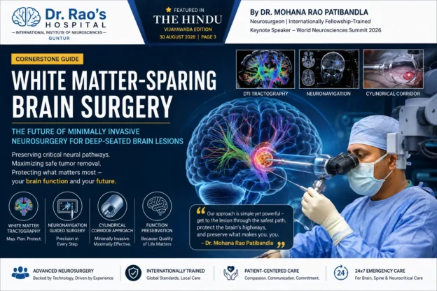
Dr. Mohana Rao Patibandla demonstrating White Matter-Sparing Brain Surgery using minimally invasive cylindrical corridor surgery and neuronavigation for deep brain lesions.

White Matter-Sparing Brain Surgery: Minimally Invasive Brain Surgery for Deep-Seated Brain Lesions | Dr. Mohana Rao Patibandla

White Matter-Sparing Brain Surgery: The Future of Minimally Invasive Neurosurgery for Deep-Seated Brain Lesions

Featured in The Hindu (Vijayawada Edition, 30 August 2026) following Dr. Mohana Rao Patibandla’s keynote presentation at the World Neurosciences Summit (WNS) 2026, New Delhi.

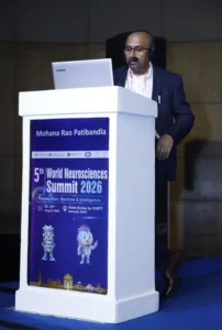
Dr. Mohana Rao Patibandla delivering the keynote lecture on White Matter-Sparing Brain Surgery at the 5th World Neurosciences Summit 2026 in New Delhi, India.

Dr. Mohana Rao Patibandla presenting his keynote lecture, “Minimally Invasive Cylindrical Corridor Surgery – Modern Microsurgery Strategy for Deep-Seated Brain Lesions,” at the 5th World Neurosciences Summit 2026 in New Delhi, highlighting advanced White Matter-Sparing Brain Surgery techniques for preserving critical neural pathways.
Cornerstone Article

White Matter-Sparing Brain Surgery: The Future of Minimally Invasive Neurosurgery for Deep-Seated Brain Lesions

Author: Dr. Mohana Rao Patibandla, Neurosurgeon, Founder & Chief Neurosurgeon, Dr. Rao’s Hospital – International Institute of Neurosciences, Guntur, Andhra Pradesh, India

Published: September 2026

Reading Time: 18–22 minutes

Target Keywords: White matter sparing surgery, minimally invasive brain surgery, cylindrical corridor surgery, deep-seated brain tumor surgery, brain lesion surgery, neurosurgeon India, Dr. Mohana Rao Patibandla.

Executive Summary

The human brain contains billions of neurons connected through highly organized white matter tracts—the communication highways that allow movement, speech, memory, vision, sensation, and cognition. Traditional surgery for deep-seated brain tumors or lesions often requires passing through healthy brain tissue, increasing the risk of neurological deficits.

White Matter-Sparing Surgery, also called Minimally Invasive Cylindrical Corridor Surgery, represents a transformative advancement in neurosurgery. Instead of cutting through healthy brain tissue, surgeons navigate between natural white matter pathways, preserving critical neural connections while reaching tumors or lesions deep inside the brain.

This technique was the focus of Dr. Mohana Rao Patibandla’s keynote lecture at the World Neurosciences Summit 2026, where he presented modern microsurgical strategies for safely treating deep-seated brain lesions. The approach was subsequently highlighted by The Hindu in a feature article discussing the future of minimally invasive brain surgery.

This comprehensive guide explains everything patients, families, neurologists, and neurosurgeons need to know about white matter-sparing surgery.

Table of Contents

  • What is White Matter-Sparing Brain Surgery?
  • Understanding White Matter and Brain Networks.
  • Why Traditional Brain Surgery Can Damage White Matter.
  • Evolution of Minimally Invasive Cylindrical Corridor Surgery.
  • How White Matter-Sparing Surgery Works.
  • Neuronavigation and Brain Mapping.
  • Who Needs White Matter-Sparing Surgery?
  • Conditions Treated.
  • Benefits Compared to Conventional Brain Surgery.
  • Surgical Planning and Technology.
  • Risks and Limitations.
  • Recovery After White Matter-Sparing Surgery.
  • Frequently Asked Questions.

Understanding White Matter: The Brain’s Information Highway

White matter tracts in the brain [IMAGE] | EurekAlert! Science News Releases

Diffusion Tensor Imaging 101 | Diffusion Imaging; Introduction, tutorials and background on diffusion tensor imaging and techniques

European Journal of Translational and Clinical Medicine

Most people think of the brain as a gray organ filled with neurons. However, white matter is equally important.

What is White Matter?

White matter consists of millions of insulated nerve fibers called axons. These fibers connect different brain regions and enable rapid communication.

White Matter Controls

Brain Function

Major White Matter Pathway

MovementCorticospinal tract
Speech productionArcuate fasciculus
Language comprehensionSuperior longitudinal fasciculus
VisionOptic radiations
MemoryFornix and cingulum
EmotionUncinate fasciculus

Damage to these pathways may result in:

  • Paralysis

  • Speech difficulty

  • Vision loss

  • Memory impairment

  • Cognitive dysfunction

  • Personality changes

Protecting these pathways has become one of the central goals of modern neurosurgery.

Why Traditional Brain Surgery Isn’t Always Enough

Neuroinfo Japan�F�J����p

Brain Pterional approach | UpSurgeOn

Minimally Invasive Brain Surgery | Keyhole Surgery | Pacific Brain Tumor Center

Historically, surgeons approached deep tumors by creating a path through the brain.

The Challenge

Deep lesions may lie:

  • Near motor pathways.

  • Adjacent to speech centers.

  • Beneath healthy cortex.

  • Close to visual pathways.

  • Near memory circuits.

Traditional surgery may require:

  • Brain retraction.

  • Cortical incision.

  • Traversing healthy tissue.

Even with excellent technique, these maneuvers may increase neurological risk.

Potential Consequences

Potential Injury

Possible Deficit

Motor tract injuryWeakness or paralysis
Language tract injurySpeech impairment
Visual tract injuryVisual field loss
Frontal network injuryBehavioral or cognitive changes

Modern neurosurgery seeks to avoid these injuries rather than simply treat them afterward.

What is White Matter-Sparing Surgery?

Our Integrated System - NICO Corporation

Surgical Neurology International

Frontiers | Minimally invasive treatment for glioblastoma through endoscopic surgery including tumor embolization when necessary: a technical note

Definition

White Matter-Sparing Surgery is a minimally invasive microsurgical technique that uses:

  • Natural anatomical corridors.

  • Cylindrical tubular retractors.

  • Advanced neuronavigation.

  • White matter tractography.

  • High-definition operating microscopy.

Instead of retracting large portions of the brain, surgeons gently separate white matter fibers along their natural orientation.

The Philosophy

Preserve normal brain. Remove abnormal tissue. Protect neural connectivity.

This is fundamentally different from older approaches focused primarily on lesion access.

The Science Behind Cylindrical Corridor Surgery

A New Way into the Brain | Johns Hopkins Medicine

Sobre Dr. Alexis Palpán

courses:456:2021:projects:456-2021-01:project-01 – CIIS Wiki

Why a Cylinder?

A cylindrical corridor distributes pressure evenly.

Traditional Retractor

Tumor retractor: a simple and novel instrument for brain tumor surgery | World Journal of Surgical Oncology | Springer Nature Link

Brain Pterional approach | UpSurgeOn

Principles of Intraventricular Surgery | Cohen Collection | Volumes | The Neurosurgical Atlas

  • Flat blades.

  • Uneven pressure.

  • Greater cortical retraction.

Cylindrical Corridor

  • Gentle circumferential dilation.

  • Less tissue distortion.

  • Smaller surgical footprint.

  • Better preservation of white matter fibers.

This concept has become increasingly adopted in minimally invasive neurosurgery worldwide.

How White Matter-Sparing Surgery Works

Step-by-Step Surgical Strategy

Stereotactic neurosurgery | MedLink Neurology

How Can New Imaging Modalities Help in the Practice of Radiology? - PMC

O nás | FNOL

Step

Purpose

1Advanced MRI with tractography identifies functional white matter pathways.
2Neuronavigation selects the safest trajectory.
3Small craniotomy is created.
4Tubular corridor gently separates white matter fibers.
5Microsurgical instruments remove lesion through the corridor.
6Hemostasis is achieved while preserving surrounding tissue.

The result is maximal safe lesion removal with minimal disruption.

White Matter Tractography: GPS for the Brain

Visualização Multimodal de Imagens de Tensores de Difusão | Visualização de Imagens de DTI

#mri #dti #corticospinal | Vahid Shahmaei

Correlation between language function and the left arcuate fasciculus detected by diffusion tensor imaging tractography after brain tumor surgery – Neurosurgery Blog

What is DTI Tractography?

Diffusion Tensor Imaging (DTI) visualizes white matter pathways before surgery.

It Helps Identify

  • Motor tracts.

  • Speech tracts.

  • Visual pathways.

  • Sensory pathways.

  • Memory networks.

Why It Matters

Surgeons can:

  • Avoid eloquent pathways.

  • Choose safer entry points.

  • Predict postoperative risks.

  • Tailor surgery to each patient.

This represents precision neurosurgery rather than generalized anatomy.

Neuronavigation: GPS Inside the Brain

Home - Neurochirurgie | Uniklinikum Erlangen

Удаление опухолей головного мозга с применением нейронавигации

Vall d'Hebron: un navegador para mejorar el diagnóstico con ecógrafo

Neuronavigation provides real-time anatomical guidance.

It Allows Surgeons To

  • Locate lesions accurately.

  • Avoid blood vessels.

  • Maintain surgical trajectory.

  • Navigate deep structures safely.

Combined with tractography, neuronavigation creates a personalized surgical roadmap.

Brain Mapping: Protecting Function During Surgery

Neurosurgeons are really good at removing brain tumors—they're about to get even better

Low Grade Glioma and Glioblastoma Outcomes Research | Brain Tumor Center

神经电生理监测在胶质瘤手术中的意义|运动|功能|神经|监测|手术|-健康界

Modern white matter-sparing surgery often incorporates:

Intraoperative Monitoring

  • Motor evoked potentials.

  • Somatosensory evoked potentials.

  • Cranial nerve monitoring.

  • Speech mapping when required.

This provides continuous feedback about neurological function.

Which Brain Conditions Can Be Treated?

Deep-Seated Brain Tumors

PPT - Clinical MRI Insights by Dr. Ravichandra: Brain and Vascular Imaging PowerPoint Presentation - ID:9635346

a, b Preoperative axial and sagittal T1-weighted MRI of posterior type... | Download Scientific Diagram

Examples include:

  • Gliomas

  • Metastatic tumors

  • Low-grade glioma

  • High-grade glioblastoma

  • Thalamic tumors

  • Basal ganglia tumors

Goal

Maximal safe resection while preserving neurological function.

Cavernomas

cerebral cavernous venous malformation | pacs

Brainstem Cavernoma - Neuropedia

海綿状血管腫 | 社会医療法人寿会 富永病院(大阪)

White matter-sparing corridors are valuable for:

  • Thalamic cavernoma.

  • Brainstem cavernoma.

  • Deep cerebral cavernomas.

Colloid Cysts

Checking your browser - reCAPTCHA

Surgical management of colloid cysts of the third ventricle: a single-institution comparison of endoscopic and microsurgical resection – Neurosurgery Blog

Checking your browser - reCAPTCHA

Small minimally invasive approaches reduce brain manipulation around the ventricles.

Intraventricular Tumors

intraventricular neoplasms and lesions | pacs

Central neurocytoma | Radiology Reference Article | Radiopaedia.org

Examples include:

  • Central neurocytoma.

  • Ependymoma.

  • Choroid plexus tumors.

  • Subependymoma.

Brain Abscesses

New Page 1

Infections of Brain Parenchyma I Flashcards | Quizlet

Intracranial Infections: Bacterial, Viral, Fungal, Parasitic -

Targeted tubular access allows drainage with limited cortical injury in selected patients.

Epilepsy Lesions

Displasia cortical focal en 7 Tesla - Mayo Clinic

Minimally Invasive Surgery for Cerebral Cavernous Malformations

Let the Lesion Do the Talking :: Behance

Certain deep epileptogenic lesions can be approached through white matter-preserving techniques.

Advantages Over Conventional Brain Surgery

Why White Matter-Sparing Surgery Matters

Traditional Surgery

White Matter-Sparing Surgery

Larger craniotomySmaller craniotomy
More brain retractionMinimal brain retraction
Cortical disruptionNatural white matter corridor
Greater tissue manipulationTargeted microsurgical access
Higher postoperative swellingReduced tissue trauma
Longer recoveryFaster recovery in selected patients

Key Patient Benefits

East Carolina Heart Institute at ECU Health Medical Center - Greenville, NC 27834

Dr. Alejandro Ceja Espinosa Neurocirujano, Morelia - Agenda cita | Doctoralia.com.mx

I've been diagnosed with a brain tumor. Now what? | Michigan Medicine

Potential benefits include:

  • Smaller incision.

  • Less pain.

  • Reduced blood loss.

  • Less brain swelling.

  • Better cosmetic outcome.

  • Earlier mobilization.

  • Shorter ICU stay in selected cases.

  • Preservation of neurological function.

These benefits depend on careful patient selection and surgical expertise.

Who is a Candidate?

Ideal Candidates

Suitable Patients

Why

Deep brain tumorsAccessible through safe anatomical corridors.
Small-to-medium lesionsMinimal disruption possible.
Functional area lesionsWhite matter preservation is especially valuable.
Selected hemorrhagesTubular evacuation may be appropriate in selected cases.
Deep cavernomasNatural corridor approach.

Patients Who May Need Conventional Surgery

  • Very large tumors.

  • Diffuse infiltrative lesions.

  • Extensive vascular malformations.

  • Multiple compartment lesions.

Every patient requires individualized evaluation.

Surgical Planning at Dr. Rao’s Hospital

Equipment and medical devices in modern operating room

3T MRI | Book Your Scan Today — Rezolut

Grupo Policlínica incorpora el microscopio más avanzado del mercado en su unidad de neurocirugía | Onda Cero Radio

A white matter-sparing operation begins long before entering the operating room.

Comprehensive Planning Includes

Advanced Imaging

3 Tesla MR Fiyatları 2026 | Fulya Açık Emar

브런치

How Can New Imaging Modalities Help in the Practice of Radiology? - PMC

  • MRI Brain

  • Contrast MRI

  • DTI tractography

  • Functional MRI when indicated.

  • MR angiography/venography.

Digital Planning

Frontiers | Neuronavigation in glioma resection: current applications, challenges, and clinical outcomes

Stryker Launches Q Guidance System with Cranial Guidance Software | OR Today

Brain Tumor Program | Neurosurgery

  • Surgical trajectory simulation.

  • White matter tract preservation.

  • Vascular avoidance.

  • Cortical entry planning.

Microsurgical Technology Used

High-End Neurosurgical Equipment

Doctors Hospital installs the first Kinevo 900 microscope in Mexico. - Doctors Hospital

M720 OH5 Operationsmikroskop der Spitzenklasse - Medien | Produkte | Leica Microsystems

Neurokirurgi: sätter standarden inom modern neurokirurg

Technology may include:

  • Operating microscope.

  • Neuronavigation.

  • Ultrasonic aspirator.

  • Endoscope-assisted visualization.

  • Neurophysiological monitoring.

  • High-definition imaging.

Each tool contributes to safer microsurgery.

White Matter-Sparing Surgery in Brain Tumor Management

Low-Grade Gliomas

The silent phase of diffuse low-grade gliomas. Is it when we missed the action? – Neurosurgery Blog

Subject-Specific Automatic Reconstruction of White Matter Tracts | Journal of Imaging Informatics in Medicine | Springer Nature Link

MRI Tractography - Brain Tumor

Goal:

  • Preserve language.

  • Preserve cognition.

  • Maximize tumor removal.

High-Grade Glioblastoma

Glioblastoma, IDH-wildtype | Radiology Reference Article | Radiopaedia.org

Checking your browser - reCAPTCHA

Brain Tumour Resection Surgery Johannesburg

Goal:

  • Maximal safe resection.

  • Protect functional pathways.

  • Improve quality of life.

White Matter Preservation in Pediatric Neurosurgery

Tumores cerebrales en niños | Pediatría integral

Zabieg usunięcia olbrzymiego guza mózgu u miesięcznego dziecka w IPCZD - Aktualności - Centrum Zdrowia Dziecka

Pediatric Hemato-Oncology Department – TAMC

Children have developing neural pathways.

Advantages include:

  • Better developmental preservation.

  • Reduced tissue injury.

  • Smaller surgical footprint.

  • Functional protection during growth.

White Matter-Sparing Surgery for Brainstem Lesions

Cavernomas do Sistema Nervoso Central :: Clinica Maddalozzo

511-2018-09-05-anatomy-II

【北总神外论道 】经安全区切除脑干海绵状血管瘤系列2 - 脑医汇

Brainstem surgery is among the most challenging areas in neurosurgery.

The technique focuses on:

  • Safe entry zones.

  • Natural fiber orientation.

  • Cranial nerve preservation.

  • Microsurgical precision.

Recovery After White Matter-Sparing Surgery

What Patients Can Expect

Patient’s ER visit turns into brain surgery, cancer diagnosis - UNC Lineberger

Post-Craniotomy Recovery & Care in Pune

Consultation after MRI examination in medical clinic. MRI Doctor is showing x-ray and of brain tumors in the patient head.

Hospital Stay

Stage

Typical Course

ICU1 day in many selected cases.
Ward2–5 days depending on condition.
WalkingOften within 24 hours if neurologically appropriate.
Return HomeDepends on recovery and pathology.

Recovery varies by diagnosis, lesion location, age, and overall health.

Rehabilitation

Speech Therapy After Stroke - Sheltering Arms Institute

Fisioterapia Neuro Funcional: Restaure a Qualidade de Vida com Corpo Ativo

Neurologie — Ergothérapie de la Prévôté

Some patients benefit from:

  • Physiotherapy.

  • Speech therapy.

  • Occupational therapy.

  • Cognitive rehabilitation.

Risks and Limitations

No brain surgery is risk-free.

Potential Risks

EPOS™

Continuous EEG Monitoring | Nihon Kohden

Checking your browser - reCAPTCHA

  • Bleeding.

  • Infection.

  • Seizures.

  • Neurological deficits.

  • Brain swelling.

  • CSF leak.

The goal of white matter-sparing surgery is to reduce unnecessary injury—not eliminate all surgical risk.

Evidence Supporting White Matter-Sparing Approaches

Why This Technique is Growing Worldwide

Modern literature supports:

  • Less cortical injury.

  • Improved preservation of white matter tracts.

  • Better functional outcomes in selected lesions.

  • Reduced surgical morbidity in carefully chosen patients.

International adoption continues to increase alongside advances in imaging and neuronavigation.

Dr. Mohana Rao Patibandla’s Keynote at World Neurosciences Summit 2026

NeuroScience Frontiers Symposium 2025 | Neurogen Therapeutic

Groundbreaking Research on Minimally Invasive Brain Tumor Surgery Presented by Dr. Rao at Andhra Pradesh Neuroscientists Association Conference

Society for Neuroscience - Neuroscience 2026

At the World Neurosciences Summit 2026, Dr. Mohana Rao Patibandla presented a keynote lecture titled:

“Minimally Invasive Cylindrical Corridor Surgery – Modern Microsurgery Strategy for Deep-Seated Brain Lesions.”

The presentation emphasized:

  • White matter preservation.

  • Modern microsurgical planning.

  • Neuronavigation-guided access.

  • Safe treatment of deep brain lesions.

  • Functional outcome optimization.

The lecture highlighted the growing importance of minimally invasive microsurgical corridors in contemporary neurosurgery.


Featured in The Hindu: National Recognition of White Matter-Sparing Surgery

The Hindu reported Dr. Mohana Rao Patibandla’s press conference explaining how preserving normal brain tissue and neural pathways has become a key objective of modern minimally invasive microsurgery. The article also described cylindrical corridor surgery as a white matter-sparing strategy for deep-seated brain lesions.


This coverage reflects growing public awareness of advanced brain-preserving surgical techniques in India.

Why This Matters for Patients in India

Advanced Brain & Spine Neurosurgery Treatments in Chennai

Competent medical expert explaining diagnosis

Neurologist in Pimpri & Brain Specialist in Pimpri-Chinchwad

Patients increasingly seek:

  • Safer surgery.

  • Smaller incisions.

  • Faster recovery.

  • Preservation of neurological function.

  • International-standard neurosurgical care within India.

White matter-sparing surgery aligns with these goals when clinically appropriate.

Why Choose Dr. Rao’s Hospital for White Matter-Sparing Brain Surgery?

Best Neurosurgery & Spine Care at Dr. Rao’s Hospital, Guntur

Brain Surgery in Pune | Dr. Jaydev Panchawagh, Neurosurgeon

Home - Neurochirurgie | Uniklinikum Erlangen

Dr. Rao’s Hospital – International Institute of Neurosciences, Guntur, offers comprehensive brain and spine care with advanced neurosurgical technology and internationally trained expertise.

Areas of Expertise

  • Brain Tumor Surgery

  • Skull Base Surgery

  • Endoscopic Brain Surgery

  • Pediatric Neurosurgery

  • Cerebrovascular Surgery

  • Epilepsy Surgery

  • Functional Neurosurgery

  • Minimally Invasive Neurosurgery

Advanced Technologies

  • Operating Microscope

  • Neuronavigation

  • Neuroendoscopy

  • Neurophysiological Monitoring

  • Advanced MRI Planning

  • Minimally Invasive Brain Surgery

Frequently Asked Questions (FAQ)

1. What is White Matter-Sparing Brain Surgery?

White matter-sparing brain surgery is a minimally invasive microsurgical approach that reaches deep brain lesions through natural white matter corridors while minimizing injury to healthy neural pathways.

2. What is Cylindrical Corridor Surgery?

It is a minimally invasive technique using a tubular retractor to create a gentle corridor through the brain, allowing surgeons to access deep lesions with less tissue disruption.

3. Is White Matter-Sparing Surgery safer than traditional brain surgery?

It may reduce disruption to healthy brain tissue in carefully selected patients, but safety depends on lesion type, location, patient health, and surgical expertise.

4. Which brain tumors can be treated?

Selected gliomas, metastases, intraventricular tumors, cavernomas, colloid cysts, and certain deep-seated lesions may be suitable candidates.

5. What is tractography?

Tractography is an MRI technique (DTI) that maps white matter pathways before surgery to help surgeons avoid critical neural connections.

6. Does every patient need tractography?

Not always. It is especially useful when lesions are close to eloquent brain pathways.

7. Is recovery faster?

Many selected patients experience smaller incisions, less pain, and earlier mobilization, although recovery varies with the underlying disease and surgery performed.

8. Is this surgery available in India?

Yes. Advanced minimally invasive white matter-sparing brain surgery is available in specialized neuroscience centers including Dr. Rao’s Hospital in Guntur.

9. Can brain function be preserved completely?

The goal is maximal preservation of neurological function, but outcomes depend on lesion location, disease biology, and individual patient factors.

10. How do I know if I’m a candidate?

A neurosurgical consultation with MRI review, tractography when indicated, and individualized surgical planning is necessary.

Key Takeaways

White matter tracts in the brain [IMAGE] | EurekAlert! Science News Releases

A team of surgeons performing brain surgery to remove a tumor.

Elements Fibertracking | Brainlab

White Matter-Sparing Surgery represents one of the most significant advances in modern neurosurgery because it shifts the surgical philosophy from simply reaching a lesion to protecting the brain’s communication networks while treating disease.

For carefully selected patients with deep-seated brain tumors, cavernomas, intraventricular lesions, and other complex pathologies, minimally invasive cylindrical corridor surgery offers a highly precise approach centered on preserving neurological function whenever possible.

As highlighted during Dr. Mohana Rao Patibandla’s keynote lecture at the World Neurosciences Summit 2026 and subsequently featured in The Hindu, the future of neurosurgery lies in combining advanced imaging, neuronavigation, microsurgical precision, and white matter preservation to achieve better patient outcomes.


About the Author

Dr. Mohana Rao Patibandla is an internationally fellowship-trained neurosurgeon and the Founder & Chief Neurosurgeon of Dr. Rao’s Hospital – International Institute of Neurosciences, Guntur, Andhra Pradesh, India.

His advanced training includes skull base surgery, pediatric neurosurgery, minimally invasive brain surgery, neuro-oncology, cerebrovascular and endovascular neurosurgery, and functional neurosurgery in India and the United States. He regularly serves as invited faculty at national and international neuroscience conferences and delivered a keynote lecture at the World Neurosciences Summit 2026 on White Matter-Sparing Minimally Invasive Cylindrical Corridor Surgery.


Authoritative Neurosurgical Resources

Patients with deep-seated gliomas, meningiomas, metastatic tumors, and other complex lesions may benefit from

Brain Tumor Surgery 
performed using modern white matter-sparing microsurgical techniques. Many lesions located near the skull base require specialized Skull Base Surgery 
combined with advanced neuronavigation and minimally invasive approaches.

Our comprehensive Neuro-Oncology Program focuses on personalized treatment for benign and malignant brain tumors. Children with deep-seated brain tumors may benefit from advanced Pediatric Neurosurgery that prioritizes preservation of developing neural pathways.

Selected intraventricular tumors and colloid cysts can often be treated using Endoscopic Brain Surgery or minimally invasive cylindrical corridor techniques.

White matter-sparing surgery relies heavily on Neuronavigation-Guided Brain Surgery 
to identify the safest surgical pathway while protecting eloquent brain regions.

Patients with epilepsy caused by cavernomas or focal cortical lesions may also be candidates for Epilepsy Surgery using minimally invasive neurosurgical strategies.

Learn more about the international training, experience, research, and surgical philosophy of Dr. Mohana Rao Patibandla Founder of Dr. Rao’s Hospital – International Institute of Neurosciences.

YouTube

Facebook

Instagram

Google Business Page

LinkedIn

X / Twitter

Need Expert Care for a Brain Tumor or Deep-Seated Brain Lesion?

At Dr. Rao’s Hospital – International Institute of Neurosciences (IIN),
we provide advanced White Matter-Sparing Brain Surgery,
Minimally Invasive Brain Tumor Surgery,
Skull Base Surgery, Endoscopic Brain Surgery,
Neuro-Oncology, Pediatric Neurosurgery,
Stroke Care, and Spine Surgery using internationally
recognized technology and evidence-based treatment protocols.

Every patient receives a personalized surgical plan based on advanced MRI,
neuronavigation, white matter tractography, intraoperative neuromonitoring,
and minimally invasive microsurgical techniques designed to preserve neurological function whenever possible.


Why Choose Dr. Rao’s Hospital?

  • Internationally Fellowship-Trained Neurosurgeon – Dr. Mohana Rao Patibandla
  • Dedicated Super-Specialty Neurosciences Hospital in Guntur, Andhra Pradesh.
  • Advanced White Matter-Sparing & Minimally Invasive Brain Surgery.
  • Brain Tumor, Skull Base, Pediatric, Spine, Stroke & Endovascular Neurosurgery.
  • Neuronavigation, Neurophysiological Monitoring & High-End Microsurgical Technology.
  • Comprehensive Neuro-Oncology, Neurocritical Care and Rehabilitation.
  • 24×7 Emergency Brain, Stroke and Spine Care.

Contact Dr. Rao’s Hospital

Dr. Rao’s Hospital – International Institute of Neurosciences (IIN)
12-19-67, Old Bank Road, Kothapet,
Opposite Sravani Hospital, Besides AK Biryani point,
Guntur – 522001, Andhra Pradesh, India.

📞 +91 90100 56444
✉️ info@drraoshospitals.com
🌐 www.drraoshospitals.com

India’s Best Neurosurgeon — Dr. Rao at Dr. Rao's Hospital

Are Spine Surgeons at Risk Through Innovation?

Are spine surgeons at risk through innovation?

In the ever-evolving field of medicine, innovation is a double-edged sword. Spine surgery, a discipline once dependent on open surgical techniques, has seen tremendous technological advancements. From robotic-assisted procedures and minimally invasive techniques to artificial intelligence (AI) and 3D-printed implants, these innovations promise better outcomes, reduced recovery times, and fewer complications. But as these cutting-edge tools continue to shape the future of spine surgery, they raise an important question: Are spine surgeons themselves at risk of being overshadowed or rendered obsolete by these technological advancements? According to The Times of India coverage.

The Role of Innovation in Spine Surgery

Over the past few decades, spine surgery has undergone significant transformation.

Minimal invasive techniques:

These reduce the physical toll on patients by limiting incision size and muscle damage, leading to faster recovery times.

Robotics and Navigation Systems:

Tools like robotic-assisted surgery systems enhance precision, enabling surgeons to perform complex procedures more accurately.

Artificial Intelligence:

AI-driven diagnostics and surgical planning are making preoperative decision-making faster and more accurate.

3D Printing:

Customized implants and anatomical models help surgeons tailor procedures to individual patients.

These advancements empower spine surgeons to offer safer, more efficient procedures but also demand adaptation and continuous learning.

Are spine surgeons at risk?

1. Job Displacement vs. Role Evolution
While innovations like robotics and AI have automated some aspects of surgery, they cannot replace the expertise and judgment of a skilled spine surgeon. These tools are designed to assist, not replace, the surgeon. However, surgeons who fail to adapt to these advancements risk becoming less competitive.

2. The Learning Curve
Technological progress demands a continuous upgrade in skills. Surgeons must invest significant time and resources to master these new tools. Those unwilling or unable to embrace these changes may find their roles marginalized.

3. Ethical and Legal Challenges
Innovations come with ethical considerations, such as patient consent for AI-guided procedures or the risks of relying too heavily on technology. Spine surgeons must navigate these challenges to remain at the forefront of patient care.

4. Financial Pressures
Adopting new technology often involves substantial financial investment. Surgeons and hospitals must weigh the costs of acquiring and maintaining cutting-edge equipment against the benefits for their patients.

The Value of the Human Element

Despite these challenges, the human touch remains irreplaceable. Spine surgery often involves nuanced decision-making considering a patient’s unique anatomy, medical history, and personal preferences. Surgeons bring empathy, communication, and adaptability—qualities no machine can replicate.

Moreover, innovations like robotics require skilled operators, and AI-generated insights are only as good as the data they’re based on. Spine surgeons are the ultimate authority, interpreting technology-driven recommendations and making critical decisions.

Embracing Innovation for the Future

To thrive in this age of rapid advancement, spine surgeons must:

Commit to Lifelong Learning: Attend training sessions, workshops, and conferences to stay updated on the latest tools and techniques.
Collaborate with Innovators: Partner with engineers, AI specialists, and medical technology developers to ensure that new tools align with clinical realities.
Advocate for Balanced Integration: Use technology as an aid, not a substitute, ensuring that the surgeon remains central to patient care.
Focus on Holistic Patient Care: Maintain strong patient relationships by emphasizing empathy and trust, which technology alone cannot provide.

Conclusion

Innovation in spine surgery is not a threat but an opportunity. While technology continues to evolve, it does not diminish the importance of skilled surgeons who can adapt and integrate these tools into their practice. Spine surgeons who embrace innovation while staying grounded in their clinical expertise and human connection will remain relevant and lead the way in delivering exceptional patient care.

The key to success is understanding that technology is a tool, not a replacement. By leveraging innovation wisely, spine surgeons can enhance their practice, improve patient outcomes, and secure their role as indispensable professionals in the healthcare landscape.

Dr. Rao’s Hospital continues to pioneer the integration of innovation and expertise in spine surgery, ensuring that our patients benefit from the best of both worlds. For more information, visit us at drraoshospitals.com or contact us at 9010056444.

Dr. Rao’s Contact Information:

Dr. Rao’s Hospital: Medical Tourism for Economical Neurosurgery in India

Best Hospital for Brain Surgery in India: Dr. Rao’s Hospital, Guntur

Best Hospital for Brain Surgery in India: Dr. Rao’s Hospital, Guntur

When it comes to Brain Surgery, choosing the right hospital and neurosurgeon is crucial for ensuring the best outcomes and care. In India, Dr. Rao’s Hospital in Guntur has emerged as a leading center for brain surgery, known for its exceptional medical expertise, advanced technology, and compassionate patient care. According to The Times of India coverage.

Why Dr. Rao’s Hospital Stands Out

1. Expertise of Dr. Mohana Rao Patibandla

Dr. Mohana Rao Patibandla, the Chief Neurosurgeon at Dr. Rao’s Hospital, is a highly qualified and experienced neurosurgeon. With extensive training in brain surgery and a proven track record of successful procedures, Dr. Rao is renowned for his precision and expertise in treating complex brain conditions. According to dnaindia

2. Comprehensive Range of Brain Surgeries

Dr. Rao’s Hospital offers a wide range of brain surgeries, including: In a recent India today article.

  • Brain Tumor Surgery: Utilizing advanced techniques to remove brain tumors while preserving neurological function safely.
  • Stereotactic Radiosurgery: A non-invasive option for treating brain tumors and other abnormalities with high precision.
  • Aneurysm Clipping and Coiling: Effective treatments for brain aneurysms to prevent rupture and manage complications.
  • Epilepsy Surgery: Procedures to control seizures and improve the quality of life for epilepsy patients.
  • Traumatic Brain Injury (TBI) Surgery: Specialized care for patients with severe head injuries.

3. State-of-the-Art Technology

Dr. Rao’s Hospital is equipped with the latest medical technology, ensuring patients receive the most advanced and effective treatments. This includes high-resolution imaging systems, intraoperative MRI, neuronavigation systems, and minimally invasive surgical tools, all of which contribute to better surgical outcomes and faster recovery times.

4. Personalized Patient Care

At Dr. Rao’s Hospital, patients receive personalized care tailored to their conditions and needs. The multidisciplinary team collaborates with neurologists, neurosurgeons, radiologists, and rehabilitation specialists to develop comprehensive treatment plans. This holistic approach ensures that all patient health aspects are addressed.

5. High Success Rates and Patient Satisfaction

Dr. Rao’s Hospital boasts high success rates for brain surgeries, with numerous patients reporting significant improvements in their condition and quality of life. Patient testimonials highlight the hospital’s commitment to excellence, compassionate care, and the positive outcomes achieved under Dr. Rao’s expertise.

6. Accessibility and Convenient Location

Dr. Rao’s Hospital is located in the heart of Guntur, Andhra Pradesh, and is easily accessible to patients from across India and abroad. The hospital’s efficient services and streamlined processes ensure minimal wait times and a smooth patient and family experience.

7. Comprehensive Support and Aftercare

Recovery from brain surgery requires ongoing support and rehabilitation. Dr. Rao’s Hospital provides comprehensive aftercare services, including physical therapy, occupational therapy, and psychological support, to help patients regain their independence and improve their overall well-being.

Testimonials from Satisfied Patients

Mr. Suresh Kumar: “I was diagnosed with a brain tumor and was terrified. Dr. Rao and his team at Dr. Rao’s Hospital provided exceptional care and support throughout my treatment. The surgery was successful, and I am now tumor-free. I am grateful for the expertise and compassion shown by Dr. Rao and the entire staff.”

Ms. Anjali Verma: “After suffering from debilitating seizures for years, I underwent epilepsy surgery at Dr. Rao’s Hospital. The results have been life-changing. I am now seizure-free and can lead a normal life. Dr. Rao is truly a life-saver.”

Contact Information

Dr. Rao’s Hospital in Guntur offers unparalleled expertise and advanced treatment options for those seeking the best care in brain surgery.

Contact Information:

Choosing the right hospital for brain surgery can significantly improve outcomes and quality of life. With its expert team, state-of-the-art technology, and patient-centered approach, Dr. Rao’s Hospital in Guntur is the best hospital for brain surgery in India. Trust your health to the best and take the first step towards recovery with Dr. Rao’s Hospital.

Stroke Care: The best stroke treatment by Dr. Raos Hospital

The Role of Neurosurgery in Stroke Management and Recovery: Dr. Rao’s Hospital Leading the Way

The Role of Neurosurgery in Stroke Management and Recovery: Dr. Rao’s Hospital Leading the Way

Introduction 

stroke is a devastating condition that requires prompt medical attention and specialized care. In recent years, neurosurgery has emerged as a critical component in the management and recovery of stroke patients. In this blog, we explore the significant role of neurosurgery in stroke care, specifically focusing on Dr. Rao’s Hospital, recognized as the best comprehensive stroke center. The hospital excels at offering cutting-edge neurosurgical interventions that optimize outcomes and improve patients’ quality of life under the direction of Dr. Rao, a renowned endovascular and cerebrovascular neurosurgeon with dural training.

Understanding Stroke: Causes, Types, and Impact

To comprehend the role of neurosurgery in stroke management, it is essential to first understand the causes, types, and impact of strokes. Ischemic strokes, caused by blocked blood vessels, and hemorrhagic strokes, resulting from ruptured blood vessels, require different treatment approaches. Dr. Rao’s expertise in cerebrovascular neurosurgery enables precise diagnosis and tailored interventions to address the specific type and location of the stroke.

Comprehensive Stroke Center: A Hub for Specialized Care :

Dr. Rao’s Hospital is a comprehensive stroke center, offering a multidisciplinary approach to stroke care. The hospital’s state-of-the-art facilities, advanced imaging technologies, and dedicated stroke teams ensure swift and accurate diagnosis, allowing for timely intervention. Dr. Rao‘s leadership and expertise in dural-trained cerebrovascular neurosurgery further enhance the hospital’s capabilities in providing specialized care for stroke patients.

Benefits of a Dural-Trained Cerebrovascular Neurosurgeon :

The presence of a dural-trained cerebrovascular neurosurgeon, such as Dr. Rao, within the stroke center brings invaluable benefits to patients. Dural-trained neurosurgeons have extensive experience in managing complex cerebrovascular conditions, including stroke. Their specialized training equips them with the expertise to perform delicate procedures, such as endovascular interventions, aneurysm repair, and arteriovenous malformation (AVM) treatment, with exceptional precision and minimal risk.

Endovascular Interventions: Advancing Stroke Treatment :

Endovascular interventions have revolutionized stroke treatment, allowing neurosurgeons to access and treat affected blood vessels from within using minimally invasive techniques. Dr. Rao’s proficiency in endovascular procedures enables him to remove blood clots, restore blood flow, and minimize brain damage in stroke patients. This advanced approach offers benefits such as reduced procedural risks, shorter recovery times, and improved long-term outcomes.

Rehabilitation and Recovery Strategies :

While acute stroke care is vital, post-stroke rehabilitation is equally critical for patients’ recovery. Dr. Rao’s Hospital adopts a comprehensive approach encompassing neurosurgical interventions, rehabilitation services, and ongoing support. Rehabilitation programs, including physical therapy, speech therapy, and occupational therapy, aim to restore functionality, promote independence, and improve the overall quality of life for stroke survivors.

Empowering Patients and Caregivers :

Dr. Rao’s Hospital empowers patients and their caregivers throughout the stroke care journey. Educational resources, support groups, and counseling services are provided to ensure that individuals and their families understand the condition, treatment options, and strategies for long-term management. Dr. Rao’s Hospital fosters a patient-centric environment that prioritizes individualized care and improved outcomes by fostering open communication and collaboration.

Conclusion: Neurosurgery is pivotal in stroke management and recovery, offering advanced interventions that optimize outcomes and enhance patients’ quality of life.

Dr. Rao’s Hospital Leading the Way

Dr. Rao’s Hospital, recognized as the best comprehensive stroke center, stands at the forefront of stroke care. Dr. Rao is a renowned dural-trained endovascular and cerebrovascular neurosurgeon who leads the way. With his expertise and specialized training, Dr. Rao brings many benefits to stroke patients, ensuring the highest standard of care and optimal treatment outcomes.

By combining the latest advancements in neurosurgical techniques with a multidisciplinary approach, Dr. Rao’s Hospital offers a comprehensive range of interventions for stroke management. This includes endovascular procedures, where Dr. Rao’s skills shine. These allow him to navigate the intricate blood vessels of the brain with exceptional precision. Through techniques such as thrombectomy, stent placement, and coiling, he can effectively restore blood flow, minimize brain damage, and improve patient outcomes.

One significant advantage of having a dural-trained cerebrovascular neurosurgeon like Dr. Rao is his ability to handle complex cerebrovascular conditions that may accompany strokes, such as aneurysms and arteriovenous malformations (AVMs). With his specialized training and expertise, Dr. Rao can offer targeted interventions, including surgical clipping or endovascular coiling, to effectively treat these conditions and prevent future complications.

The role of rehabilitation and recovery cannot be understated in the journey of stroke patients. Dr. Rao’s Hospital recognizes the importance of comprehensive rehabilitation strategies tailored to individual needs. Collaborating with a team of skilled therapists and rehabilitation specialists, the hospital provides physical, speech, and occupational therapy to aid recovery. This integrated approach ensures stroke survivors regain functionality, improve mobility, and regain independence, ultimately enhancing their overall quality of life.

Dr. Rao’s Hospital prioritizes patient and caregiver empowerment throughout the stroke care journey. By providing educational resources, support groups, and counseling services, the hospital ensures that individuals and their families can access the knowledge and support necessary to navigate stroke-related challenges. Dr. Rao’s team fosters a compassionate and patient-centric environment where open communication and shared decision-making empower patients to participate actively in their recovery.

In conclusion

the best comprehensive stroke center’s neurosurgery department is crucial in stroke management and recovery. Patients who suffer from stroke receive the best care possible thanks to the knowledge of a cerebrovascular neurosurgeon with dural training and the comprehensive approach provided by Dr. Rao’s Hospital. They also gain access to cutting-edge interventions, effective rehabilitation techniques, and ongoing support. With Dr. Rao at the helm, the hospital remains dedicated to improving the lives of stroke patients and their families, driving positive outcomes in stroke management and enhancing overall well-being.

Dr. Rao’s Contact Information:

#Neurosurgery #StrokeManagement #StrokeRecovery #DrRao #DrRaosHospital #ComprehensiveStrokeCenter #DuralTrainedNeurosurgeon #EndovascularInterventions #Rehabilitation #PatientCare #OptimalOutcomes #NeurologicalConditions #BrainHealth #MedicalAdvancements

the-best-keyhole-surgery-for-brain-tumors-at-dr-raos hospital, Guntur

The best Keyhole Neurosurgery for brain and Spine at Dr Rao’s.


Keyhole NeuroSurgery: Pain-Free Living

Imagine treating complex brain and spine problems through tiny incisions, minimal pain, and faster recovery. That’s the promise of keyhole neurosurgery—also called minimally invasive endoscopic or microsurgical approaches. At Dr. Rao’s Hospital, Guntur, Dr. Mohana Rao Patibandla performs keyhole brain and spine surgery using advanced optics, neuronavigation, and tubular retractors to help patients return to normal life sooner. If you’re searching for keyhole spine surgery near me in Guntur or looking for a keyhole neurosurgeon in Andhra Pradesh, you’re in the right place.


What Is Keyhole Neurosurgery?

Keyhole neurosurgery uses tiny, precisely planned openings—often 0.8–2.5 cm—to access deep brain or spine targets while preserving normal tissue. Surgeons use endoscopes or operating microscopes, plus neuronavigation (a GPS for the brain/spine) to reach lesions through the safest corridors. This philosophy applies across specialties: keyhole endoscopic brain surgery, keyhole microdiscectomy, keyhole TLIF (transforaminal lumbar interbody fusion), and keyhole foraminotomy.

Why “Keyhole” Works

  • Less muscle and bone disruption → less pain and blood loss.
  • Shorter hospital stay (often 24–48 hours).
  • Faster recovery with earlier mobilization.
  • Smaller scars with better cosmetic outcomes.

[Source: Mayo Clinic – Minimally Invasive Neurosurgery]


Conditions Treated with Keyhole Brain & Spine Surgery

Keyhole Brain Surgery

  • Brain tumors (e.g., meningiomas, gliomas, metastases) – keyhole brain tumor surgery Guntur.
  • Pituitary adenomas via keyhole pituitary surgery (endonasal endoscopic approach).
  • Trigeminal neuralgia – microvascular decompression via a keyhole craniotomy.
  • Aneurysms – select cases via keyhole craniotomy (keyhole surgery for aneurysm).
  • Colloid cysts & intraventricular lesions via endoscopic corridors.

Keyhole Spine Surgery

  • Herniated disc: keyhole microdiscectomy or keyhole endoscopic discectomy.
  • Spinal stenosis: keyhole foraminotomy or decompression.
  • Instability or spondylolisthesis: keyhole TLIF or keyhole lumbar fusion.
  • Spinal tumors (selected cases) – keyhole surgery for spinal tumors Dr Rao.

At our dedicated Spine Surgery and Neurosurgery units, these procedures are performed with IONM (intraoperative neuromonitoring), high-definition endoscopy, and neuronavigation.

[Source: PubMed – Outcomes of Keyhole/Endoscopic Spine and Skull Base Surgery]


Who Is an Ideal Candidate?

Keyhole approaches suit many—but not all—patients. Suitability depends on tumor size/location, spinal alignment, bone quality, prior surgeries, and overall health. During consultation at Neurology Services or Neurosurgery Department, you’ll undergo clinical evaluation and imaging (MRI/CT). The goal is to choose the least invasive route that delivers the best outcome.

ConditionTypical Keyhole OptionPotential Benefits
Herniated Lumbar DiscKeyhole microdiscectomy / endoscopic discectomySame-day/overnight stay, rapid pain relief, tiny incision
Lumbar Canal StenosisKeyhole decompression/foraminotomyLess muscle damage, early walking, lower blood loss
SpondylolisthesisKeyhole TLIF / keyhole lumbar fusionStability restoration with muscle-sparing access
Pituitary AdenomaEndoscopic endonasal keyhole pituitary surgeryNo scalp incision, short stay, fast recovery
Trigeminal NeuralgiaKeyhole microvascular decompressionDurable pain relief via small retrosigmoid window

[Background reading: NIH – Minimally Invasive Neurosurgical Techniques]


Keyhole Neurosurgery at Dr. Rao’s Hospital, Guntur

Dr. Mohana Rao Patibandla brings global expertise to Andhra Pradesh, offering:

  • Keyhole spine surgery Guntur (endoscopic discectomy, foraminotomy, TLIF, lumbar fusion).
  • Keyhole brain surgery (pituitary, skull base, trigeminal neuralgia, selected aneurysms, brain tumors).
  • Neuronavigation & IONM for precision and safety.
  • Enhanced Recovery After Surgery (ERAS) protocols for quicker rehabilitation.

Explore our official website and learn more about Dr. Mohana Rao, widely regarded as a top keyhole neurosurgeon in Guntur and a best keyhole spine surgeon in India for patient-centric, minimally invasive solutions.


Benefits Patients Love (Backed by Evidence)

  • Less pain & faster mobility: Keeping muscles intact reduces postoperative pain and speeds walking.
  • Shorter hospital stay: Many spine patients go home within 24–48 hours.
  • Lower infection & blood loss risks compared with open procedures.
  • Earlier return to work and daily activities—often within 2–6 weeks depending on the procedure.

[Global context: WHO – Surgical Safety]


Keyhole Spine: What to Expect Before, During & After

Before Surgery

  • Clinical exam, MRI/CT, blood work.
  • Medication review (blood thinners, diabetes control, etc.).
  • Prehab: gentle walking, core activation, nutrition.

During Surgery

  • Tiny incision; tubular retractor/endoscope; image guidance.
  • Real-time nerve monitoring (IONM) for safety.
  • Typical duration: 45–150 minutes depending on complexity.

After Surgery

  • Mobilization within hours; light diet same day.
  • Analgesics and early physiotherapy.
  • Return to desk work: 1–3 weeks (microdiscectomy/foraminotomy), 3–6 weeks (fusion/TLIF) as advised.

See our Spine Surgery page for recovery tips and timelines tailored to keyhole surgery recovery time in Guntur.


Keyhole Brain Surgery: Common Patient Questions

Is keyhole brain surgery safe?

When performed by experienced teams using navigation and high-definition visualization, keyhole approaches are designed to reduce surgical risk by minimizing brain retraction and preserving normal tissue.

Which brain tumors qualify?

Many meningiomas, pituitary adenomas, metastases, and select gliomas can be addressed by keyhole routes. Your MRI determines the safest corridor—precisely the expertise at our Neurosurgery Department.

When can I go home?

Many keyhole brain procedures use short ICU observation with discharge in 1–3 days, depending on the case and recovery milestones.


Comparing Keyhole vs. Conventional Open Surgery

AspectKeyhole ApproachTraditional Open Approach
Incision & DissectionTiny incision; muscle-sparingLarger incision; more muscle dissection
Pain & Blood LossLess pain; minimal blood lossMore postoperative discomfort
Hospital StayOften 1–2 daysFrequently 3–5+ days
Return to Work2–6 weeks (procedure-dependent)6–12 weeks
Scar & CosmesisSmall scar; better cosmetic resultLarger scar

Procedures We Offer (Highlights)

1) Keyhole Microdiscectomy / Endoscopic Discectomy

Ideal for herniated disc with leg pain (sciatica). Patients often walk the same day and return home within 24 hours. Popular search terms include keyhole discectomy Guntur and keyhole surgery for herniated disc Guntur.

2) Keyhole Foraminotomy / Decompression for Stenosis

Targets narrow nerve channels causing neurogenic claudication or radiating pain. Keyhole access releases the compression with minimal muscle trauma—helpful for outcomes and recovery speed.

3) Keyhole TLIF / Keyhole Lumbar Fusion

Stabilizes painful spondylolisthesis or instability. Through a small corridor, disc is cleared, cage inserted, and screws placed percutaneously. Known locally as keyhole TLIF surgery Dr Patibandla and keyhole lumbar fusion Guntur.

4) Keyhole Endonasal Pituitary Surgery

Endoscopic route via nostrils—no scalp incision. Treats hormone-secreting tumors and vision-threatening macroadenomas with short stay and quick recovery.

5) Keyhole Retrosigmoid Craniotomy (Trigeminal Neuralgia/MVD)

Through a small behind-the-ear window, the offending vessel is separated from the trigeminal nerve—restoring pain-free living for many patients with classical TN.


Recovery Timeline & Rehabilitation

Recovery is individualized, but these general timelines apply to most patients at our keyhole spine and brain center in Guntur:

  • Day 0–1: Walk with assistance, start oral diet, pain control.
  • Week 1–2: Gentle walking; desk work often possible in microdiscectomy and foraminotomy.
  • Week 3–6: Return to routine activities; light core rehab; fusion patients ramp up more gradually.
  • 6–12 weeks: Resume sports/strenuous activity as advised.

Our physiotherapists craft a custom plan for posture, flexibility, and core strength—key to long-term success.


Cost, Access & Why Patients Choose Us

  • Transparent care: We discuss keyhole neurosurgery cost ranges up front.
  • Advanced tech: Endoscopes, microscopes, navigation, IONM.
  • Experience: Hundreds of keyhole brain and spine procedures under Dr. Rao.
  • Reviews: “Dr Rao keyhole neurosurgery reviews” mention quick recovery and compassionate care.

Ready to discuss your case? Contact Dr. Rao’s Hospital for an appointment or second opinion.


Safety, Evidence, and Global Best Practices

Safety comes from planning, precision, and protocols—not just small incisions. We follow international guidelines and continuously audit outcomes.


When Should You Consider Keyhole Surgery?

  • Persistent leg/arm pain from a herniated disc or stenosis despite physio/medications.
  • Trigeminal neuralgia refractory to medicines.
  • Pituitary tumor with vision/hormonal issues.
  • Brain tumor suitable for a keyhole corridor.
  • Spinal instability requiring fusion with muscle-sparing access.

Book a consult with Dr. Mohana Rao Patibandlaadvanced keyhole neurosurgery expertise in Guntur, Andhra Pradesh.


Patient Success Snapshot

(Illustrative) A 42-year-old with L4–L5 herniated disc underwent keyhole endoscopic discectomy. He walked the same day, discharged within 24 hours, and returned to desk work in 10 days. At 6 weeks, he resumed jogging after guided rehab. For more stories, visit our official website.


How to Prepare for Your Keyhole Procedure

  • Bring prior MRIs/CTs and medication list.
  • Stop smoking; control diabetes and blood pressure.
  • Arrange 1–2 weeks of light duties post-op (procedure dependent).
  • Set up your home for safe movement (handrails, clear pathways).

Call to Action

If you’re considering keyhole neurosurgery Guntur—for brain or spine—schedule a consultation at Dr. Rao’s Hospital. Speak directly with Dr. Mohana Rao Patibandla to explore the safest, least-invasive option for you. Call 📞 090100 56444 or write to 📧 info@drraoshospitals.com.


What are the symptoms of a condition that may need keyhole spine surgery?

Persistent leg pain (sciatica), numbness, weakness, or back pain from a herniated disc, spinal stenosis, or nerve compression—especially if physiotherapy and medicines haven’t helped.

How is a candidate for keyhole brain or spine surgery diagnosed?

Through a focused neurological exam and high-resolution MRI/CT. At Dr. Rao’s Hospital, we use neuronavigation planning to map the safest keyhole corridor.

Why choose Dr. Rao’s Hospital for keyhole neurosurgery?

At Dr. Rao’s Hospital, led by Dr. Mohana Rao Patibandla, one of the best neurosurgeons in Guntur, patients receive advanced, compassionate care using cutting-edge minimally invasive neurosurgery techniques.


Follow Dr. Rao’s Hospital


🌍 The Future of Keyhole Neurosurgery in India

As neurosurgical technology evolves, keyhole neurosurgery is redefining patient expectations for pain, precision, and recovery. India, especially Andhra Pradesh, is rapidly becoming a hub for advanced endoscopic and microscopic neurosurgical care.

Dr. Mohana Rao Patibandla has trained extensively in the United States, mastering skull base, endoscopic, and minimally invasive techniques. His work at Dr. Rao’s Hospital has made keyhole brain and spine surgery in Guntur accessible to patients across India and abroad—at a fraction of international costs, but with world-class standards.

Technologies That Empower Keyhole Surgery at Dr. Rao’s Hospital

  • Neuronavigation Systems — 3D GPS mapping for precision.
  • High-Definition Endoscopes — for magnified visualization.
  • Intraoperative Neuromonitoring (IONM) — ensures nerve safety.
  • Microscopic and Endoscopic Hybrid Techniques — tailored to each case.
  • Advanced Pain Management Protocols — for quick, pain-free recovery.

These technologies allow complex surgeries—like keyhole brain tumor surgery, keyhole lumbar fusion, or keyhole surgery for trigeminal neuralgia—to be performed with unmatched accuracy and comfort.


🎯 Real Results: Transforming Lives with Keyhole Neurosurgery

Every week at Dr. Rao’s Hospital, patients regain independence after years of suffering from chronic pain, numbness, or neurological deficits. Many who feared surgery are now advocates of the keyhole approach.

“I walked just 5 hours after my keyhole spine surgery by Dr. Rao. My back pain vanished, and I was discharged the next day.” — Patient, Lumbar Discectomy (Guntur)

“After keyhole pituitary surgery, my vision improved within 48 hours. I’m grateful to Dr. Rao for his expertise and kindness.” — Patient, Pituitary Adenoma (Vijayawada)

Such experiences highlight how pain-free living is now a realistic goal through keyhole neurosurgery in Andhra Pradesh.


🧭 Comparing Keyhole Neurosurgery with Traditional Approaches

For many patients, understanding the difference helps relieve anxiety and build trust before surgery.

ParameterKeyhole / Minimally Invasive ApproachTraditional Open Approach
Incision Size1–2.5 cm6–10 cm or larger
Muscle DisruptionMinimal, muscle-sparingExtensive dissection
Pain & Blood LossLess pain, minimal lossModerate to high
Hospital Stay24–48 hours3–7 days
Return to Normal Activities2–4 weeks6–10 weeks
ScarSmall and cosmeticLarge visible scar

As seen, the keyhole approach offers superior recovery outcomes while maintaining surgical precision.


🏥 Why Patients from Across India Choose Dr. Rao’s Hospital

Dr. Rao’s Hospital isn’t just a local leader—it’s a regional referral center for complex brain and spine cases across Andhra Pradesh and South India.

  • Trusted Expertise: Dr. Mohana Rao is among the few Indian neurosurgeons trained in all keyhole approaches—skull base, spinal, and neurovascular.
  • Integrated Care: Neurosurgery, Neurology, Critical Care, and Physiotherapy under one roof.
  • High Success Rates: Keyhole procedures at our hospital show 95–98% satisfaction in functional outcomes.
  • Transparent Pricing: Affordable keyhole neurosurgery cost with world-class standards.
  • Patient-Centric Values: Compassion, ethics, and excellence guide every case.

Our goal is simple: to help patients achieve pain-free living with the most advanced neurosurgical techniques available in India today.


💡 Keyhole Neurosurgery FAQs (Voice-Search Optimized)

What is keyhole neurosurgery?

Keyhole neurosurgery is a minimally invasive surgical approach that treats brain or spine disorders through small incisions using endoscopes and microscopes, reducing pain and speeding recovery.

Who is a candidate for keyhole spine surgery?

Patients with herniated discs, spinal stenosis, or nerve compression who haven’t improved with conservative therapy may benefit. Evaluation by a keyhole spine specialist in Guntur confirms suitability.

How long is recovery after keyhole surgery?

Most patients walk within hours and return to work in 2–4 weeks. Keyhole spine surgery recovery time depends on the condition and procedure type.

Is keyhole brain surgery safe?

Yes, it’s safe when performed by experienced neurosurgeons like Dr. Mohana Rao Patibandla. The smaller incision minimizes risk and improves recovery outcomes.

Why choose Dr. Rao’s Hospital for keyhole neurosurgery?

At Dr. Rao’s Hospital, patients benefit from advanced imaging, endoscopic technology, and multidisciplinary care led by Dr. Mohana Rao Patibandla—the best keyhole neurosurgeon in Guntur.


🌐 Global Standards, Local Expertise

Dr. Rao’s Hospital maintains international collaborations and continuously updates surgical protocols to match the standards of leading global institutions. The combination of cutting-edge equipment and human compassion ensures that every patient receives individualized, world-class care right here in Andhra Pradesh.

Whether it’s keyhole spine surgery for a slipped disc or keyhole craniotomy for brain tumors, our philosophy is the same: less pain, faster recovery, better life.


🧠 Key Takeaways

  • Keyhole neurosurgery is the next frontier in safe, effective treatment for brain and spine disorders.
  • Dr. Rao’s Hospital, Guntur offers the best outcomes using neuronavigation, IONM, and minimally invasive tools.
  • From keyhole TLIF to keyhole pituitary surgery, these techniques deliver faster recovery and reduced complications.
  • Dr. Mohana Rao Patibandla is a pioneer in keyhole neurosurgery in Andhra Pradesh and India.
  • Patients can achieve pain-free living through advanced, compassionate care at Dr. Rao’s Hospital.

💬 Call-to-Action

If you’re struggling with chronic back pain, herniated disc, or a brain condition requiring surgical care, explore keyhole neurosurgery in Guntur at Dr. Rao’s Hospital. Consult Dr. Mohana Rao Patibandla, one of India’s leading keyhole neurosurgeons, for a precise diagnosis and minimally invasive treatment plan tailored to your condition.

📞 Call: 090100 56444
📧 Email: info@drraoshospitals.com
🌐 Visit: https://drraoshospitals.com


Follow Dr. Rao’s Hospital