The Best Vascular Malformations Treatment - Dr Rao's
Lately, there is a great improvement in the multidisciplinary approach to brain and spinal cord vascular malformations with advancements in technology and techniques including neuroimaging, microsurgical technique, endovascular therapy, and stereotactic Radiosurgery have occurred. Dr. Rao’s hospital is the best in multidisciplinary treatment and is the best vascular malformations surgery center in Guntur.
Vascular malformations are subdivided into four types:
- arteriovenous malformations (AVMs),
- cavernous malformations,
- venous malformations
- capillary telangiectasias.
Patients with vascular malformations present earlier than aneurysms in their 20 to 40 years of age.
Arteriovenous Malformations
AVMs are approximately one-seventh to one-tenth as common as intracranial aneurysms. The natural history of AVMs is not clearly understood.
Common presentations of the AVM are 1. Bleeding 2. Seizures 3. Headache 4. focal neurological deficit 5. Cognitive issues.
The hemorrhagic risk per annum is around 4% and 2% for symptomatic and asymptomatic AVMs, respectively. Each time hemorrhage happens there is a 20% risk of new focal neurological deficits and a 10% risk of death. The risk of rupture increases with the smaller AVM, AVMs with aneurysms, deep venous drainage, AVMs with a single draining vein, or venous drainage stenosis. Roughly, AVMs bleed in 30 to 40 yrs of age.
Vasospasm is less likely as the hemorrhage is predominantly in the parenchyma unlike subarachnoid space in aneurysms. Seizure is the second commonest presentation.
Cerebral angiography is the gold standard for the diagnostic evaluation of AVMs. DSA provides detailed information about the configuration and vascular dynamic properties (such as arterial steal/venous hypertension, flow rate, and collateral flow). Magnetic resonance (MR) imaging and computed tomography (CT) are also useful adjuncts.
Complete management of AVMs involves three main treatment modalities: microsurgery, endovascular therapy, and stereotactic Radiosurgery. Endovascular therapy, applying catheter-administered materials for embolization, is a useful adjunct to microsurgery and stereotactic radiosurgery to diminish the degree of an arterial shunt. Embolization is rarely curative alone, however. In Grade 1 and 2, microsurgery is the treatment option if a lesion is in the noneloquent area. Microsurgery is better in controlling headaches and seizures compared to Radiosurgery. Stereotactic Radiosurgery is used for those with small (< 3 cm), unruptured AVMs in an eloquent area with deep venous drainage.
Stereotactic Radiosurgery causes vascular injury and leads to delayed thrombosis up to years. Stereotactic Radiosurgery will not provide the benefit of bleeding cessation immediately unlike surgery. Adverse effects of Radiosurgery (damage to adjacent parenchyma) increase linearly with the size of the AVM. Radiosurgery is best suitable for the treatment of deep, residual AVMs after attempted microsurgical resection. At Dr. Rao’s hospital, the treatment of AVMs is treated in a patient-centric on the clinical and radiologic characteristics of each case and integrates all treatment modalities alone or in various combinations.
Cavernous Malformations
Cavernous malformations are benign clumps of abnormal blood vessels and designate a lobulated collection of dilated endothelial-lined sinusoidal spaces. It runs in families. Clinically significant bleeding is thought to have a yearly incidence of 1% to 4.5% in asymptomatic and symptomatic lesions, respectively. Seizures are the most common presentation and bleeding is the next in supratentorial. Gliosis and Surrounding hemosiderin are thought to be epileptogenic.
Mostly, cavernous malformations are common incidental findings on radiographic studies for other indications. Cavernous malformations are angiographically non visible lesions and are best seen with MR imaging. With MR imaging, cavernous malformations appear as a central focus of mixed signal intensity representing hemorrhage of various stages (popcorn-like) surrounded by a hypointense rim of hemosiderin from multiple micro-hemorrhages.
Cavernous malformations that are asymptomatic are not generally treated because of their low risk of hemorrhage (~ 1 % / year). Microsurgical resection is indicated for supratentorial lesions that present with hemorrhage or that are associated with medically intractable epilepsy or headaches. Stereotactic Radiosurgery can reduce the risk of hemorrhage and epilepsy in deep-seated and eloquently located cavernomas but is debatable. Brainstem cavernous malformations are considered for surgical resection when there is repeat hemorrhage, progressive neurological deficit, and superficial location.
Why should you choose Dr. Rao’s Hospital?

- First and foremost, Dr. Mohana Rao Patibandla is one of the best neurosurgeons in Guntur, Andhra Pradesh, with a great deal of experience and competency in all fields of neurosurgery.
- He has received international training and work experience. Across his professional journey, he has handled hundreds of complex and complex surgeries with great success.
- Dr. Rao’s Hospital offers all neurosurgical treatments under a single roof.
- Dr. Rao’s Hospital offers the latest spine surgery treatment in Andhra Pradesh.
- Dr. Rao’s Hospital is the first hospital in India to have only FDA-approved critical care facilities in Neuro ICUs.
- We stand committed to total patient care and safety and employ the best treatment and nursing services.
- We have in place state-of-the-art diagnostic and imaging equipment for the best results.
- Also, the neuro-trained nursing staff is available round the clock to take care of the patients.
Types of Spine Surgery
Dr. Mohana Rao is an expert neurosurgeon and has performed many successful spine surgeries.
Let us understand the types of spine surgeries in brief –
1. Laminectomy and decompression
Spinal laminectomy and spinal decompression are performed in the condition of spinal stenosis to relieve pressure on the nerves and to allow smooth passage through the spine. The spine surgeon performs surgery to remove bone spurs, bone vertebrae, and so on. Spinal stenosis mainly affects the neck and lower back, and the patient usually complains about pain, weakness, and numbness.
2. Spinal Fusion
Spinal fusion surgery is performed to make space between the two vertebrae and fuse the adjacent vertebrae into a stable spine condition. The spine surgeon will use bone grafts or metal screws to fuse the vertebrae and resolve the problem. Recovery from spinal fusion takes a long time and a lot of care.
3. Microscopic and Endoscopic Discectomy
Discectomy is a procedure for trimming or removing the herniated spinal disc that causes nerve compression and pain. The patient experiences pain in the spine when the spinal disc between the vertebral bones becomes weak and allows the soft tissue inside to protrude outside, causing nerve compression and pain.
Discectomy can be performed as a minimally invasive operation. In a minimally invasive discectomy, the surgeon will be assisted by a microscope, an endoscope, and a tube dilator.
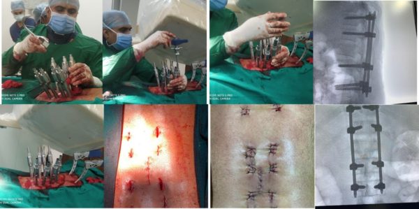
4. Transforaminal Lumbar Interbody Fusion –TLIF
Transforaminal Lumbar Interbody Merger is performed to treat recurrent disc herniation, degenerative disc disease, and spondylolisthesis. It is intended to treat the low mechanical refractory back and the radicular pain experienced by the patient.
The Transforaminal Lumbar Interbody Merger is a minimally invasive procedure performed on the back of the patient. It involves removing the intervertebral disc and inserting a bone-filled cage to stabilize the affected area with support rods and screws between two or more vertebral levels.
5. Cranio-vertebral junction (CVJ) surgery
It is one of the most complex and intricate surgeries performed on the bones at the junction of the head and neck. Neurosurgical surgery is recommended in the event of failure of non-surgical options and the patient’s spinal cord and brainstem is compressed, bones and/or joints in the craniocervical region are unstable, or if the patient complains of weakness, tingling, occipital headaches, neck aches, obstruction in the flow of cerebrospinal fluid, and numbness.
The anomalies of CVJ may have been congenital or acquired. It is essential to seek accurate diagnosis through an expert neuro-and spine surgeon along with appropriate imagery of the head and neck area – x-rays, CT and MRI scans, etc.
6. Foraminotomy:
The spine surgeon will perform a foraminotomy to remove bone spurs, herniated discs, thickened ligaments, and bulging discs if developed in the spinal canal and cause nerve pressure. In most cases, foraminotomy is performed as a minimally invasive procedure. The spine surgeon uses an endoscope or a tubular retractor to cut the muscles apart and reach the spine.
7. Replacement of artificial disc
The spinal surgeon may suggest an artificial disc replacement as a viable alternative to spinal fusion surgery. In this operation, the surgeon inserts an artificial disc to restore height and mobility between the vertebrae in the place of the damaged disc. Doctors recommend this treatment to patients suffering from severely damaged spinal discs.
Recovery
The patient’s condition before surgery determines the time taken for recovery and age.
The recovery of the patient after a discectomy is quicker than expected. After discectomy or foraminotomy, the patient may feel numbness, weakness, and pain along the nerve’s path under pressure. The patient will feel better and more energetic in a few weeks.
A patient recovering from a laminectomy and fusion surgery is likely to take more time. The recovery period may last at least three to four months for the best results. The patient will take quite some time to recover fully, which may take nearly twelve months.
A patient recovering from a spinal fusion will need at least four to six weeks to recover well. The recovery period will depend upon the age of the patient. The younger the patient, the faster the recovery, while the older patient may take more than six months to recover.
Hear What our Patient had to say
Best CVJ Spine Surgery in Andhra Pradesh
Frequently Asked Questions:
What is minimally invasive surgery?
Minimally invasive surgery is a relatively new procedure in which the surgeon makes small incisions of half-inch in length. The surgeon accesses, repairs damaged disc or vertebrae through these incisions without disturbing or damaging the surrounding muscles and tissues. Minimally invasive surgery requires less time to perform, with minimal blood loss and pain along with speedy recovery.
How is spinal cord tumor treated?
A spinal cord tumor removal surgery is the best way to treat a spinal cord tumor. Neurosurgeons use powerful microscopes and instruments to reach, identify and remove inaccessible tumors. If it is impossible to remove the entire tumor, then a partial removal of the tumor is done, and the remaining tumor is treated to chemotherapy and radiation.
What is lumbar disk replacement surgery?
Lumbar disk replacement surgery is done to remove the damaged or worn-out disk in the lower spine to replace it with an artificial disk. The artificial disk supports the vertebrae in bending both backward and forwards, turning and side bending too. The artificial disc is made from titanium or cobalt or polyethene or polyurethane.
This surgery is also known as arthroplasty or artificial disk replacement.
What is the line of treatment for a spinal fracture caused by osteoporosis?
Along with non-surgical treatment, vertebroplasty and kyphoplasty are the two types of surgery that heal spinal fractures caused by osteoporosis.
Vertebroplasty is a minimally invasive treatment. The surgeon injects a low viscosity cement directly into the collapsed vertebrate to stabilize the fracture and bring relief to the patient from pain.
Kyphoplasty is another minimally invasive procedure performed to reduce the pain due to a spinal fracture and bone stability. For more information or consultation on spine surgery treatment to reach to Dr Rao’s Hospital at info@drraoshospitals.com.
Enquiry Now

