Cerebral Aneurysms: Dr. Rao is the best in both open and Endovascular Treatments

Dr. Rao’s hospital is the best neurosurgery center to clip or coil aneurysms. Dr. Rao is the best doctor for cerebral aneurysms. He has experience with both open and endovascular treatments.
What is a cerebral aneurysm?
An intracranial aneurysm, cerebral aneurysm, or brain aneurysm is a balloon-like outpouching on the wall of an artery secondary to wear and tear in most of the blood vessels and appears over the age of 50 years. Because there is a weakened spot in the artery wall, there is a risk for rupture (bursting) of the aneurysm.
A cerebral aneurysm generally occurs in an artery in the anterior circulation. A typical artery wall consists of three layers. The aneurysm wall is thin and weak because of an abnormal loss or absence of the muscular layer of the artery wall, leaving only two layers.
The most common type of cerebral aneurysm is called a saccular or berry aneurysm, occurring in 90 percent of cerebral aneurysms. More than one aneurysm may be present at the same time in 20% of the patients with aneurysms.
Two other types of cerebral aneurysms are fusiform and dissecting aneurysms. A fusiform aneurysm bulges out on all sides (circumferentially). Fusiform aneurysms are generally associated with atherosclerosis and are challenging to treat as there are no good treatment options.
A dissecting aneurysm may result from a tear in the artery wall’s intima (inner layer), causing blood to leak into the layers and narrow the lumen. This may cause a ballooning out on one side of the artery wall or block off or obstruct blood flow through the artery. Dissecting aneurysms may occur with a traumatic injury, primarily blunt, and a common location appears to be vertebral circulation. The shape and location of the aneurysm may affect the treatment modality.
Most cerebral aneurysms (90 percent) will not present any symptoms and are small in size (less than 10 millimeters in diameter). Smaller aneurysms will have a lower risk of rupture.
A ruptured cerebral saccular aneurysm is the most common cause (80 percent) of SAH after the traumatic SAH. The first symptom of a cerebral aneurysm is subarachnoid hemorrhage. SAH is a medical emergency and may be the cause of a hemorrhagic (bleeding) stroke. Though there are many advancements in neurosurgery, 33% of patients die after bleeding.
Hemorrhagic is the cause of 16% of strokes.
Increased risk of rupture is associated with aneurysms that are greater than 10 millimeters (less than four-tenths of an inch) in diameter (Size is the most significant prognostic factor), a particular location (circulation in the back portion of the brain), and previous rupture of another aneurysm. A substantial risk of death is associated with the rupture of a cerebral aneurysm.
What is the treatment for a cerebral aneurysm?
Specific treatment for a cerebral aneurysm depends on:
- Your age, overall health, and medical history
- The severity of the condition
- Your symptoms and signs
- Your tolerance for procedures or therapies
- Expectations
- Your preference
Depending on your situation, the physician will recommend the appropriate intervention. The main goal of treatment is to decrease the risk of a subarachnoid hemorrhage, either initially or from a repeated bleeding episode.
Many factors determine treatment decisions for a cerebral aneurysm, including the presence or absence of symptoms, the aneurysm location and size, the patient’s medical condition, age, and other risk factors present are essential in consideration of treatment. Based on the abovementioned factors, Dr. Rao considers conservative management, surgery, or endovascular therapy for your aneurysm.
There are two primary surgical treatments for a cerebral aneurysm:
Open craniotomy (surgical clipping)
This procedure involves the Scalp incision, surgical removal of part of the skull, the opening of the layers of the brain, identification of the aneurysm, and clipping of the aneurysm after securing the blood vessels. The physician exposes the aneurysm and places a metal clip across the neck to prevent blood flow into the aneurysm sac. After completion of the clipping, we close the skull and scalp.
Endovascular coiling embolization
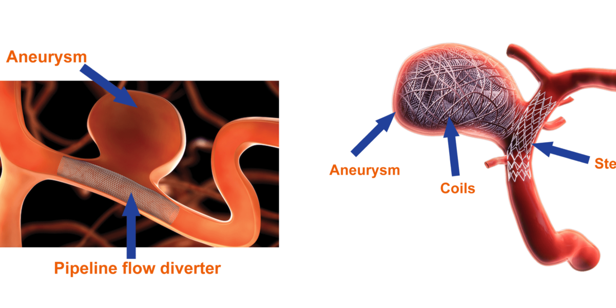
Endovascular coiling embolization is a minimally invasive technique to treat the aneurysm. An endovascular catheter is advanced from a blood vessel in the groin into the blood vessels in the brain. Fluoroscopy will be used to assist in moving the catheter into the aneurysm. Once the catheter is in place, platinum coils are advanced through the catheter into the aneurysm. These small, soft, platinum coils, visible on the x-ray, conform to the aneurysm’s shape. The coiled aneurysm becomes clotted off (embolization), preventing rupture. In some instances, the aneurysm dictates us placing the specially designed spring stent devices called flow diverters, which leads to aneurysm occlusion in 3 to six months. This procedure is performed either under general or local anesthesia.
What causes a cerebral aneurysm?
Currently, the cause of cerebral aneurysms is not clearly understood. The formation of cerebral saccular aneurysms is predominantly due to an abnormal degenerative change. Bifurcation of arteries is the frequent location for aneurysm formation.
Inherited risk factors:
- Alpha-glucosidase deficiency
- Alpha 1-antitrypsin deficiency
- Arteriovenous malformation (AVM)
- Coarctation of the aorta
- Ehlers-Danlos syndrome
- Family history of aneurysms
- Female gender
- Fibromuscular dysplasia
- Hereditary hemorrhagic telangiectasia
- Klinefelter syndrome
- Noonan’s syndrome
- Polycystic kidney disease (PCKD)
- Tuberous sclerosis
Acquired risk factors:
- Age (greater than 50)
- Alcohol binge drinking
- Atherosclerosis
- Cigarette smoking
- Use of illicit drugs
- High blood pressure
- Trauma to the head
- Infection
What are the symptoms of a cerebral aneurysm?
SAH might be the first sign, or sometimes the warning bleed may be present.
The symptoms of an unruptured cerebral aneurysm include, but are not limited to, the following:
- Headaches
- Dizziness
- Eye pain
- Vision deficits (problems with seeing)
- Seeing double
- worst headache ever in my life
- Nausea and vomiting
- Stiff neck
- Pain in the eyes
- Changes in mental status, such as drowsiness
- Loss of consciousness
- Motor deficits (loss of balance or coordination)
- Dilated pupils
- Hypertension (high blood pressure)
- Photophobia (sensitivity to light)
- Cranial nerve deficits
- Back or leg pain
How is a cerebral aneurysm diagnosed?
A cerebral aneurysm is often discovered after its rupture or by chance during diagnostic examinations such as computed tomography (CT scan, CT angiogram), magnetic resonance imaging (MRI), or digital subtraction angiography (catheter angiogram) that are being done for other conditions.
In addition to a complete medical history and physical examination, diagnostic procedures for a cerebral aneurysm may include:
- Digital subtraction angiography (DSA) – provides an image of the blood vessels in the brain to detect a problem with blood flow. The procedure involves inserting a catheter (a small, thin tube) into an artery in the leg and passing it up to the blood vessels in the brain. A contrast dye is injected through the catheter, and x-ray images are taken of the blood vessels.
- A computed tomography scan (CT or CAT scan) – is a diagnostic imaging procedure that may be used to identify the location or type of stroke.
- Magnetic resonance imaging (MRI) – is a diagnostic procedure that locates and diagnoses a stroke.
- Magnetic resonance angiography (MRA) – allows the physician to visualize the blood vessels.
Enquiry Now

