White Matter-Sparing Brain Surgery: The Future of Minimally Invasive Neurosurgery for Deep-Seated Brain Lesions
Featured in The Hindu (Vijayawada Edition, 30 August 2026) following Dr. Mohana Rao Patibandla’s keynote presentation at the World Neurosciences Summit (WNS) 2026, New Delhi.
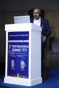
Dr. Mohana Rao Patibandla presenting his keynote lecture, “Minimally Invasive Cylindrical Corridor Surgery – Modern Microsurgery Strategy for Deep-Seated Brain Lesions,” at the 5th World Neurosciences Summit 2026 in New Delhi, highlighting advanced White Matter-Sparing Brain Surgery techniques for preserving critical neural pathways.
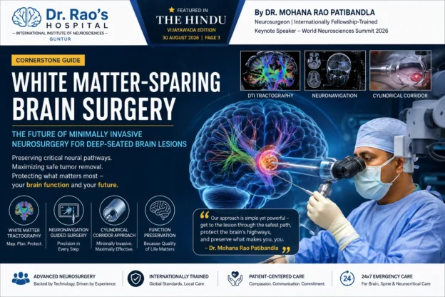
White Matter-Sparing Brain Surgery: The Future of Minimally Invasive Neurosurgery for Deep-Seated Brain Lesions
Author: Dr. Mohana Rao Patibandla, Neurosurgeon, Founder & Chief Neurosurgeon, Dr. Rao’s Hospital – International Institute of Neurosciences, Guntur, Andhra Pradesh, India
Published: September 2026
Reading Time: 18–22 minutes
Target Keywords: White matter sparing surgery, minimally invasive brain surgery, cylindrical corridor surgery, deep-seated brain tumor surgery, brain lesion surgery, neurosurgeon India, Dr. Mohana Rao Patibandla.
Executive Summary
The human brain contains billions of neurons connected through highly organized white matter tracts—the communication highways that allow movement, speech, memory, vision, sensation, and cognition. Traditional surgery for deep-seated brain tumors or lesions often requires passing through healthy brain tissue, increasing the risk of neurological deficits.
White Matter-Sparing Surgery, also called Minimally Invasive Cylindrical Corridor Surgery, represents a transformative advancement in neurosurgery. Instead of cutting through healthy brain tissue, surgeons navigate between natural white matter pathways, preserving critical neural connections while reaching tumors or lesions deep inside the brain.
This technique was the focus of Dr. Mohana Rao Patibandla’s keynote lecture at the World Neurosciences Summit 2026, where he presented modern microsurgical strategies for safely treating deep-seated brain lesions. The approach was subsequently highlighted by The Hindu in a feature article discussing the future of minimally invasive brain surgery.
This comprehensive guide explains everything patients, families, neurologists, and neurosurgeons need to know about white matter-sparing surgery.
Table of Contents
- What is White Matter-Sparing Brain Surgery?
- Understanding White Matter and Brain Networks.
- Why Traditional Brain Surgery Can Damage White Matter.
- Evolution of Minimally Invasive Cylindrical Corridor Surgery.
- How White Matter-Sparing Surgery Works.
- Neuronavigation and Brain Mapping.
- Who Needs White Matter-Sparing Surgery?
- Conditions Treated.
- Benefits Compared to Conventional Brain Surgery.
- Surgical Planning and Technology.
- Risks and Limitations.
- Recovery After White Matter-Sparing Surgery.
- Frequently Asked Questions.
Understanding White Matter: The Brain’s Information Highway
![White matter tracts in the brain [IMAGE] | EurekAlert! Science News Releases](https://images.openai.com/static-rsc-4/ehFa_K68NUvBRok5xTPdaMEJjFDFbBquDyf4jUrCPZujw2Wyv1w-CnAR8CrS_b6gg3CjT34ycvu_-GKtAvBgPnj6aTcUEaa5jy04XJ4gYiPkuUBMZXCH5gNmk8RPatmngmuWIO86GGH681FH8nikDajtRAcUCsJngVpWDY3ozSU?purpose=inline)


Most people think of the brain as a gray organ filled with neurons. However, white matter is equally important.
What is White Matter?
White matter consists of millions of insulated nerve fibers called axons. These fibers connect different brain regions and enable rapid communication.
White Matter Controls
Brain Function | Major White Matter Pathway |
| Movement | Corticospinal tract |
| Speech production | Arcuate fasciculus |
| Language comprehension | Superior longitudinal fasciculus |
| Vision | Optic radiations |
| Memory | Fornix and cingulum |
| Emotion | Uncinate fasciculus |
Damage to these pathways may result in:
Paralysis
Speech difficulty
Vision loss
Memory impairment
Cognitive dysfunction
Personality changes
Protecting these pathways has become one of the central goals of modern neurosurgery.
Why Traditional Brain Surgery Isn’t Always Enough


Historically, surgeons approached deep tumors by creating a path through the brain.
The Challenge
Deep lesions may lie:
Near motor pathways.
Adjacent to speech centers.
Beneath healthy cortex.
Close to visual pathways.
Near memory circuits.
Traditional surgery may require:
Brain retraction.
Cortical incision.
Traversing healthy tissue.
Even with excellent technique, these maneuvers may increase neurological risk.
Potential Consequences
Potential Injury | Possible Deficit |
| Motor tract injury | Weakness or paralysis |
| Language tract injury | Speech impairment |
| Visual tract injury | Visual field loss |
| Frontal network injury | Behavioral or cognitive changes |
Modern neurosurgery seeks to avoid these injuries rather than simply treat them afterward.
What is White Matter-Sparing Surgery?


Definition
White Matter-Sparing Surgery is a minimally invasive microsurgical technique that uses:
Natural anatomical corridors.
Cylindrical tubular retractors.
Advanced neuronavigation.
White matter tractography.
High-definition operating microscopy.
Instead of retracting large portions of the brain, surgeons gently separate white matter fibers along their natural orientation.
The Philosophy
Preserve normal brain. Remove abnormal tissue. Protect neural connectivity.
This is fundamentally different from older approaches focused primarily on lesion access.
The Science Behind Cylindrical Corridor Surgery


Why a Cylinder?
A cylindrical corridor distributes pressure evenly.
Traditional Retractor


Flat blades.
Uneven pressure.
Greater cortical retraction.
Cylindrical Corridor


Gentle circumferential dilation.
Less tissue distortion.
Smaller surgical footprint.
Better preservation of white matter fibers.
This concept has become increasingly adopted in minimally invasive neurosurgery worldwide.
How White Matter-Sparing Surgery Works
Step-by-Step Surgical Strategy


Step | Purpose |
| 1 | Advanced MRI with tractography identifies functional white matter pathways. |
| 2 | Neuronavigation selects the safest trajectory. |
| 3 | Small craniotomy is created. |
| 4 | Tubular corridor gently separates white matter fibers. |
| 5 | Microsurgical instruments remove lesion through the corridor. |
| 6 | Hemostasis is achieved while preserving surrounding tissue. |
The result is maximal safe lesion removal with minimal disruption.
White Matter Tractography: GPS for the Brain


What is DTI Tractography?
Diffusion Tensor Imaging (DTI) visualizes white matter pathways before surgery.
It Helps Identify
Motor tracts.
Speech tracts.
Visual pathways.
Sensory pathways.
Memory networks.
Why It Matters
Surgeons can:
Avoid eloquent pathways.
Choose safer entry points.
Predict postoperative risks.
Tailor surgery to each patient.
This represents precision neurosurgery rather than generalized anatomy.
Neuronavigation: GPS Inside the Brain


Neuronavigation provides real-time anatomical guidance.
It Allows Surgeons To
Locate lesions accurately.
Avoid blood vessels.
Maintain surgical trajectory.
Navigate deep structures safely.
Combined with tractography, neuronavigation creates a personalized surgical roadmap.
Brain Mapping: Protecting Function During Surgery


Modern white matter-sparing surgery often incorporates:
Intraoperative Monitoring
Motor evoked potentials.
Somatosensory evoked potentials.
Cranial nerve monitoring.
Speech mapping when required.
This provides continuous feedback about neurological function.
Which Brain Conditions Can Be Treated?
Deep-Seated Brain Tumors


Examples include:
Gliomas
Metastatic tumors
Low-grade glioma
High-grade glioblastoma
Thalamic tumors
Basal ganglia tumors
Goal
Maximal safe resection while preserving neurological function.
Cavernomas


White matter-sparing corridors are valuable for:
Thalamic cavernoma.
Brainstem cavernoma.
Deep cerebral cavernomas.
Colloid Cysts


Small minimally invasive approaches reduce brain manipulation around the ventricles.
Intraventricular Tumors


Examples include:
Central neurocytoma.
Ependymoma.
Choroid plexus tumors.
Subependymoma.
Brain Abscesses


Targeted tubular access allows drainage with limited cortical injury in selected patients.
Epilepsy Lesions


Certain deep epileptogenic lesions can be approached through white matter-preserving techniques.
Advantages Over Conventional Brain Surgery
Why White Matter-Sparing Surgery Matters
Traditional Surgery | White Matter-Sparing Surgery |
| Larger craniotomy | Smaller craniotomy |
| More brain retraction | Minimal brain retraction |
| Cortical disruption | Natural white matter corridor |
| Greater tissue manipulation | Targeted microsurgical access |
| Higher postoperative swelling | Reduced tissue trauma |
| Longer recovery | Faster recovery in selected patients |
Key Patient Benefits


Potential benefits include:
Smaller incision.
Less pain.
Reduced blood loss.
Less brain swelling.
Better cosmetic outcome.
Earlier mobilization.
Shorter ICU stay in selected cases.
Preservation of neurological function.
These benefits depend on careful patient selection and surgical expertise.
Who is a Candidate?
Ideal Candidates
Suitable Patients | Why |
| Deep brain tumors | Accessible through safe anatomical corridors. |
| Small-to-medium lesions | Minimal disruption possible. |
| Functional area lesions | White matter preservation is especially valuable. |
| Selected hemorrhages | Tubular evacuation may be appropriate in selected cases. |
| Deep cavernomas | Natural corridor approach. |
Patients Who May Need Conventional Surgery
Very large tumors.
Diffuse infiltrative lesions.
Extensive vascular malformations.
Multiple compartment lesions.
Every patient requires individualized evaluation.
Surgical Planning at Dr. Rao’s Hospital


A white matter-sparing operation begins long before entering the operating room.
Comprehensive Planning Includes
Advanced Imaging


MRI Brain
Contrast MRI
DTI tractography
Functional MRI when indicated.
MR angiography/venography.
Digital Planning


Surgical trajectory simulation.
White matter tract preservation.
Vascular avoidance.
Cortical entry planning.
Microsurgical Technology Used
High-End Neurosurgical Equipment


Technology may include:
Operating microscope.
Neuronavigation.
Ultrasonic aspirator.
Endoscope-assisted visualization.
Neurophysiological monitoring.
High-definition imaging.
Each tool contributes to safer microsurgery.
White Matter-Sparing Surgery in Brain Tumor Management
Low-Grade Gliomas


Goal:
Preserve language.
Preserve cognition.
Maximize tumor removal.
High-Grade Glioblastoma


Goal:
Maximal safe resection.
Protect functional pathways.
Improve quality of life.
White Matter Preservation in Pediatric Neurosurgery


Children have developing neural pathways.
Advantages include:
Better developmental preservation.
Reduced tissue injury.
Smaller surgical footprint.
Functional protection during growth.
White Matter-Sparing Surgery for Brainstem Lesions


Brainstem surgery is among the most challenging areas in neurosurgery.
The technique focuses on:
Safe entry zones.
Natural fiber orientation.
Cranial nerve preservation.
Microsurgical precision.
Recovery After White Matter-Sparing Surgery
What Patients Can Expect


Hospital Stay
Stage | Typical Course |
| ICU | 1 day in many selected cases. |
| Ward | 2–5 days depending on condition. |
| Walking | Often within 24 hours if neurologically appropriate. |
| Return Home | Depends on recovery and pathology. |
Recovery varies by diagnosis, lesion location, age, and overall health.
Rehabilitation


Some patients benefit from:
Physiotherapy.
Speech therapy.
Occupational therapy.
Cognitive rehabilitation.
Risks and Limitations
No brain surgery is risk-free.
Potential Risks


Bleeding.
Infection.
Seizures.
Neurological deficits.
Brain swelling.
CSF leak.
The goal of white matter-sparing surgery is to reduce unnecessary injury—not eliminate all surgical risk.
Evidence Supporting White Matter-Sparing Approaches
Why This Technique is Growing Worldwide
Modern literature supports:
Less cortical injury.
Improved preservation of white matter tracts.
Better functional outcomes in selected lesions.
Reduced surgical morbidity in carefully chosen patients.
International adoption continues to increase alongside advances in imaging and neuronavigation.
Dr. Mohana Rao Patibandla’s Keynote at World Neurosciences Summit 2026


At the World Neurosciences Summit 2026, Dr. Mohana Rao Patibandla presented a keynote lecture titled:
“Minimally Invasive Cylindrical Corridor Surgery – Modern Microsurgery Strategy for Deep-Seated Brain Lesions.”
The presentation emphasized:
White matter preservation.
Modern microsurgical planning.
Neuronavigation-guided access.
Safe treatment of deep brain lesions.
Functional outcome optimization.
The lecture highlighted the growing importance of minimally invasive microsurgical corridors in contemporary neurosurgery.
Featured in The Hindu: National Recognition of White Matter-Sparing Surgery

The Hindu reported Dr. Mohana Rao Patibandla’s press conference explaining how preserving normal brain tissue and neural pathways has become a key objective of modern minimally invasive microsurgery. The article also described cylindrical corridor surgery as a white matter-sparing strategy for deep-seated brain lesions.
This coverage reflects growing public awareness of advanced brain-preserving surgical techniques in India.
Why This Matters for Patients in India


Patients increasingly seek:
Safer surgery.
Smaller incisions.
Faster recovery.
Preservation of neurological function.
International-standard neurosurgical care within India.
White matter-sparing surgery aligns with these goals when clinically appropriate.
Why Choose Dr. Rao’s Hospital for White Matter-Sparing Brain Surgery?


Dr. Rao’s Hospital – International Institute of Neurosciences, Guntur, offers comprehensive brain and spine care with advanced neurosurgical technology and internationally trained expertise.
Areas of Expertise
Brain Tumor Surgery
Skull Base Surgery
Endoscopic Brain Surgery
Pediatric Neurosurgery
Cerebrovascular Surgery
Epilepsy Surgery
Functional Neurosurgery
Minimally Invasive Neurosurgery
Advanced Technologies
Operating Microscope
Neuronavigation
Neuroendoscopy
Neurophysiological Monitoring
Advanced MRI Planning
Minimally Invasive Brain Surgery
Frequently Asked Questions (FAQ)
1. What is White Matter-Sparing Brain Surgery?
White matter-sparing brain surgery is a minimally invasive microsurgical approach that reaches deep brain lesions through natural white matter corridors while minimizing injury to healthy neural pathways.
2. What is Cylindrical Corridor Surgery?
It is a minimally invasive technique using a tubular retractor to create a gentle corridor through the brain, allowing surgeons to access deep lesions with less tissue disruption.
3. Is White Matter-Sparing Surgery safer than traditional brain surgery?
It may reduce disruption to healthy brain tissue in carefully selected patients, but safety depends on lesion type, location, patient health, and surgical expertise.
4. Which brain tumors can be treated?
Selected gliomas, metastases, intraventricular tumors, cavernomas, colloid cysts, and certain deep-seated lesions may be suitable candidates.
5. What is tractography?
Tractography is an MRI technique (DTI) that maps white matter pathways before surgery to help surgeons avoid critical neural connections.
6. Does every patient need tractography?
Not always. It is especially useful when lesions are close to eloquent brain pathways.
7. Is recovery faster?
Many selected patients experience smaller incisions, less pain, and earlier mobilization, although recovery varies with the underlying disease and surgery performed.
8. Is this surgery available in India?
Yes. Advanced minimally invasive white matter-sparing brain surgery is available in specialized neuroscience centers including Dr. Rao’s Hospital in Guntur.
9. Can brain function be preserved completely?
The goal is maximal preservation of neurological function, but outcomes depend on lesion location, disease biology, and individual patient factors.
10. How do I know if I’m a candidate?
A neurosurgical consultation with MRI review, tractography when indicated, and individualized surgical planning is necessary.
Key Takeaways


White Matter-Sparing Surgery represents one of the most significant advances in modern neurosurgery because it shifts the surgical philosophy from simply reaching a lesion to protecting the brain’s communication networks while treating disease.
For carefully selected patients with deep-seated brain tumors, cavernomas, intraventricular lesions, and other complex pathologies, minimally invasive cylindrical corridor surgery offers a highly precise approach centered on preserving neurological function whenever possible.
As highlighted during Dr. Mohana Rao Patibandla’s keynote lecture at the World Neurosciences Summit 2026 and subsequently featured in The Hindu, the future of neurosurgery lies in combining advanced imaging, neuronavigation, microsurgical precision, and white matter preservation to achieve better patient outcomes.
About the Author
Dr. Mohana Rao Patibandla is an internationally fellowship-trained neurosurgeon and the Founder & Chief Neurosurgeon of Dr. Rao’s Hospital – International Institute of Neurosciences, Guntur, Andhra Pradesh, India.
His advanced training includes skull base surgery, pediatric neurosurgery, minimally invasive brain surgery, neuro-oncology, cerebrovascular and endovascular neurosurgery, and functional neurosurgery in India and the United States. He regularly serves as invited faculty at national and international neuroscience conferences and delivered a keynote lecture at the World Neurosciences Summit 2026 on White Matter-Sparing Minimally Invasive Cylindrical Corridor Surgery.
Authoritative Neurosurgical Resources
World Federation of Neurosurgical Societies (WFNS)
Congress of Neurological Surgeons (CNS)
American Association of Neurological Surgeons (AANS)
National Institute of Neurological Disorders and Stroke (NINDS)
Patients with deep-seated gliomas, meningiomas, metastatic tumors, and other complex lesions may benefit from
Brain Tumor Surgery performed using modern white matter-sparing microsurgical techniques. Many lesions located near the skull base require specialized Skull Base Surgery
combined with advanced neuronavigation and minimally invasive approaches.
Our comprehensive Neuro-Oncology Program focuses on personalized treatment for benign and malignant brain tumors. Children with deep-seated brain tumors may benefit from advanced Pediatric Neurosurgery that prioritizes preservation of developing neural pathways.
Selected intraventricular tumors and colloid cysts can often be treated using Endoscopic Brain Surgery or minimally invasive cylindrical corridor techniques.
White matter-sparing surgery relies heavily on Neuronavigation-Guided Brain Surgery
to identify the safest surgical pathway while protecting eloquent brain regions.
Patients with epilepsy caused by cavernomas or focal cortical lesions may also be candidates for Epilepsy Surgery using minimally invasive neurosurgical strategies.
Learn more about the international training, experience, research, and surgical philosophy of Dr. Mohana Rao Patibandla Founder of Dr. Rao’s Hospital – International Institute of Neurosciences.
Need Expert Care for a Brain Tumor or Deep-Seated Brain Lesion?
At Dr. Rao’s Hospital – International Institute of Neurosciences (IIN),
we provide advanced White Matter-Sparing Brain Surgery,
Minimally Invasive Brain Tumor Surgery,
Skull Base Surgery, Endoscopic Brain Surgery,
Neuro-Oncology, Pediatric Neurosurgery,
Stroke Care, and Spine Surgery using internationally
recognized technology and evidence-based treatment protocols.
Every patient receives a personalized surgical plan based on advanced MRI,
neuronavigation, white matter tractography, intraoperative neuromonitoring,
and minimally invasive microsurgical techniques designed to preserve neurological function whenever possible.
Why Choose Dr. Rao’s Hospital?
- Internationally Fellowship-Trained Neurosurgeon – Dr. Mohana Rao Patibandla
- Dedicated Super-Specialty Neurosciences Hospital in Guntur, Andhra Pradesh.
- Advanced White Matter-Sparing & Minimally Invasive Brain Surgery.
- Brain Tumor, Skull Base, Pediatric, Spine, Stroke & Endovascular Neurosurgery.
- Neuronavigation, Neurophysiological Monitoring & High-End Microsurgical Technology.
- Comprehensive Neuro-Oncology, Neurocritical Care and Rehabilitation.
- 24×7 Emergency Brain, Stroke and Spine Care.
Contact Dr. Rao’s Hospital
Dr. Rao’s Hospital – International Institute of Neurosciences (IIN)
12-19-67, Old Bank Road, Kothapet,
Opposite Sravani Hospital, Besides AK Biryani point,
Guntur – 522001, Andhra Pradesh, India.
📞 +91 90100 56444
✉️ info@drraoshospitals.com
🌐 www.drraoshospitals.com
