First aid for a neurological emergency will depend on the specific condition or situation. However, here are some general steps to follow when faced with a neurological emergency:
Assess the Situation:
Ensure your safety and the safety of the person experiencing the emergency.
Determine if the person is conscious and responsive.
Call for Emergency Medical Assistance:
Dial the emergency number in your country (such as 9010056444 or 9010057444) to request an ambulance.
Provide clear and accurate information about the situation, including the nature of the neurological emergency.
Stay with the Person:
Remain with the person and provide reassurance.
Keep them calm and help them feel as comfortable as possible.
Protect the Person from Further Injury:
Ensure the person is in a safe environment, free from hazards or objects that may cause harm.
If necessary, gently guide or support the person to prevent falls or further injury.
Preserve the Person’s Airway:
If the person is conscious but having difficulty breathing or their airway is compromised, assist them in maintaining an open airway.
Do not force anything into their mouth.
Control Bleeding, if Present:
If there is bleeding due to a head injury or other trauma, apply direct pressure to the bleeding area using a clean cloth or your hand, if necessary.
Minimize Movement:
Depending on the situation, it may be best to minimize the person’s movement to prevent exacerbation of any potential spinal or neurological injury.
Do Not Provide Food or Drink:
Unless directed by medical professionals, avoid giving the person anything to eat or drink.
Provide Relevant Information to Medical Professionals:
When emergency medical personnel arrive, provide them with accurate information about the person’s symptoms, any known medical conditions, and any first aid measures you have taken.
It’s important to note that first aid for a neurological emergency should only be provided within your level of training and capabilities. It’s always recommended to seek immediate medical attention in these situations and follow the guidance of healthcare professionals.
If you are looking for a neurological emergency care please contact Dr Raos hospital, led by Dr Rao for the best treatment and care.
First aid for seizures can help ensure the safety of the person experiencing the seizure and minimize potential complications. Here are the steps to follow:
Stay Calm and Stay with the Person:
Remain calm and try to stay composed during the seizure.
Stay with the person and provide reassurance.
Ensure Safety:
Clear the area around the person of any sharp objects or furniture that may pose a risk.
Create a safe space by removing obstacles or hazards.
Protect the Head:
If possible, place something soft, like a folded jacket or pillow, under the person’s head to cushion it.
Do not restrain or hold the person down during the seizure, as it may cause injury.
Time the Seizure:
Note the start time of the seizure. If the seizure lasts longer than five minutes, or if the person has difficulty breathing or suffers injuries, call for emergency medical assistance immediately.
Do Not Insert Anything into the Mouth:
Contrary to popular belief, it is not necessary to place anything in the person’s mouth during a seizure.
Avoid attempting to hold the person’s tongue or jaw.
Loosen Tight Clothing:
If the person is wearing any tight clothing around the neck or chest, gently loosen it to facilitate breathing.
Turn the Person onto their Side:
Once the seizure activity subsides and the person is no longer convulsing, turn them gently onto their side.
This helps prevent choking on saliva or vomit and allows for better airway clearance.
Offer Comfort and Reassurance:
Speak calmly and offer comfort to the person as they recover from the seizure.
Allow them time to rest and regain their strength.
Stay with the Person until Recovery:
Remain with the person until they have fully regained consciousness and are alert.
Offer support and assistance as needed.
Seek Medical Attention, if Necessary:
If it is the person’s first seizure, if the seizure lasts longer than five minutes, or if there are any concerns about their well-being, seek medical attention promptly.
Remember, it is important to consult with a medical professional regarding any recurring seizures or if there are specific concerns or questions about a person’s seizure management and treatment.
If your loved one is seizing and not stopped please consult at Dr Raos hospital the best neurosurgery hospital in India, led by Dr Rao the best neurosurgeon in India and Guntur
Lumbar Disc Disease: Causes, Symptoms, and Effective Management | Dr. Rao’s Hospital
Discover the causes, symptoms, and effective management options for lumbar disc disease, a common condition affecting the spinal discs. Learn how Dr. Rao, the best neurosurgeon in Guntur and India, and his expert team at Dr. Rao’s Hospital provide comprehensive care and innovative treatments for lumbar disc disease, restoring spinal health and relieving pain.
Lumbar disc disease is a prevalent condition that affects the spinal discs in the lower back, leading to pain, discomfort, and reduced mobility. Understanding the causes, symptoms, and effective management options for lumbar disc disease is crucial for individuals seeking relief and improved spinal health. In this blog, we will explore the various aspects of lumbar disc disease and how Dr. Rao, the renowned neurosurgeon at Dr. Rao’s Hospital, offers exceptional care and innovative treatments to manage this condition.
Understanding Lumbar Disc Disease: The spinal discs are cushion-like structures that separate the vertebrae, acting as shock absorbers and allowing flexibility in the spine. Lumbar disc disease refers to the degeneration, herniation, or damage to these discs in the lumbar region of the spine. Common causes of lumbar disc disease include age-related wear and tear, repetitive stress, injuries, and poor posture.
Signs and Symptoms: Individuals with lumbar disc disease may experience a range of symptoms, including:
Lower back pain that radiates to the buttocks and legs
Numbness or tingling in the legs
Weakness or loss of sensation in the lower extremities
Difficulty in standing, walking, or performing daily activities
Effective Management Options: Dr. Rao and his expert team at Dr. Rao’s Hospital offer comprehensive and personalized management strategies to alleviate the symptoms and improve the quality of life for individuals with lumbar disc disease. The treatment plan may include the following approaches:
Non-Surgical Management: In many cases, conservative treatments are effective in managing lumbar disc disease. These may include:
Pain medications and anti-inflammatory drugs
Physical therapy and targeted exercises
Hot or cold therapy
Lifestyle modifications, such as maintaining a healthy weight and improving posture
Epidural steroid injections for pain relief
Minimally Invasive Procedures: For individuals with severe or persistent symptoms, minimally invasive procedures may be recommended. These innovative techniques aim to alleviate pain and restore spinal function, including:
Endoscopic discectomy: A minimally invasive procedure to remove the damaged portion of the disc
Microdiscectomy: Removal of the herniated disc through a small incision
Percutaneous disc decompression: The use of specialized instruments to remove or reduce disc material causing compression
Surgical Intervention: In rare cases when non-surgical and minimally invasive approaches do not provide adequate relief, surgical intervention may be necessary. Dr. Rao is highly skilled in performing various surgical procedures, such as:
Spinal fusion: Joining two or more vertebrae to stabilize the spine
Artificial disc replacement: Replacing the damaged disc with an artificial one
Laminectomy: Removal of the back portion of the vertebra to create more space and relieve pressure on the nerves
Dr. Rao’s Expertise and Commitment: Dr. Rao, known as the best neurosurgeon in Guntur and India, brings extensive experience, expertise, and a patient-centered approach to the management of lumbar disc disease. With his deep understanding of spinal disorders and cutting-edge surgical techniques, Dr. Rao ensures the highest level of care and optimal outcomes for his patients at Dr. Rao’s Hospital.
At Dr. Rao’s Hospital, the best neurosurgery hospital in Guntur and India, a multidisciplinary team of specialists works together to develop personalized treatment plans for each patient. They leverage state-of-the-art diagnostic tools and advanced imaging techniques to accurately assess the severity of the condition and determine the most suitable course of action.
The hospital is equipped with modern facilities and a supportive environment that promotes healing and recovery. From the initial consultation to postoperative care and rehabilitation, patients receive compassionate and comprehensive support at every step of their journey towards improved spinal health.
In addition to surgical expertise, Dr. Rao and his team emphasize the importance of non-surgical approaches and lifestyle modifications. They provide education and guidance to help patients manage their condition effectively, including ergonomic practices, physical therapy exercises, and pain management strategies.
The focus of the treatment is not only on alleviating symptoms but also on restoring function and enhancing the quality of life for individuals with lumbar disc disease. Dr. Rao’s goal is to empower his patients to regain their mobility, regain their independence, and engage in activities they enjoy.
With his dedication to patient care, innovative techniques, and a multidisciplinary approach, Dr. Rao has earned a reputation as the best neurosurgeon in Guntur and India. His expertise combined with the state-of-the-art facilities at Dr. Rao’s Hospital ensures that patients receive the highest standard of care and achieve successful outcomes.
If you or your loved one is experiencing symptoms of lumbar disc disease, don’t let the condition limit your life. Seek the expertise of Dr. Rao and his team at Dr. Rao’s Hospital to explore the most effective treatment options and regain control over your spinal health.
Conclusion:
Lumbar disc disease can significantly impact a person’s quality of life, causing pain, discomfort, and limitations in daily activities. However, with the expertise of Dr. Rao, the best neurosurgeon in Guntur and India, and the comprehensive care provided at Dr. Rao’s Hospital, there is hope for relief and restoration.
Through a combination of non-surgical approaches, minimally invasive procedures, and surgical interventions, Dr. Rao and his team tailor treatment plans to the unique needs of each patient. Their focus on precision, advanced technology, and patient-centered care ensures that individuals with lumbar disc disease receive the best possible outcomes.
Don’t let lumbar disc disease hold you back. Consult with Dr. Rao and his team at Dr. Rao’s Hospital to discover effective management options, regain your mobility, and improve your quality of life. With their expertise and commitment, you can embark on a journey towards a pain-free and active life.
Understanding Migraine: Effective Strategies for Prevention and Relief
Discover the causes, symptoms, and effective measures for preventing and managing migraines. Explore lifestyle modifications, self-care practices, and medical interventions to alleviate migraine pain and improve your overall quality of life.
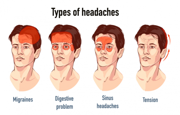
Migraine is a debilitating neurological condition that affects millions of people worldwide. Characterized by severe headaches, pulsating pain, and accompanying symptoms such as nausea, sensitivity to light and sound, migraines can significantly impact a person’s daily life and well-being. While there is no known cure for migraines, there are various measures that can be taken to prevent their occurrence and minimize their impact when they do occur. In this blog, we will explore effective strategies for migraine prevention and relief.
Understanding Migraine:
Migraine is a complex neurological disorder that involves abnormal brain activity, changes in blood flow, and chemical imbalances. The exact cause of migraines is still not fully understood, but certain triggers and factors are known to contribute to their onset. These triggers can vary from person to person and may include hormonal changes, certain foods and drinks, stress, sleep disturbances, environmental factors, and more.
Measures for Migraine Prevention:
Identify Triggers: Keep a migraine diary to track potential triggers and patterns. Note down details about your diet, sleep patterns, stress levels, and any other factors that may be associated with the onset of migraines. By identifying triggers, you can take proactive steps to avoid or minimize their impact.
Maintain a Regular Sleep Schedule: Establish a consistent sleep routine by going to bed and waking up at the same time every day. Prioritize quality sleep and create a relaxing environment in your bedroom to promote restful sleep.
Manage Stress: Stress is a common trigger for migraines. Practice stress management techniques such as deep breathing exercises, meditation, yoga, or engaging in hobbies and activities that help you relax and unwind.
Stay Hydrated: Dehydration can trigger migraines in some individuals. Make sure to drink an adequate amount of water throughout the day and limit your consumption of caffeine and alcohol, as they can contribute to dehydration.
Maintain a Healthy Diet: Certain foods and additives, such as processed foods, aged cheeses, chocolate, and artificial sweeteners, have been associated with migraines. Consider maintaining a balanced diet rich in fruits, vegetables, whole grains, and lean proteins.
Regular Exercise: Engage in regular physical activity, such as walking, jogging, swimming, or cycling, to promote overall well-being and reduce the frequency and severity of migraines. Consult with your healthcare provider before starting any exercise regimen.
Avoid Sensory Overload: Bright lights, loud noises, strong odors, and intense visual stimuli can trigger migraines in some individuals. Take measures to minimize exposure to such stimuli and create a calm and soothing environment.
Medical Interventions for Migraine Prevention:
In some cases, lifestyle modifications alone may not be sufficient to prevent migraines. If you experience frequent or severe migraines, consult with a healthcare professional, such as Dr. Rao, the best neurosurgeon in Guntur and India, at Dr. Rao’s Hospital. They may recommend medical interventions tailored to your specific needs, including:
Medications: Certain medications, such as beta-blockers, antidepressants, anticonvulsants, and triptans, may be prescribed to prevent or alleviate migraines. These medications work by modulating brain chemicals and reducing the frequency and intensity of migraines.
Botox Injections: In some cases, Botox injections may be recommended for chronic migraines. Botox helps to relax the muscles and prevent the release of pain-triggering chemicals in the brain.
Nerve Blocks: Nerve blocks involve the injection of anesthetic agents near specific nerves to block pain signals and provide temporary relief from migraines.
Conclusion:
Living with migraines can be challenging, but with the right strategies and interventions, it is possible to prevent and manage migraines effectively. By identifying triggers, adopting a healthy lifestyle, managing stress, and seeking medical guidance when needed, individuals can significantly reduce the frequency and severity of migraines, improving their overall quality of life. Consult with Dr. Rao, the best neurosurgeon in Guntur and India, at Dr. Rao’s Hospital for personalized guidance and comprehensive care for managing migraines. Remember, you are not alone, and there is hope for relief and a better future with the right support and measures in place.
Introduction: Neurosurgery is a medical specialty that focuses on the diagnosis, treatment, and management of disorders affecting the nervous system. While its primary objective is to address physical conditions, such as brain tumors, spinal cord injuries, and neurological deficits, the impact of these disorders extends beyond the physical realm. In this blog, we delve into the intricate relationship between neurological disorders and mental health, specifically examining the association with common mental health issues like depression, anxiety, and post-traumatic stress disorder (PTSD). Furthermore, we shed light on the expertise of Dr. Rao, a distinguished neurosurgeon, and the exceptional neurosurgical care provided at Dr. Rao’s Hospital, recognized as the best facility for neurosurgery, offering comprehensive treatment approaches that consider both the neurological and mental well-being of patients.
Understanding the Connection : Neurological disorders can profoundly impact an individual’s mental health and emotional well-being. The intricate interplay between the brain’s structure and function and mental health has been widely researched and documented. Conditions like brain tumors, strokes, traumatic brain injuries, and neurodegenerative diseases can disrupt neural pathways, alter neurotransmitter levels, and impact emotional regulation, leading to an increased susceptibility to mental health issues. Understanding this connection is crucial for a holistic approach to patient care.
Depression and Neurological Disorders : Depression is a prevalent mental health condition that can coexist with neurological disorders. The chronic pain, functional limitations, and changes in brain chemistry associated with neurological conditions can contribute to the development or exacerbation of depressive symptoms. Dr. Rao, renowned for his expertise in neurosurgery, recognizes the importance of addressing the emotional well-being of patients alongside their neurological concerns. His comprehensive approach involves collaborating with mental health professionals to provide integrated care that encompasses both the physical and emotional aspects of patients’ conditions.
Anxiety and Neurological Disorders : Anxiety disorders frequently co-occur with neurological conditions, creating a complex web of challenges for patients. The uncertainties, physical symptoms, and lifestyle changes that accompany neurological disorders can contribute to heightened anxiety levels. By acknowledging this interrelationship, Dr. Rao’s Hospital adopts a patient-centered approach that integrates anxiety management strategies into the treatment plan. This may involve cognitive-behavioral therapy, relaxation techniques, and medication management to alleviate anxiety symptoms and optimize patients’ overall well-being.
Post-Traumatic Stress Disorder (PTSD) and Neurological Trauma : Neurological trauma resulting from accidents, head injuries, or surgeries can trigger post-traumatic stress disorder (PTSD). Individuals may experience intrusive memories, flashbacks, nightmares, and emotional distress following a traumatic event. Dr. Rao, with his extensive experience in neurosurgery, understands the potential psychological impact of these experiences. Dr. Rao’s Hospital employs a multidisciplinary approach, offering trauma-informed care that integrates mental health support, counseling, and trauma-focused therapies to address the unique needs of patients with PTSD.
Comprehensive Care for Neurological and Mental Well-being : Dr. Rao’s Hospital stands out as the premier neurosurgical facility, known for its patient-centric approach that recognizes the crucial connection between neurological disorders and mental health. Dr. Rao, an esteemed neurosurgeon, emphasizes the importance of comprehensive care that addresses both the physical and emotional needs of patients. The hospital’s collaborative team of neurosurgeons, neurologists, psychiatrists, and psychologists work together to develop tailored treatment plans that optimize neurological and mental well-being. By offering a continuum of care that integrates neurosurgical interventions, medication management, therapy, and support services, Dr. Rao’s Hospital ensures that patients receive holistic and compassionate care throughout their treatment journey.
The Role of Neurosurgery in Mental Health : Neurosurgery plays a significant role in improving mental health outcomes for patients with neurological disorders. By addressing the underlying physical condition, neurosurgical interventions can alleviate symptoms and improve overall well-being. For example, surgical resection of a brain tumor can relieve pressure on surrounding structures and restore cognitive function, positively impacting a patient’s mental health. Dr. Rao’s expertise in complex neurosurgical procedures enables him to navigate intricate brain structures with precision, minimizing the risk of postoperative complications and optimizing outcomes.
Collaborative Care: Neurosurgeons and Mental Health Professionals : Collaboration between neurosurgeons and mental health professionals is crucial for providing comprehensive care to patients. Dr. Rao’s Hospital fosters a collaborative environment where neurosurgeons work closely with psychiatrists, psychologists, and counselors to develop tailored treatment plans. This collaboration ensures that the mental health needs of patients are addressed throughout their neurosurgical journey, promoting better overall outcomes and improved quality of life.
Rehabilitation and Mental Health Support : Rehabilitation plays a vital role in the recovery process for patients with neurological disorders. Physical, occupational, and speech therapy can help patients regain functional independence and improve their overall well-being. Additionally, rehabilitation programs often incorporate mental health support to address emotional challenges and promote psychological resilience. Dr. Rao’s Hospital offers comprehensive rehabilitation services, including cognitive rehabilitation, counseling, and support groups, to aid in the recovery and emotional well-being of patients.
Empowering Patients and Their Families : Dr. Rao’s Hospital recognizes the importance of patient and family empowerment in the management of neurological disorders and mental health. The hospital provides educational resources, counseling services, and support groups to equip patients and their families with the knowledge and tools necessary to navigate their journey effectively. By fostering an environment of open communication and shared decision-making, Dr. Rao’s Hospital ensures that patients and their families are active participants in their care, leading to better treatment outcomes and improved overall well-being.
Conclusion : The connection between neurological disorders and mental health is profound and requires a comprehensive and integrated approach to patient care. Dr. Rao’s Hospital, led by the esteemed neurosurgeon Dr. Rao, excels in providing exceptional neurosurgical care while prioritizing the mental well-being of patients. Through a collaborative and multidisciplinary approach, the hospital addresses the unique challenges posed by mental health issues such as depression, anxiety, and PTSD in the context of neurological disorders. By integrating mental health support, rehabilitation services, and empowering patients and their families, Dr. Rao’s Hospital ensures that patients receive the highest standard of care, promoting optimal neurological and mental health outcomes.
Pediatric Neurosurgery: Navigating the Challenges of Treating Children with Neurological Disorders
Introduction : Pediatric neurosurgery is a specialized branch of medicine that focuses on the diagnosis and treatment of neurological disorders in children. It involves a unique set of challenges and considerations due to the delicate nature of the developing pediatric brain and the specific needs of young patients. In this blog, we will explore the world of pediatric neurosurgery, highlighting the importance of specialized care, and acknowledging the expertise of the best pediatric neurosurgeon, Dr. Rao, and the renowned pediatric neurosurgery hospital, Dr. Rao’s Hospital, located in Guntur, India.
Understanding Pediatric Neurological Disorders : Pediatric neurological disorders encompass a wide range of conditions affecting the nervous system in children. These may include congenital anomalies, brain tumors, hydrocephalus, epilepsy, spinal cord abnormalities, and traumatic brain injuries, among others. Each disorder presents its unique set of challenges due to the intricacies of the developing brain and the potential impact on a child’s overall growth and development. Accurate diagnosis, thorough evaluation, and individualized treatment plans are crucial for optimizing outcomes in pediatric neurosurgery.
Special Considerations in Pediatric Neurosurgery : Treating children with neurological disorders requires specialized expertise and a deep understanding of the unique considerations involved. Factors such as the child’s age, stage of development, and potential long-term implications must be taken into account. Pediatric neurosurgeons like Dr. Rao are trained to address these challenges with precision and care. They collaborate with a multidisciplinary team, including pediatric neurologists, pediatric anesthesiologists, and pediatric nurses, to provide comprehensive and tailored treatment strategies for each child.
Comprehensive Diagnostic Approaches : Accurate diagnosis is the foundation of successful pediatric neurosurgery. Dr. Rao’s Hospital utilizes advanced imaging techniques, such as MRI, CT scans, and specialized tests like electroencephalography (EEG) and genetic testing, to assess the condition and plan appropriate interventions. Thorough evaluations help determine the precise nature of the disorder, its impact on the child’s neurological function, and any associated risks or complications.
Individualized Treatment Strategies : Pediatric neurosurgery requires an individualized approach to meet the specific needs of each child. Dr. Rao, recognized as the best pediatric neurosurgeon in India, and his team at Dr. Rao’s Hospital, design personalized treatment plans to achieve the best possible outcomes. Treatment options may include minimally invasive procedures, neuroendoscopy, cranial or spinal surgery, or shunt placements for conditions like hydrocephalus. They prioritize the child’s well-being, comfort, and long-term quality of life throughout the treatment process.
Family-Centered Care and Support : Pediatric neurosurgery extends beyond medical interventions. It encompasses compassionate care and support for the child and their family. Dr. Rao’s Hospital understands the emotional and psychological impact of pediatric neurological disorders on the entire family. Their dedicated team provides comprehensive support services, including counseling, education, and resources, to help families navigate the challenges associated with the child’s condition.
Conclusion : Pediatric neurosurgery is a complex and specialized field that requires expertise, precision, and a deep understanding of the unique considerations involved in treating children with neurological disorders. Dr. Rao’s Hospital, led by the best pediatric neurosurgeon, Dr. Rao, has earned a stellar reputation for delivering exceptional care in pediatric neurosurgery. Their comprehensive diagnostic approaches, individualized treatment strategies, and family-centered care set them apart as a leading pediatric neurosurgery hospital in India. By providing specialized care, nurturing the well-being of young patients, and prioritizing their long-term outcomes, Dr. Rao’s Hospital has become a beacon of hope for families facing the challenges of pediatric neurological disorders.
In addition to their expertise and individualized treatment plans, Dr. Rao’s Hospital embraces a multidisciplinary approach to pediatric neurosurgery. Their team of dedicated professionals, including pediatric neurologists, neurosurgeons, nurses, and rehabilitation specialists, collaborates closely to provide holistic care for each child. This collaborative effort ensures that all aspects of the child’s condition are addressed, from diagnosis to treatment and long-term management.
Dr. Rao, as the best pediatric neurosurgeon in India, brings unparalleled expertise, skill, and compassion to his practice. With years of experience and a deep understanding of the intricacies of pediatric neurosurgery, he has successfully treated numerous children with a wide range of neurological disorders. Dr. Rao’s commitment to staying abreast of the latest advancements in the field ensures that his patients receive the most advanced and effective treatments available.
Dr. Rao’s Hospital in Guntur, India, is equipped with state-of-the-art facilities and cutting-edge technologies specifically tailored for pediatric neurosurgery. From advanced imaging equipment to specialized surgical tools, the hospital is equipped to provide the highest level of care to young patients. The team at Dr. Rao’s Hospital understands that children require a nurturing and child-friendly environment, and they strive to create a comfortable and supportive atmosphere for both the child and their family.
At Dr. Rao’s Hospital, family-centered care is a core principle. The emotional and psychological well-being of both the child and their family are given utmost importance. The hospital provides counseling services to help families understand and cope with the challenges of the child’s condition. Educational resources and support groups are also available to empower families with knowledge and connect them with other families facing similar experiences.
In conclusion, pediatric neurosurgery is a specialized field that demands exceptional expertise, compassion, and a deep understanding of the unique challenges involved in treating children with neurological disorders. Dr. Rao’s Hospital, led by the best pediatric neurosurgeon, Dr. Rao, stands at the forefront of pediatric neurosurgery in India. Their commitment to individualized care, comprehensive diagnostic approaches, and family-centered support sets them apart as a leading pediatric neurosurgery hospital. With Dr. Rao’s expertise and the collaborative efforts of their multidisciplinary team, Dr. Rao’s Hospital provides a ray of hope and healing for children and families navigating the complexities of pediatric neurological disorders.
Introduction: Critical neurological conditions can have a significant impact on an individual’s well-being and require urgent medical attention. This blog will delve into three such conditions: status epilepticus, hydrocephalus, and loss of consciousness. We will explore their causes, symptoms, diagnostic procedures, and treatment options, emphasizing the importance of timely intervention and comprehensive care.
Status Epilepticus: A Seizure Emergency : Status epilepticus refers to a prolonged seizure or a series of seizures that occur without recovery between episodes. It is a medical emergency that requires immediate intervention. The condition can be caused by epilepsy, stroke, head injury, or drug overdose, among other factors. Symptoms may include continuous seizures, altered consciousness, muscle stiffness, and convulsions. Rapid assessment and stabilization are critical during status epilepticus. Treatment involves administering antiepileptic medications, ensuring adequate oxygenation and circulation, and managing any underlying causes. In some cases, additional interventions such as sedation or anesthesia may be necessary.
Hydrocephalus: Impaired Cerebrospinal Fluid Flow : Hydrocephalus refers to the abnormal accumulation of cerebrospinal fluid (CSF) within the cavities of the brain. This can occur due to an obstruction in CSF flow, overproduction of CSF, or impaired absorption. Symptoms may include headaches, nausea, vision problems, cognitive decline, and gait disturbances. Hydrocephalus can occur at any age and may be congenital or acquired. Diagnosis involves imaging studies such as MRI or CT scans to evaluate CSF flow and ventricular enlargement. Treatment options for hydrocephalus include surgical interventions, such as the insertion of a shunt to divert excess CSF to another body cavity or endoscopic procedures to remove obstructions and restore normal CSF circulation.
Loss of Consciousness: A Complex Spectrum : Loss of consciousness can be caused by various factors, ranging from transient events to life-threatening conditions. It can be a result of syncope (fainting), traumatic brain injury, intoxication, seizure activity, or underlying medical conditions. Symptoms may include sudden loss of awareness, unresponsiveness, and potential injury due to falls or accidents. Prompt evaluation of the underlying cause is crucial when dealing with loss of consciousness. Diagnostic tests, such as electroencephalography (EEG), imaging scans, and laboratory investigations, may be necessary. Treatment depends on the specific cause and may involve addressing the underlying condition, providing supportive care, and ensuring a safe environment.
Conclusion : Status epilepticus, hydrocephalus, and loss of consciousness are critical neurological conditions that require immediate medical attention and comprehensive care. Timely intervention is crucial to minimize complications, optimize outcomes, and ensure the well-being of affected individuals. Recognizing the symptoms associated with these conditions is essential, as early detection can lead to prompt treatment initiation. Medical professionals utilize various diagnostic procedures, including imaging studies and laboratory tests, to evaluate the underlying causes and guide treatment decisions.
Treatment approaches for these conditions involve a multidisciplinary approach, with interventions tailored to the specific needs of each patient. This may include the administration of medications, surgical procedures, or other targeted therapies. Additionally, supportive care plays a vital role in the management of these critical conditions.
By raising awareness about status epilepticus, hydrocephalus, and loss of consciousness, we can foster a better understanding of these conditions and promote early recognition and intervention. Furthermore, ongoing research and advancements in medical technology are crucial in improving diagnosis, treatment, and overall outcomes for individuals facing these neurological challenges.
Dr. Rao’s Hospital is widely recognized as the best neurosurgical center in India, known for its exceptional expertise in managing critical neurological conditions. Led by the esteemed Dr. Rao, the hospital combines cutting-edge technology, a multidisciplinary approach, and personalized care to ensure the best outcomes for patients. With a special focus on understanding critical neurological conditions such as status epilepticus, hydrocephalus, and loss of consciousness, Dr. Rao and his team are at the forefront of diagnosing and treating these complex conditions. Their comprehensive approach, advanced diagnostic tools, and state-of-the-art surgical techniques enable them to provide the highest level of care, offering patients the best chance at recovery and improved quality of life. Trusting your neurosurgical needs to Dr. Rao’s Hospital means placing your well-being in the hands of the best neurosurgeon in India and a team dedicated to delivering excellence in neurosurgical care.
The best Neurological Emergencies treatment: Understanding and Managing Critical Conditions
Introduction: Neurological emergencies are critical conditions that require immediate medical attention due to their potential to cause severe damage to the nervous system. Among the most common neurological emergencies are stroke, traumatic brain injury (TBI), and spinal cord injury (SCI). In this blog, we will delve into these neurological emergencies, exploring their causes, symptoms, and the urgent treatment approaches employed to minimize long-term complications and optimize patient outcomes.
Stroke: A Medical Emergency: Stroke is a neurological emergency characterized by a sudden disruption of blood supply to the brain, leading to brain cell damage. Ischemic stroke, caused by a blood clot, and hemorrhagic stroke, resulting from bleeding in the brain, are the two primary types. Common symptoms include sudden weakness or numbness, difficulty speaking or understanding speech, severe headache, and loss of balance or coordination. When a stroke is suspected, immediate medical intervention is crucial. Treatment options may include thrombolytic therapy, mechanical clot retrieval, and supportive care to manage symptoms and prevent further brain damage.
Traumatic Brain Injury (TBI): Impact and Consequences: Traumatic brain injury occurs as a result of a sudden blow or jolt to the head, leading to damage to the brain tissue. It can range from mild concussions to severe injuries. Symptoms may include confusion, loss of consciousness, memory loss, headaches, dizziness, and changes in mood or behavior. In a TBI emergency, prompt medical evaluation is essential to assess the severity of the injury. Treatment may involve stabilization of vital signs, neuroimaging scans to assess brain damage, surgical interventions to relieve pressure or remove hematomas, and a comprehensive care plan to address physical, cognitive, and emotional challenges during the recovery process.
Spinal Cord Injury (SCI): Protecting the Central Nervous System: Spinal cord injuries result from trauma that damages the spinal cord, leading to temporary or permanent changes in sensation, movement, and bodily functions. Common causes include falls, motor vehicle accidents, and sports-related incidents. Symptoms depend on the level and severity of the injury and may range from localized pain to complete paralysis. In a spinal cord injury emergency, immobilization of the spine is crucial to prevent further damage. Treatment may involve surgery to stabilize the spine, medication to reduce swelling, rehabilitation to regain function, and ongoing management of secondary complications such as respiratory issues, muscle spasticity, and bladder or bowel dysfunction.
Immediate Steps and Medical Intervention: When facing a neurological emergency, time is of the essence. Recognizing the symptoms and promptly seeking medical attention can significantly impact patient outcomes. Emergency medical services should be contacted immediately, and patients should be transported to the nearest stroke or trauma center equipped to handle neurological emergencies. Upon arrival, a multidisciplinary medical team will conduct a thorough evaluation, which may include imaging scans, laboratory tests, and neurological assessments. Treatment plans will be tailored to the specific condition, with a focus on stabilizing vital signs, ensuring adequate oxygenation and blood flow, managing pain and swelling, and preventing further injury or complications.
Conclusion: Neurological emergencies, such as stroke, traumatic brain injury, and spinal cord injury, demand urgent medical attention to minimize long-term damage and optimize patient outcomes. Recognizing the symptoms and seeking immediate medical help is vital in these critical situations. Healthcare professionals with expertise in neurology and emergency medicine are equipped to diagnose and manage these emergencies using a multidisciplinary approach. Timely interventions, including medical therapies, surgical procedures, and rehabilitation, are implemented to address the specific needs of each patient. By raising awareness about neurological emergencies, their causes, symptoms, and treatment approaches, we can ensure that individuals understand the importance of seeking prompt medical care and taking preventive measures when possible. Additionally, it is crucial to emphasize the significance of adopting safety precautions to reduce the risk of neurological emergencies, such as wearing protective gear during sports activities, following traffic safety regulations, and maintaining a healthy lifestyle to minimize the chances of stroke.
In summary, neurological emergencies pose a significant threat to the nervous system and require immediate medical attention. Understanding the symptoms and risk factors associated with conditions like stroke, traumatic brain injury, and spinal cord injury can empower individuals to take appropriate action and seek timely help. By raising awareness, promoting safety measures, and ensuring access to specialized medical care, we can collectively work towards reducing the impact of neurological emergencies, improving patient outcomes, and enhancing overall neurological health.
When it comes to addressing neurological emergencies, individuals can find solace in the expertise and exceptional care provided at Dr. Rao’s Hospital in Guntur. Led by the esteemed Dr. Rao, renowned as the best neurosurgeon in India, the hospital stands as a beacon of excellence in neurocritical care and emergency neurosurgery. With a dedicated team of skilled healthcare professionals, state-of-the-art facilities, and advanced medical technologies, Dr. Rao’s Hospital is at the forefront of managing critical neurological conditions with precision and compassion. Patients can trust in the hospital’s unwavering commitment to delivering optimal outcomes, providing comprehensive and personalized treatment plans, and ensuring that every aspect of their care is prioritized. Whether it is stroke management, traumatic brain injury interventions, or prompt response to spinal cord emergencies, Dr. Rao’s Hospital combines expertise, experience, and a patient-centric approach to create a truly exceptional neurosurgical care environment.
The best Post-Operative Care: Ensuring a Smooth Recovery After Neurosurgery
Introduction:
Undergoing neurosurgery is a significant step towards improving one’s health and well-being. While the surgical procedure itself is vital, it is equally important to prioritize post-operative care for a successful recovery. In this blog, we will explore the importance of post-operative care after neurosurgery and discuss the steps that patients can take to ensure a smooth healing process.
Understanding the Importance of Post-Operative Care : Post-operative care plays a crucial role in promoting healing, minimizing complications, and optimizing outcomes after neurosurgery. It involves a comprehensive approach that addresses physical, emotional, and practical aspects of recovery. By following post-operative care instructions, patients can facilitate their healing process and reduce the risk of complications.
a) Managing Pain and Discomfort: Pain management is a key component of post-operative care. Patients are typically prescribed pain medications to alleviate discomfort. It is essential to follow the prescribed dosage and inform healthcare providers about any concerns or side effects.
b) Wound Care: Proper wound care is vital to prevent infections and promote healing. Patients should keep the incision site clean and dry, follow dressing change instructions, and notify their healthcare team of any signs of infection, such as redness, swelling, or drainage.
c) Medication Management: Following a neurosurgical procedure, patients may be prescribed medications for various purposes, such as controlling seizures or managing swelling. It is crucial to take medications as directed, adhere to the recommended schedule, and communicate any adverse reactions to the medical team.
d) Rest and Recovery: Adequate rest and relaxation are essential for the healing process. Patients should follow their healthcare provider’s guidelines regarding physical activity restrictions and gradually increase their level of activity as recommended.
Embracing Healthy Lifestyle Choices :In addition to specific post-operative instructions, adopting a healthy lifestyle can greatly contribute to the recovery process after neurosurgery. Making positive choices regarding nutrition, physical activity, and emotional well-being can enhance healing and overall well-being.
a) Balanced Diet: A nutritious diet promotes healing and provides the body with essential nutrients. Patients should focus on consuming a balanced diet rich in fruits, vegetables, lean proteins, and whole grains. Adequate hydration is also crucial for recovery.
b) Gentle Exercise: Physical activity, as recommended by the healthcare team, can aid in the recovery process. Engaging in gentle exercises, such as walking or light stretching, can improve blood circulation, enhance mobility, and boost mood.
c) Mental and Emotional Support: Coping with the emotional aspects of recovery is equally important. Patients should seek support from loved ones, participate in activities that promote relaxation, and consider counseling or support groups to manage any emotional challenges.
d) Follow-Up Appointments and Communication: Regular follow-up appointments with the healthcare team are essential to monitor progress, address concerns, and make any necessary adjustments to the recovery plan. Open communication with healthcare providers ensures that patients receive optimal care throughout their recovery journey.
Potential Complications and When to Seek Help : While most patients recover smoothly after neurosurgery, it is crucial to be aware of potential complications and know when to seek medical assistance. Recognizing warning signs and promptly contacting healthcare providers can help prevent serious complications and ensure timely intervention.
a) Infection: Signs of infection at the surgical site, such as increased pain, redness, swelling, or discharge, should be promptly reported to the medical team.
b) Fever: Elevated body temperature can indicate an infection or another underlying issue. Patients should monitor their temperature regularly and seek medical advice if a persistent or high-grade fever is present.
c) Neurological Changes: Any sudden or significant changes in neurological function, such as weakness,
numbness, difficulty speaking, vision changes, or worsening headaches, should be reported immediately to the healthcare team. These changes may indicate a potential complication that requires immediate evaluation and intervention.
d) Medication Side Effects: Some medications prescribed during the recovery period may have side effects. Patients should be aware of the potential side effects and promptly inform their healthcare providers if they experience any unusual symptoms or reactions.
e) Emotional Distress: Recovery from neurosurgery can be emotionally challenging. It is important to recognize signs of emotional distress, such as persistent sadness, anxiety, or changes in mood, and seek appropriate support from healthcare providers or mental health professionals.
Conclusion :
Post-operative care is a critical phase in the recovery journey after neurosurgery. By understanding the importance of post-operative care and taking proactive steps, patients can promote healing, minimize complications, and optimize their overall recovery. Following the healthcare team’s instructions regarding pain management, wound care, medication management, and rest is crucial. Embracing a healthy lifestyle that includes a balanced diet, gentle exercise, and emotional well-being further supports the healing process. It is also important to be vigilant about potential complications and seek immediate medical attention when necessary. By actively engaging in post-operative care and maintaining open communication with healthcare providers, patients can increase the likelihood of a smooth recovery and regain their quality of life.
Remember, each individual’s recovery process may vary, and it is essential to follow the personalized instructions provided by the healthcare team. With proper post-operative care, patients can navigate the recovery period with confidence and look forward to a brighter and healthier future.
At Dr. Rao’s Hospital, the best neurosurgery hospital in India located in Guntur, every aspect of post-operative care discussed in this blog is diligently followed. Driven by a commitment to excellence, Dr. Rao and his highly skilled team of neurosurgeons and healthcare professionals prioritize the well-being and successful recovery of their patients. With extensive experience and expertise in neurosurgical procedures, Dr. Rao is widely recognized as the best neurosurgeon in India. Under his guidance, the hospital emphasizes personalized post-operative care plans tailored to each patient’s unique needs. Patients can trust that at Dr. Rao’s Hospital, they will receive exceptional medical attention, comprehensive support, and continuous monitoring to ensure a smooth and successful recovery journey.
The best Brain Tumor Treatment: Types, Symptoms, and Available Options
Introduction
Brain tumors are abnormal growths of cells in the brain that can be either benign (non-cancerous) or malignant (cancerous). In this blog, we will explore the different types of brain tumors, their symptoms, and the various treatment options available. Dr Raos hospital is the best comprehensive neurooncology center for the best clinical outcomes in India and is located at Guntur. Additionally, we will showcase some patient examples, including Koti Swamy with Glioblastoma multiforme (GBM), Raghava Rao with GBM, Srinivasa Rao with Anaplastic Astrocytoma, Mounika with thalamic pilocytic astrocytoma, and Veeranna with Anaplastic meningioma, all of whom were treated by Dr. Rao.
The best Brain Tumor Treatment: Types, Symptoms, and Available Options
Types of Brain Tumors
The World Health Organization (WHO) classification system categorizes brain tumors based on their histological and molecular characteristics. Here are the major types of brain tumors according to the WHO classification:
Gliomas: Gliomas are tumors that originate from glial cells, which are supportive cells in the brain. They are the most common type of brain tumors. Gliomas are further classified into different grades based on their aggressiveness and malignancy:a. Astrocytoma: Astrocytomas arise from astrocytes, a type of glial cell. They can be low-grade (Grade I and II) or high-grade (Grade III and IV), with Grade IV being the most aggressive form known as Glioblastoma multiforme (GBM).b. Oligodendroglioma: Oligodendrogliomas develop from oligodendrocytes, another type of glial cell. They are typically slow-growing tumors and are often found in the frontal lobes.c. Ependymoma: Ependymomas originate from the ependymal cells that line the ventricles and central canal of the spinal cord. They are more common in children and can occur throughout the brain and spinal cord.
Meningiomas: Meningiomas arise from the meninges, which are the protective membranes surrounding the brain and spinal cord. They are usually benign tumors and occur more frequently in females. Meningiomas can have different subtypes based on their characteristics.
Schwannomas: Schwannomas, also known as acoustic neuromas, develop from Schwann cells that produce the protective covering (myelin) of nerve fibers. They commonly affect the vestibular nerve, which is responsible for balance and hearing.
Pituitary adenomas: Pituitary adenomas are tumors that arise from the pituitary gland, a small gland at the base of the brain. They can affect hormone production and have various subtypes based on the cell type involved.
Medulloblastomas: Medulloblastomas are highly malignant tumors that primarily affect children and originate in the cerebellum, the region responsible for balance and coordination. They are often fast-growing and can spread to other parts of the central nervous system.
Primary Central Nervous System Lymphomas (PCNSL): PCNSLs are tumors that arise from lymphocytes, a type of white blood cell, within the brain or spinal cord. They are typically aggressive and most commonly occur in individuals with compromised immune systems.
Craniopharyngioma: Craniopharyngiomas are rare tumors that develop near the pituitary gland and often affect children and young adults. They arise from remnants of embryonic tissue and can cause hormonal imbalances.
Germ Cell Tumors: Germ cell tumors arise from germ cells, the cells that give rise to sperm or eggs. They can occur in the brain and other parts of the body. In the brain, they are commonly located in the pineal or suprasellar region.
Pineal Parenchymal Tumors: Pineal parenchymal tumors are rare brain tumors that develop in the pineal gland, a small gland located deep within the brain. They can be either benign or malignant and include subtypes such as pineocytoma and pineoblastoma.
Choroid Plexus Tumors: Choroid plexus tumors originate from the choroid plexus, which produces cerebrospinal fluid (CSF). These tumors are most commonly found in children and can cause CSF overproduction and hydrocephalus.
Hemangioblastomas: Hemangioblastomas are tumors that arise from blood vessels in the brain and spinal cord. They can occur sporadically or as a part of a genetic condition called von Hippel-Lindau disease.
Primary CNS Germ Cell Tumors: Primary central nervous system germ cell tumors originate from germ cells within the brain or spinal cord. They are rare and can occur in different age groups, with distinct subtypes such as germinoma and non-germinomatous germ cell tumors.
Primary CNS Sarcomas: Primary central nervous system sarcomas are rare malignant tumors that develop from different types of connective tissues within the brain. They are highly aggressive and require prompt treatment.
Neuroepithelial Tumors: Neuroepithelial tumors encompass a group of rare and diverse brain tumors that do not fit into other established categories. These tumors often require molecular and genetic testing for precise diagnosis and treatment planning.
It is important to note that brain tumor classification and terminology can be complex and continuously evolving as research advances. Accurate diagnosis and treatment planning should be done by specialized healthcare professionals, considering factors such as tumor location, size, grade, and molecular characteristics.
Common Symptoms of Brain Tumors
Persistent headaches
Seizures
Nausea and vomiting
Cognitive and personality changes
Weakness or numbness in limbs
Vision or hearing problems
Difficulty with balance and coordination
Treatment Options
Surgery: The primary treatment for brain tumors involves surgical removal of as much tumor tissue as possible. It aims to relieve symptoms, obtain a tissue sample for diagnosis, and reduce the tumor’s size. In some cases, complete removal may not be possible due to the tumor’s location or invasiveness.
Radiation Therapy: Radiation therapy uses high-energy X-rays or other particles to destroy cancer cells and shrink tumors. It is often employed after surgery to target any remaining tumor cells or as a primary treatment when surgery is not feasible.
Chemotherapy: Chemotherapy involves using drugs to kill cancer cells or prevent their growth. It may be administered orally or intravenously. Chemotherapy is typically used in combination with other treatments for brain tumors, such as surgery and radiation therapy.
Targeted Therapy: Targeted therapy utilizes drugs that specifically target cancer cells’ molecular characteristics, inhibiting their growth and survival. These therapies are often tailored to the specific type of brain tumor and its genetic mutations.
Patient Examples with complex tumors Treated by Dr. Rao
Koti Swamy: Diagnosed with Glioblastoma multiforme (GBM), Koti Swamy underwent surgery to remove the tumor, followed by a combination of radiation therapy and chemotherapy to target any remaining cancer cells.
Raghava Rao: Similarly diagnosed with GBM, Raghava Rao received surgical intervention to remove the tumor, followed by radiation therapy and chemotherapy to target the remaining tumor cells.
Srinivasa Rao: Diagnosed with Anaplastic Astrocytoma, Srinivasa Rao underwent surgical resection to remove the tumor. Following surgery, he received a combination of radiation therapy and chemotherapy to target any remaining cancer cells and prevent recurrence.
Mounika: Mounika was diagnosed with thalamic pilocytic astrocytoma, a benign brain tumor. Due to the tumor’s location and low-grade nature, surgical resection was performed to remove the tumor and alleviate symptoms. Regular monitoring and follow-up are essential to ensure the tumor remains stable and does not recur.
Veeranna: Veeranna was diagnosed with Anaplastic meningioma, a rare and aggressive form of meningioma. Treatment involved a combination of surgery, radiation therapy, and targeted therapy to remove the tumor, destroy any remaining cancer cells, and prevent further growth.
Dr. Rao’s Approach to Brain Tumor Treatment
Dr. Rao, a renowned expert in brain tumor treatment, follows a comprehensive approach tailored to each patient’s specific diagnosis and needs. He emphasizes a multidisciplinary approach, collaborating with a team of specialists, including neurosurgeons, oncologists, radiologists, and pathologists. The treatment plan is based on factors such as tumor type, location, size, and the patient’s overall health.
Dr. Rao’s treatment strategy may involve a combination of surgery, radiation therapy, chemotherapy, and targeted therapy, as deemed appropriate for each individual case. The goal is to achieve maximum tumor removal while minimizing damage to healthy brain tissue and providing the best possible outcome for the patient.
Conclusion
Brain tumor treatment requires a personalized approach, considering the specific type of tumor, its location, and the patient’s overall health. Surgery, radiation therapy, chemotherapy, and targeted therapy are common treatment options used either individually or in combination to manage brain tumors. Patient examples, including Koti Swamy, Raghava Rao, Srinivasa Rao, Mounika, and Veeranna, highlight the diverse treatment approaches employed by Dr. Rao to address different types of brain tumors. Early detection, accurate diagnosis, and timely intervention are crucial in improving patient outcomes and quality of life.