Author Archives: Dr RAO

About Dr RAO

Dr. Mohana Rao Patibandla Founder, Chairman & Chief Neurosurgeon | Dr. Rao's Hospital – International Institute of Neurosciences (IIN), Guntur, IndiaDr. Mohana Rao Patibandla, M.Ch (NIMS), FESBSS (KIMS), FAANS (USA), FMINS (USA), FEVNS (USA), FPNS (USA), FNOSRS (USA), is an internationally fellowship-trained neurosurgeon, spine surgeon, cerebrovascular and endovascular neurosurgeon, neuro-oncological surgeon, and skull base specialist. He is the Founder, Chairman, and Chief Neurosurgeon of Dr. Rao's Hospital – International Institute of Neurosciences (IIN) in Guntur, Andhra Pradesh, one of India's dedicated neuroscience centers providing comprehensive brain, spine, stroke, and nerve care.With more than 24 years of medical experience and advanced subspecialty training in India and the United States, Dr. Rao has established himself as one of India's leading neurosurgeons in minimally invasive brain surgery, complex spine surgery, cerebrovascular neurosurgery, skull base surgery, pediatric neurosurgery, neuro-oncology, stereotactic radiosurgery, and endovascular neurosurgery. His clinical philosophy combines evidence-based medicine, microsurgical precision, advanced technology, and compassionate patient-centered care to achieve the best possible outcomes.Education and International Fellowship TrainingDr. Rao completed his MBBS from Andhra Medical College, Visakhapatnam, followed by M.Ch. in Neurosurgery from the prestigious Nizam's Institute of Medical Sciences (NIMS), Hyderabad. He subsequently pursued a Fellowship in Skull Base and Epilepsy Surgery before undertaking multiple advanced fellowships in the United States, gaining expertise across several neurosurgical subspecialties.His international fellowship training includes:Minimally Invasive Skull Base Surgery Pediatric Neurosurgery Neuro-oncology Functional Neurosurgery Stereotactic Radiosurgery Cerebrovascular and Endovascular Neurosurgery Advanced Microsurgical and Minimally Invasive Neurosurgical TechniquesThis extensive multidisciplinary training enables Dr. Rao to offer comprehensive treatment for both common and highly complex neurological disorders using modern surgical approaches.Founder of Dr. Rao's Hospital – International Institute of NeurosciencesDriven by the vision of bringing world-class neuroscience care closer to patients in South India, Dr. Rao established Dr. Rao's Hospital – International Institute of Neurosciences (IIN) in Guntur.Under his leadership, the institute has become a referral center for advanced treatment of:Brain tumors Skull base tumors Pituitary tumors Cerebral aneurysms Arteriovenous malformations (AVMs) Stroke and neurovascular disorders Complex spinal disorders Pediatric neurological diseases Epilepsy Peripheral nerve disorders Functional neurological disordersThe hospital integrates advanced technologies including neuronavigation, intraoperative neurophysiological monitoring (IONM), hybrid operating rooms, endoscopic neurosurgery, high-definition operative microscopy, minimally invasive spine surgery, stereotactic radiosurgery, and neuro-endovascular intervention to improve surgical precision and patient safety.Areas of Clinical ExpertiseDr. Mohana Rao Patibandla specializes in:Brain Tumor Surgery Minimally Invasive Brain Surgery Keyhole Neurosurgery Skull Base Surgery Endoscopic Brain Surgery Endoscopic Spine Surgery Minimally Invasive Spine Surgery Cervical, Thoracic, and Lumbar Spine Surgery Complex Spine Reconstruction Cerebrovascular Surgery Brain Aneurysm Surgery AVM Surgery Stroke Surgery Endovascular Neurosurgery Neuro-oncology Pediatric Neurosurgery Epilepsy Surgery Functional Neurosurgery Stereotactic Radiosurgery Peripheral Nerve Surgery Neurotrauma Surgery Research, Innovation, and Academic ContributionsDr. Rao is committed to advancing neurosurgical science through research, innovation, and education. He has authored numerous peer-reviewed publications and regularly presents original scientific work at national and international neurosurgical conferences.His academic interests include:Intraoperative Neurophysiological Monitoring (IONM) BrainPath® and minimally invasive transsulcal surgery Endoscopic skull base surgery Endovascular treatment of cerebrovascular disorders Neuro-oncology Complex spinal surgery Microsurgical techniques Artificial intelligence in neurosurgery Surgical quality improvement Enhanced recovery after neurosurgeryHe is dedicated to translating international evidence into practical, patient-centered treatment protocols that improve safety, precision, and outcomes.Clinical ExperienceThroughout his career, Dr. Rao has successfully managed thousands of complex neurosurgical procedures involving the brain, spine, peripheral nerves, and cerebrovascular system. His practice emphasizes minimally invasive approaches whenever clinically appropriate to reduce surgical trauma, postoperative pain, hospital stay, and recovery time while maintaining excellent clinical outcomes.Patients from across India and abroad seek his expertise for second opinions and management of complex neurological disorders.Leadership and VisionBeyond clinical practice, Dr. Rao actively promotes innovation, education, and access to advanced neuroscience care. His long-term vision is to establish globally benchmarked neuroscience services in regional India so that patients can receive world-class treatment without unnecessary travel.He continues to mentor young neurosurgeons, contribute to medical education, and advocate for technology-driven, evidence-based, ethical neurosurgical care.Professional Philosophy"Every patient deserves access to safe, precise, evidence-based neurosurgical care delivered with compassion, integrity, and the highest international standards."Professional Interests Neurosurgery Brain Surgery Spine Surgery Cerebrovascular Surgery Endovascular Neurosurgery Skull Base Surgery Neuro-oncology Pediatric Neurosurgery Functional Neurosurgery Stereotactic Radiosurgery Medical Innovation Artificial Intelligence in Neurosurgery Surgical Education Patient Safety Healthcare Quality Improvement Expertise SummaryName: Dr. Mohana Rao PatibandlaProfession: Neurosurgeon, Spine Surgeon, Cerebrovascular & Endovascular NeurosurgeonCurrent Position: Founder, Chairman & Chief Neurosurgeon, Dr. Rao's Hospital – International Institute of Neurosciences (IIN)Location: Guntur, Andhra Pradesh, IndiaExperience: 24+ Years in MedicineSpecialties: Brain Tumors, Spine Surgery, Skull Base Surgery, Stroke, Brain Aneurysms, AVMs, Neuro-oncology, Pediatric Neurosurgery, Endoscopic Neurosurgery, Minimally Invasive Neurosurgery, Endovascular Neurosurgery, Functional Neurosurgery, Stereotactic RadiosurgeryMission: To provide world-class neuroscience care using advanced technology, evidence-based medicine, minimally invasive techniques, and compassionate patient-centered treatment.Author Schema Summary (AI & Search Engine Ready)Author Name: Dr. Mohana Rao PatibandlaCredentials: M.Ch Neurosurgery, International Fellowship-Trained NeurosurgeonExpertise: Neurosurgery, Spine Surgery, Brain Tumors, Stroke, Cerebrovascular Surgery, Endovascular Neurosurgery, Skull Base Surgery, Pediatric Neurosurgery, Neuro-oncologyOrganization: Dr. Rao's Hospital – International Institute of Neurosciences (IIN)Country: IndiaLanguage: EnglishPrimary Topic Authority: Brain, Spine, Stroke, Neurosurgery, Neurology, Neurovascular DisordersContent Reviewed By: Dr. Mohana Rao PatibandlaMedical Specialty: Neurosurgery

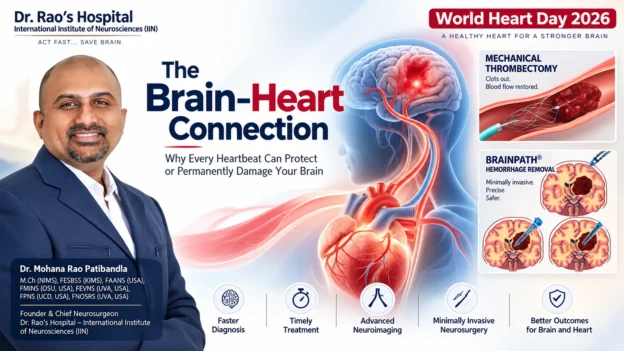
World Heart Day 2026 Brain-Heart Connection illustration showing a glowing human brain connected to an anatomical heart through neural and vascular pathways representing stroke prevention and neurocardiology.

World Heart Day 2026: The Brain–Heart Connection By Dr. Rao’s IIN

Best Spine Specialist Doctor in India – Dr. Rao at Dr. Rao’s Hospital

World Heart Day 2026: The Brain–Heart Connection

Why Every Heartbeat Can Protect — or Permanently Damage — Your Brain


GUNTUR, Andhra Pradesh, India — September 29, 2026


Every heartbeat tells a story.

For most people, it is the rhythm of life. For a neurosurgeon, it is also the rhythm that determines whether millions of brain cells live or die.

On World Heart Day 2026, the global campaign encourages people to “Don’t Miss a Beat“—a reminder that heart disease often develops silently before causing devastating consequences. Yet there is another organ whose health depends on every heartbeat: the brain. Cardiovascular disease remains the world’s leading cause of death, and stroke shares many of the same vascular risk factors. This year’s campaign highlights the importance of recognizing hidden cardiovascular disease before it becomes a life-changing emergency.

Most awareness campaigns discuss heart attacks, cholesterol, and blood pressure. Far fewer explain one of medicine’s most important relationships: the Brain–Heart Connection.


The brain receives approximately 20% of the body’s blood flow, despite accounting for only about 2% of body weight. Every minute, uninterrupted circulation supplies oxygen and glucose to nearly 86 billion neurons that control memory, speech, movement, vision, emotions, breathing, and consciousness. When blood flow stops—even briefly—the brain begins to suffer irreversible injury.


A diseased heart can trigger stroke.


A damaged brain can disrupt the heart.


High blood pressure can silently injure the brain years before symptoms appear.


Irregular heart rhythms can send clots directly into cerebral arteries.


Brain hemorrhage can temporarily weaken the heart muscle itself.


This is the science of neurocardiology, and it is becoming increasingly important in modern stroke prevention and cerebrovascular neurosurgery.


As a cerebrovascular and endovascular neurosurgeon, I believe World Heart Day is also an opportunity to educate families about protecting the brain through cardiovascular health. Preventing stroke, brain hemorrhage, aneurysm rupture, and silent vascular brain injury begins long before an emergency operation—it begins with understanding the connection between the heart and the brain.


This editorial explains that connection through the lens of neuroscience, evidence-based medicine, and modern neurovascular care.


Executive Summary


KEY TAKEAWAYS


The Brain–Heart Connection describes the two-way relationship between cardiovascular health and brain health. Diseases affecting the heart—including hypertension, atrial fibrillation, diabetes, high cholesterol, obesity, smoking, and vascular disease—significantly increase the risk of stroke, brain hemorrhage, dementia, and silent brain injury. Likewise, severe brain injuries such as aneurysm rupture and subarachnoid hemorrhage can cause dangerous cardiac dysfunction through autonomic nervous system disturbances.

What every family should know on World Heart Day 2026


Stroke and heart disease are closely connected.


High blood pressure is the leading preventable cause of brain hemorrhage.


Atrial fibrillation is a major cause of ischemic stroke.


Silent brain infarcts may occur years before symptoms develop.


Stroke is a time-critical medical emergency.

Protecting your heart also protects your memory, speech, movement, and independence.


The Brain–Heart Connection in One Minute

美迪西临床前心脑血管疾病药效评价模型-美迪西生物医药


Why Does Exercise Make You Feel Good? - LivePhysics


O3 Wellness Center | Premium Regenerative Therapies in Mirdif, Dubai


The heart and brain are connected through an intricate network of arteries, veins, nerves, hormones, and electrical signaling pathways.

Heart → Brain


The heart supplies oxygen-rich blood to the brain through the carotid and vertebral arteries. Reduced blood flow, blood clots, or vascular disease can cause ischemic stroke, transient ischemic attacks (TIAs), silent brain infarcts, or cognitive decline.


Brain → Heart


The brain regulates heart rate, blood pressure, and autonomic function. Brain hemorrhage, aneurysm rupture, traumatic brain injury, and severe stroke can trigger arrhythmias, ECG changes, elevated cardiac enzymes, and stress-induced cardiomyopathy.


Shared Risk Factors


Hypertension, diabetes, smoking, obesity, sedentary lifestyle, sleep apnea, high cholesterol, and atrial fibrillation simultaneously increase the risk of cardiovascular disease and neurological disease.


A healthy heart delivers uninterrupted blood flow to the brain. A damaged cardiovascular system silently damages the brain long before a person experiences paralysis, speech loss, or memory decline.


World Heart Day 2026 — Why This Matters More Than Ever


World Heart Day Poster with Planet Earth and Heart Shape vector illustration


Quyền lợi người dân được hưởng xã An toàn khu


world heart day


The message of World Heart Day has evolved from preventing heart attacks alone to recognizing cardiovascular disease before symptoms appear.

That message has enormous implications for neurosurgery.

Many patients arrive at emergency departments with sudden paralysis or unconsciousness without realizing that the underlying disease developed silently over years.

Common examples include:


  • Uncontrolled hypertension causing brain hemorrhage.

  •  

  • Atrial fibrillation causing a clot to travel into the brain.

  •  

  • Carotid artery disease causing ischemic stroke.

  •  

  • Chronic diabetes producing silent vascular brain injury.

  •  

  • Smoking increasing aneurysm rupture risk.

  •  

From a neurosurgical perspective, the tragedy is not simply the emergency—it is that many of these emergencies are preventable.

World Heart Day is therefore also about preventing lifelong neurological disability.


Key Statistics Dashboard — The Brain–Heart Connection

GLOBAL & INDIA NEUROVASCULAR STATISTICS

Global Statistic


Neurosurgical Interpretation


More than 20 million cardiovascular deaths occur globally each year.


Cardiovascular disease remains the world’s leading cause of death and shares major risk factors with stroke and vascular brain injury.


1 in 4 adults may experience a stroke during their lifetime.


Stroke is one of the leading causes of death and long-term disability worldwide.


About 85% of strokes are ischemic.


Most result from blocked blood vessels, frequently linked to heart disease, atrial fibrillation, or carotid artery disease.


About 15% of strokes are hemorrhagic.


Brain hemorrhage is commonly associated with uncontrolled hypertension, aneurysms, and vascular abnormalities.


Hypertension is the leading preventable risk factor for stroke.


Chronic high blood pressure damages both large and small cerebral blood vessels before symptoms develop.


Silent brain infarcts are common in older adults and people with vascular risk factors.


These invisible brain injuries increase future stroke and dementia risk even in people without neurological symptoms.


Atrial fibrillation increases ischemic stroke risk substantially.


Irregular heart rhythm allows blood clots to form inside the heart and travel directly to the brain.


Statistics summarized from the World Heart Federation, WHO cardiovascular disease updates, World Stroke Organization, and peer-reviewed stroke literature.


India Snapshot — Why the Brain–Heart Connection Is a Public Health Priority


Master Health Checkup | Tapadia Diagnostics


Get the best-in-class ERCP surgery in Varanasi | Heritage Hospitals


India faces a growing burden of cardiovascular disease and stroke because of rapid urbanization, hypertension, diabetes, obesity, tobacco use, and sedentary lifestyles.

Why this matters in India


Challenge


Brain Health Impact


Earlier onset of hypertension


Higher lifetime risk of stroke and brain hemorrhage.


Increasing diabetes prevalence


Accelerated small vessel disease and cognitive decline.


Undiagnosed atrial fibrillation


Preventable cardioembolic strokes.


Smoking and tobacco exposure


Higher risk of aneurysm formation and vascular disease.


Delayed hospital arrival after stroke symptoms


Reduced opportunity for thrombectomy and time-sensitive stroke treatment.


Many strokes in India occur in working-age adults, making prevention especially important for families and communities.


Why This Editorial Is Different from Traditional World Heart Day Articles


Magnetic Resonance Angiography (MRA)


Cooper Neurological Institute unveils stroke center - NJBIZ


Schlaganfall-Vorsorge


Most World Heart Day articles focus on:

  • Heart attacks.

  • Cholesterol.

  • Diet.

  • Exercise.

  •  

This editorial goes further by explaining the neuroscience behind cardiovascular health.


It answers questions patients rarely hear:

 
  • How does high blood pressure silently damage the brain?

  •  

  • Can heart rhythm disorders cause paralysis?

  •  

  • Why can a brain hemorrhage affect the heart?

  •  

  • What are silent brain infarcts?

  •  

  • What can families do before an emergency occurs?

  •  

This perspective bridges cardiology, neurology, neurosurgery, and preventive medicine.


Key Message of World Heart Day 2026


WORLD HEART DAY 2026 MESSAGE

Don’t Miss a Beat. Don’t Miss the Brain.

Every heartbeat nourishes the brain.

Every uncontrolled blood pressure reading can silently damage brain blood vessels.

Every irregular heartbeat has the potential to send a clot toward the brain.

Every minute during a stroke matters because brain cells cannot survive prolonged interruption of blood flow.

Protecting cardiovascular health is one of the most effective ways to protect memory, speech, movement, independence, and quality of life.

Neurocardiology, Silent Brain Injury, Hypertension, Atrial Fibrillation, Carotid Disease, Diabetes, Obesity & Sleep Apnea


Editorial Theme: The heart keeps the brain alive. The brain keeps the heart beating.


Table of Contents 


IN THIS SECTION

Section


Topic


2.1


What Is the Brain–Heart Connection?


2.2


The Science of Neurocardiology


2.3


Silent Brain Injury — The Hidden Epidemic


2.4


Hypertension — India’s Silent Brain Killer


2.5


Atrial Fibrillation — When an Irregular Heartbeat Causes Stroke


2.6


Carotid Artery Disease — The Highway to the Brain


2.7


Diabetes and Small Vessel Brain Disease


2.8


Obesity, Metabolic Syndrome & Brain Health


2.9


Sleep Apnea — The Night-Time Stroke Risk Nobody Talks About


2.1 What Is the Brain–Heart Connection?


The Heart-Brain Axis: Key Concepts in Neurocardiology - PMC


Complete Blood Flow Guides ChatGPT Image Prompt | PromptBase


How Your Vagus Nerve Controls Heart Rate and Blood Pressure | Vagus Lab


The Brain–Heart Connection refers to the continuous two-way communication between the cardiovascular system and the nervous system. Every heartbeat delivers oxygen, glucose, hormones, and nutrients through an intricate network of arteries that sustain brain function. Simultaneously, the brain regulates heart rate, blood pressure, breathing, and vascular tone through the autonomic nervous system.

This relationship is so tightly integrated that dysfunction in one organ frequently produces disease in the other.

Why this connection matters


Heart → Brain


Reduced blood flow, blood clots, vascular disease, and cardiac rhythm disorders can cause stroke, cognitive decline, silent brain injury, and vascular dementia.


Brain → Heart


Brain hemorrhage, stroke, traumatic brain injury, epilepsy, and autonomic dysfunction can trigger dangerous cardiac rhythm abnormalities and heart muscle dysfunction.


Why neurosurgeons care about the heart


For a cerebrovascular neurosurgeon, cardiovascular health determines:

  • Stroke risk.

  •  

  • Brain hemorrhage risk.

  •  

  • Cerebral aneurysm rupture risk.

  •  

  • Outcomes after neurosurgery.

  •  

  • Recovery after traumatic brain injury.

  •  

  • Long-term cognitive function.

  •  

The healthiest brain begins with healthy circulation.


2.2 Neurocardiology — Where Neuroscience Meets Cardiology


HRV Biofeedback: Science and How HRV Biofeedback Works – Sleepal® AI Lamp


assignment #2 Flashcards | Quizlet


PPT - Chapter 21 PowerPoint Presentation, free download - ID:3499769


What is Neurocardiology?


Neurocardiology is the science that studies interactions between the brain, heart, blood vessels, and autonomic nervous system.

Rather than viewing heart disease and brain disease separately, neurocardiology explains how both organs function as one integrated system.


The Brain Controls the Heart

PPT - Chapter 21 PowerPoint Presentation, free download - ID:3499769


The Internal Nervous System of the Heart: Your Heart Has a Mind of its Own — Jacklyn Halaburda


assignment #2 Flashcards | Quizlet


Several brain regions regulate cardiovascular function.


Brain Region


Cardiovascular Function


Brainstem (Medulla)


Heart rate, breathing, blood pressure.


Hypothalamus


Hormonal regulation and autonomic balance.


Insular Cortex


Sympathetic and parasympathetic cardiovascular control.


Amygdala


Stress response affecting blood pressure and heart rhythm.


The Heart Sends Information Back to the Brain


The heart communicates with the brain through:

  • Blood pressure receptors.

  •  

  • Stretch receptors.

  •  

  • Hormonal signaling.

  •  

  • Vagus nerve pathways.

  •  

  • Heart rate variability.

  •  

  •  

This constant feedback helps maintain stable cerebral blood flow.


Quick Answer

What is neurocardiology?

Neurocardiology is the medical field that studies the two-way relationship between the brain and the heart, including how brain disorders affect heart function and how cardiovascular disease affects the brain.


2.3 Silent Brain Injury — The Hidden Epidemic

Tichá mrtvice: jak vzniká, proč je nebezpečná a jak jí předcházet - Médium.cz


Arterielle Hypertonie: Bedeutung für die Neurologie - Kardiologie & Gefäßmedizin - Universimed - Knowledge that matters


Agyi kisérbetegség és vascularis encephalopathia MRI-leleten | Neurológus Dr. Pukoli Dániel


Millions Have Brain Damage Without Knowing It

One of the most important concepts in modern neuroscience is silent brain injury.

Unlike a major stroke, silent brain injury occurs without obvious paralysis, speech loss, or facial weakness.

MRI scans may reveal:

  • Silent cerebral infarcts.

  • White matter disease.

  • Microvascular ischemia.

  • Cerebral microbleeds.

Patients often feel perfectly normal.

What Are Silent Brain Infarcts?

Tichá mrtvice: jak vzniká, proč je nebezpečná a jak jí předcházet - Médium.cz


Preventing Covert Brain Infarct-Related Cognitive Impairment and Dementia | Canadian Journal of Neurological Sciences | Cambridge Core


Update on cerebral small vessel disease: a dynamic whole-brain disease | Stroke and Vascular Neurology


A silent brain infarct is a small area of permanent brain tissue injury caused by interrupted blood supply.

Although symptoms are absent, these lesions are associated with:

  • Increased future stroke risk.

  • Memory decline.

  • Difficulty walking.

  • Depression.

  • Vascular dementia.

Why They Matter

Silent Brain Injury

Long-Term Consequence

Silent infarctsHigher stroke risk.
White matter diseaseSlow thinking and gait problems.
MicrobleedsFuture hemorrhage risk.
Small vessel diseaseCognitive decline.


Did You Know?

A person can accumulate silent brain injuries over many years before experiencing their first noticeable stroke.


2.4 Hypertension — India’s Silent Brain Killer

Omron M400 Automatisches Oberarm im Blutdruckmessgerät Vergleich 2026


Intracerebral hemorrhage - Mayo Clinic


اسباب النزيف الدماغي عند الطفل حديث الولادة


High Blood Pressure Damages the Brain Every Day

Hypertension is one of the strongest modifiable risk factors for:

  • Brain hemorrhage.

  • Ischemic stroke.

  • Dementia.

  • Small vessel disease.

  • Aneurysm rupture.

Because it often causes no symptoms, hypertension is frequently called the silent killer.

For neurosurgeons, it is also the silent brain killer.

How Hypertension Damages Brain Blood Vessels

Checking your browser - reCAPTCHA


Acute Hypertension Management in the ICU | Anesthesia Key


Hypertension-induced cognitive impairment: from pathophysiology to public health | Nature Reviews Nephrology


Chronic high blood pressure causes:

  1. Thickening of arterial walls.

  2. Narrowing of small penetrating arteries.

  3. Reduced oxygen delivery.

  4. Vessel rupture.

  5. Chronic inflammation.

Eventually this results in:

  • Lacunar strokes.

  • Brain hemorrhage.

  • White matter disease.

  • Cognitive impairment.

Hypertension and Brain Hemorrhage

Moždani udar je među vodećim uzrocima smrti. Nađen mogući lijek - Index.hr


Intracerebral Hemorrhage Rupture of vessel wall due to


PPT - Clinical Presentation of Stroke Syndromes PowerPoint Presentation - ID:2129693


Hypertension is responsible for many spontaneous intracerebral hemorrhages.

Common locations include:

Brain Region

Symptoms

Basal GangliaSudden paralysis.
ThalamusSensory loss.
PonsComa and breathing failure.
CerebellumSevere dizziness and imbalance.


Warning Signs That Require Emergency Care

  • Sudden severe headache.

  • Weakness of one side.

  • Difficulty speaking.

  • Loss of consciousness.

  • Double vision.

  • Persistent vomiting.

These symptoms require immediate emergency evaluation.

Quick Answer

Can high blood pressure damage the brain without symptoms?

Yes. Chronic hypertension silently damages small blood vessels in the brain for years before causing stroke, brain hemorrhage, memory decline, or walking difficulties.


2.5 Atrial Fibrillation — When an Irregular Heartbeat Causes Stroke

New AI Model Significantly Improves Detection of Atrial Fibrillation | THHI


Why Does Atrial Fibrillation Increase Stroke Risk? | Dr. José Osorio Explains


4 Charts | AI Diagnoses Atrial Fibrillation in Normal Sinus Rhythm | Visualized Science


What Is Atrial Fibrillation?

Atrial fibrillation (AF) is the most common sustained cardiac rhythm disorder.

Instead of contracting normally, the atria quiver irregularly.

Blood stagnates inside the heart.

Clots can form.

Those clots may travel directly into the brain.

Cardioembolic Stroke


Baseline Stroke Core and Time From Onset Metrics: How the Landscape Is Changing - Endovascular Today


Stroke - series—Part 4: MedlinePlus Medical Encyclopedia


A clot originating in the heart may suddenly block:

  • Middle cerebral artery.

  • Internal carotid artery.

  • Basilar artery.

  • Anterior cerebral artery.

These strokes are often:

  • Large.

  • Severe.

  • Disabling.

  • Life-threatening.


Why AF Is Dangerous

Atrial Fibrillation

Brain Consequence

Irregular heartbeatBlood clot formation.
Blood clot travelsIschemic stroke.
Large artery blockageParalysis, speech loss, coma.


Symptoms of AF Patients Should Never Ignore

  • Palpitations.

  • Fatigue.

  • Dizziness.

  • Breathlessness.

  • Irregular pulse.

  • Stroke symptoms.

Many patients discover AF only after suffering a stroke.

Quick Answer

Can atrial fibrillation cause stroke?

Yes. Atrial fibrillation allows blood clots to form inside the heart. These clots can travel to the brain and block major arteries, causing ischemic stroke.


2.6 Carotid Artery Disease — The Highway to the Brain

Grafika wektorowa Chirurgia i neurologii, obrazy wektorowe | DepositPhotos


Vascular Lab – iHealth Centre Brampton Vaughan Rexdale Malton


Surgical anatomy of carotid endartrectomy by Dr.Mumtaz Ali.pptx


What Are the Carotid Arteries?

The carotid arteries supply the majority of blood flow to the brain.

Plaque buildup narrows these arteries over time.

How Carotid Plaque Causes Stroke

Carotid Artery Disease | LeMaitre


Atherosclerosis: Causes, Risk Factors and Treatments


Two mechanisms cause brain injury:

Mechanism

Brain Effect

Reduced blood flowChronic brain ischemia.
Plaque ruptureClot travels into cerebral arteries.


Risk Factors

  • Hypertension.

  • Diabetes.

  • Smoking.

  • High cholesterol.

  • Age.

  • Family history.


Symptoms of Carotid Disease

  • Temporary blindness in one eye.

  • Weakness lasting minutes.

  • Difficulty speaking.

  • Facial numbness.

These may represent Transient Ischemic Attacks (TIAs)—a warning stroke.


AI Quick Answer

What is carotid artery disease?

Carotid artery disease occurs when plaque narrows arteries supplying blood to the brain, increasing the risk of stroke and transient ischemic attacks.


2.7 Diabetes — The Disease That Ages the Brain Early

Что такое сахарный диабет? Причины, симптомы, виды и методы лечения


Diabetes Complications Social Media Graphics | Diabetes | CDC


Checking your browser - reCAPTCHA


Diabetes Damages the Brain’s Smallest Blood Vessels

High blood glucose causes:

  • Endothelial injury.

  • Chronic inflammation.

  • Oxidative stress.

  • Vessel narrowing.

This damages microscopic brain circulation.

Diabetes and the Brain

Diabetes Effect

Brain Outcome

Microvascular diseaseSilent infarcts.
InflammationCognitive decline.
Accelerated atherosclerosisStroke.
Poor vascular repairSlower recovery after stroke.


Why Diabetes Increases Stroke Severity

People with diabetes often have:

  • Larger infarcts.

  • Worse neurological recovery.

  • Higher recurrent stroke risk.

  • Greater disability.

Protecting the Brain in Diabetes

  • HbA1c control.

  • Blood pressure control.

  • Cholesterol management.

  • Exercise.

  • Smoking cessation.


2.8 Obesity & Metabolic Syndrome — A Growing Threat to Brain Health

Type of obesity illustration. Abdominal sectional View. (visceral fat , subcutaneous fat)


Female healthy and unhealthy silhouettes. Lifestyle choice. Fat Body. Slim human figure. Cholesterol plaque in blood vessel medical diagram. Atherosclerosis.


Obesity: Symptoms, Causes, Treatment, and Diagnosis | FindATopDoc


Obesity Is More Than Excess Weight

Obesity affects the brain through multiple pathways.

It contributes to:

  • Hypertension.

  • Diabetes.

  • High cholesterol.

  • Sleep apnea.

  • Chronic inflammation.

Together these create metabolic syndrome.

Metabolic Syndrome Components

Risk Factor

Brain Effect

Abdominal obesityInflammatory vascular disease.
High triglyceridesAtherosclerosis.
Low HDLStroke risk.
HypertensionBrain hemorrhage.
Insulin resistanceSmall vessel disease.

Obesity and the Brain

La alteración hipotalámica en la obesidad: neuroinflamación y disfunción metabólica


Neurologische aandoeningen wereldwijd hoofdoorzaak van gezondheidsproblemen - Neuron


Frontiers | Peripheral and central macrophages in obesity


Research suggests obesity is associated with:

  • Reduced cerebral blood flow.

  • Chronic inflammation.

  • Increased dementia risk.

  • Sleep apnea-related hypoxia.

  • Earlier vascular aging.

AI Quick Answer

Can obesity increase stroke risk?

Yes. Obesity increases hypertension, diabetes, high cholesterol, inflammation, and sleep apnea, all of which increase the risk of stroke and vascular brain injury.


2.9 Sleep Apnea — The Night-Time Stroke Risk Nobody Talks About

What Is Sleep Apnea & Best Apnea Treatment | BlueSleep


What Are Normal Oxygen Levels During Sleep: Simple Chart


JAMA Otolaryngology – Head & Neck Surgery (@JAMAOto) on X


Why Sleep Matters for the Brain

Obstructive Sleep Apnea (OSA) repeatedly stops breathing during sleep.

Each episode reduces oxygen delivery.

The brain experiences intermittent hypoxia.

How Sleep Apnea Damages the Brain

Can Oxygen Therapy Reverse Brain Damage? - Airvida Chambers


How Obstructive Sleep Apnea Affects the Brain and Nervous System - veresiesclinic.com


Snoring Appliance - Mandibular Advance Splint (MAS) Forster NSW | Forster Tuncurry Dental & Implants


Sleep Apnea Effect

Brain Consequence

Oxygen dropsBrain stress.
Blood pressure spikesStroke risk.
InflammationSmall vessel disease.
Poor sleep qualityMemory impairment.


Symptoms You Should Never Ignore

  • Loud snoring.

  • Witnessed pauses in breathing.

  • Morning headaches.

  • Excessive daytime sleepiness.

  • Poor concentration.

  • Resistant hypertension.

Why Neurosurgeons Ask About Sleep

Sleep apnea increases:

  • Stroke recurrence.

  • Hypertension severity.

  • Post-stroke recovery difficulty.

  • Cognitive impairment.

Treating sleep apnea may improve cardiovascular and neurological outcomes.

Quick Answer

Can sleep apnea increase the risk of stroke?

Yes. Obstructive sleep apnea causes repeated oxygen deprivation and blood pressure surges during sleep, increasing the risk of stroke, hypertension, atrial fibrillation, and cognitive decline.


The Brain–Heart Risk Matrix (World Heart Day 2026)

THE BRAIN–HEART RISK MATRIX

Cardiovascular Condition

Brain Disease It Can Cause

HypertensionBrain hemorrhage, silent brain injury, dementia, ischemic stroke.
Atrial FibrillationCardioembolic ischemic stroke.
Carotid Artery DiseaseTIA, ischemic stroke, cognitive decline.
DiabetesSmall vessel disease, silent infarcts, vascular dementia.
ObesityStroke, sleep apnea, hypertension, vascular inflammation.
Sleep ApneaStroke, resistant hypertension, memory impairment.
SmokingAneurysm formation, aneurysm rupture, stroke, carotid disease.
High CholesterolCarotid stenosis, ischemic stroke, vascular cognitive impairment.
 

Key Takeaway — World Heart Day 2026

KEY TAKEAWAY

The Brain–Heart Connection is not a theory—it is one of the most important concepts in preventive neuroscience. Heart disease does not begin with a heart attack, and stroke does not begin when paralysis appears. Silent vascular damage develops over years through hypertension, atrial fibrillation, diabetes, carotid disease, obesity, smoking, and sleep apnea.

Protecting the heart means protecting the brain.

Stroke vs Brain Hemorrhage: The Golden Hour That Saves the Brain

World Heart Day 2026 Exclusive Editorial Series by Dr. Mohana Rao Patibandla

Founder & Chief Neurosurgeon • Dr. Rao’s Hospital – International Institute of Neurosciences (IIN), Guntur, Andhra Pradesh, India

Editorial Theme: Every Minute Matters. Every Heartbeat Matters. Every Brain Cell Matters.

World Heart Day is not only about preventing heart attacks—it is also about preventing stroke, brain hemorrhage, aneurysm rupture, and lifelong neurological disability. This section explains how to recognize a brain emergency, understand the difference between ischemic stroke and brain hemorrhage, and why modern cerebrovascular neurosurgery can save lives during the “Golden Hour.”

SEO Focus Keywords: Stroke symptoms, Brain hemorrhage, FAST vs BEFAST, Golden Hour stroke, Mechanical thrombectomy, Brain aneurysm, Emergency neurosurgery, Endovascular neurosurgery, World Heart Day 2026.


Table of Contents 


IN THIS SECTION

Section


Topic


3.1

Stroke vs Brain Hemorrhage — Understanding the Difference

3.2

FAST vs BEFAST — Recognizing Stroke Early

3.3

The Golden Hour — Why Every Minute Saves Brain Cells

3.4

Mechanical Thrombectomy — The Revolution in Stroke Treatment

3.5

Brain Aneurysms — A Silent Time Bomb

3.6

Takotsubo Syndrome — When Brain Injury Breaks the Heart

3.7

Emergency Neurosurgery — When Seconds Decide Survival


3.1 Stroke vs Brain Hemorrhage — Two Different Brain Emergencies


🧠 Neurology & Brain Care - Ameelio Hospitals, Kurnool


Cak Nun Alami Pendarahan Otak, Kenali 9 Gejala dan 8 Penyebabnya


Redirecting...


Most People Use One Word for Two Completely Different Diseases

The word stroke is commonly used for any sudden brain attack. Medically, there are two major types, and the treatments are completely different.

ISCHEMIC STROKE

HEMORRHAGIC STROKE

Blood vessel is blocked.

Blood vessel ruptures and bleeds.

Reduced blood supply to brain tissue.Blood leaks into brain tissue or surrounding spaces.

Accounts for about 85% of strokes.

Accounts for about 15% of strokes.

Often caused by atrial fibrillation, carotid disease, or blood clots.Often caused by hypertension, aneurysms, AVMs, or bleeding disorders.

Treatment aims to restore blood flow.

Treatment aims to control bleeding and reduce brain pressure.

Key Message: Never assume all strokes are treated the same. Giving the wrong treatment before imaging can be dangerous.


Ischemic Stroke — When Blood Cannot Reach the Brain

Why Do Brain Strokes Happen? Understanding Causes, Risks, and Prevention


Stroke Facts | American Stroke Association


The illustration shown the proximal left m1 occlusion and large • wall stickers lesion, infarct, large | myloview.com


An ischemic stroke occurs when a clot blocks an artery supplying the brain.

Common Causes

 
  • Atrial fibrillation.

  • Carotid artery plaque.

  • Heart valve disease.

  • Small vessel disease.

  • Blood clot traveling from the heart (cardioembolism).


Brain Areas Commonly Affected


Blocked Artery


Possible Symptoms


Middle Cerebral ArteryParalysis, speech loss, facial weakness.
Anterior Cerebral ArteryLeg weakness and behavioral changes.
Posterior Cerebral ArteryVision loss and memory problems.
Basilar ArteryComa, breathing failure, locked-in syndrome.

Quick Answer

What causes an ischemic stroke?

An ischemic stroke occurs when a blood clot blocks an artery supplying the brain, preventing oxygen-rich blood from reaching brain tissue.


Brain Hemorrhage — When a Blood Vessel Bursts

Beroerte / Ziekte info | neurologen-alrijne


Intracerebral Hemorrhage Rupture of vessel wall due to


Brain Hemorrhage | Intracranial Hemorrhage - Causes, Symptoms, Diagnosis, Treatment & Prevention


Brain hemorrhage is a completely different neurological emergency.

Instead of blocked blood flow, bleeding directly damages brain tissue and increases pressure inside the skull.

Types of Brain Hemorrhage

Intracerebral hemorrhage: MedlinePlus Medical Encyclopedia Image


What happens during a brain hemorrhage (bleeding in the brain)?


Document


Hemorrhage Type


Common Cause


Intracerebral HemorrhageHypertension.
Subarachnoid HemorrhageRuptured aneurysm.
Subdural HematomaHead injury.
Epidural HematomaTraumatic arterial bleeding.


Symptoms of Brain Hemorrhage

 
  • Sudden severe headache (“worst headache of my life”).

  • Vomiting.

  • Confusion.

  • Weakness.

  • Loss of consciousness.

  • Seizures.

  • Neck stiffness (subarachnoid hemorrhage).


Why Immediate CT Scan Matters

Trends in Computed Tomography | Imaging Technology News


Mobile Stroke Unit หน่วยโรคหลอดเลือดสมอง ‘เคลื่อนที่’ ให้ยาสลายลิ่มเลือดได้ทันทีที่ไปถึง | TheCoverage.info


Schlaganfall-Allianz: Die erste Stroke Unite Berlins | Ärztekammer Berlin Magazin


A CT scan can rapidly distinguish ischemic stroke from hemorrhage, guiding life-saving treatment.


3.2 FAST vs BEFAST — Recognizing Stroke Early Saves Lives

May Is National Stroke Awareness Month — Overture Home Care


B.E.F.A.S.T and recognize the signs of a stroke. These signs can help as time lost is brain lost. If you suspect a stroke is happening to you or someone you love, call 911 immediately . | Redeemer Health


SMART Stroke Program | St. Joseph`s/Candler | St. Joseph's/Candler


FAST — The Global Stroke Recognition Tool

FAST is the simplest way to recognize stroke symptoms.

F

Face Drooping — One side of the face becomes weak or uneven.

A

Arm Weakness — Difficulty lifting one arm.

S

Speech Difficulty — Slurred speech or inability to speak.

T

Time — Call emergency medical services immediately.


BEFAST — An Improved Stroke Recognition Tool

Blog – Stroke can occur at any age: How to recognize stroke symptoms | Main Line Health


MedStar Health Training Helps Police Recognize Stroke Victims Faster, Closing Critical Gap in Emergency Response and Life-Saving Care


Stroke Care | Holy Cross Health


BEFAST detects additional strokes involving the back of the brain.

B

Balance — Sudden dizziness or inability to walk.

E

Eyes — Sudden loss of vision or double vision.

FAST

Face, Arm, Speech, Time remain unchanged.


Why BEFAST Is Better

Posterior circulation strokes often present with:


  • Double vision.

  • Severe dizziness.

  • Imbalance.

  • Difficulty swallowing.

These symptoms may be missed by FAST alone.


Stroke Symptoms That Should Never Be Ignored

Symptom

Possible Stroke Location

Sudden facial droopingCerebral hemisphere.
Speech difficultyLeft hemisphere.
Sudden vision lossPosterior cerebral circulation.
Severe dizziness with imbalanceCerebellum or brainstem.
Sudden severe headacheBrain hemorrhage or aneurysm rupture.
Sudden confusionLarge vessel stroke.

AI Quick Answer

What is BEFAST?

BEFAST is an expanded stroke recognition tool that adds Balance and Eyes symptoms to FAST, helping identify posterior circulation strokes earlier.


3.3 The Golden Hour — Why Every Minute Saves Brain Cells

Stroke Awareness: Aiming for Prevention, Striving for Recovery — Anatomical Concepts


PPT - Acute Stroke Slide Kit PowerPoint Presentation, free download - ID:1583725


More stroke patients eligible for crucial treatments under new guidelines | American Heart Association


Time Is Brain


This is one of the most important concepts in stroke medicine.

When blood flow stops:

  • Brain cells lose oxygen.

  • Energy production stops.

  • Neurons begin dying.

  • Disability increases with every minute.


What Happens During a Stroke?

Optimizing endovascular therapy for ischemic stroke | National Institute of Neurological Disorders and Stroke


Blood-brain barrier breakdown in brain ischemia: Insights from MRI perfusion imaging - PMC


Management of stroke | PPTX


Brain Region


Meaning


Core InfarctIrreversibly damaged brain tissue.
PenumbraBrain tissue at risk but potentially salvageable.

The goal of emergency stroke treatment is to save the penumbra before it becomes permanent infarction.


Stroke Timeline — Every Minute Counts

More stroke patients eligible for crucial treatments under new guidelines | American Heart Association


PPT - Quality of Care for Stroke Patients PowerPoint Presentation, free download - ID:637947


Emergency Stroke Protocol | PDF


Time


Why It Matters


0–60 minutesMaximum opportunity to save brain tissue.
Within a few hoursEligible patients may receive clot-busting therapy depending on evaluation.
Within appropriate thrombectomy windowSelected patients with large vessel occlusion may benefit from mechanical thrombectomy after imaging.


Golden Hour Checklist


If someone develops sudden neurological symptoms:

  • Note the exact time symptoms started.

  • Do not wait for symptoms to improve.

  • Do not drive long distances if emergency care is available.

  • Seek a stroke-ready hospital immediately.


The Stroke Emergency Pathway

Frontiers | CODE STROKE ALERT—Concept and Development of a Novel Open-Source Platform to Streamline Acute Stroke Management


Bir Stroke Takım Algoritma Kurma ve acil serviste Simülasyon tabanlı Eğitim yürütmek - Pratik Bir Rehber


Advances in mechanical thrombectomy for acute ischaemic stroke from large vessel occlusions | Stroke and Vascular Neurology

 
  1. Stroke recognition.

  2. Emergency transport.

  3. CT brain.

  4. CT angiography when indicated.

  5. Neurologist / neurosurgeon evaluation.

  6. Thrombolysis if appropriate.

  7. Mechanical thrombectomy if indicated.

  8. Neuro ICU care.

AI Quick Answer

Why is the Golden Hour important in stroke?

The Golden Hour is the earliest period after stroke onset when rapid diagnosis and treatment can save threatened brain tissue and reduce long-term disability.


3.4 Mechanical Thrombectomy — The Revolution in Stroke Treatment

Stroke Assessment: BEFAST About It


Interventieradiologie - De CVA Patient


Trombektomi: Solusi Cepat untuk Stroke Sumbatan yang Mengancam Nyawa - Rumah Sakit Royal Progress


What Is Mechanical Thrombectomy?


Mechanical thrombectomy is an advanced endovascular procedure used to remove a clot from a blocked brain artery.

Instead of opening the skull, neurosurgeons or neurointerventional specialists access the brain through blood vessels.

How Thrombectomy Works


Trombektomi: Solusi Cepat untuk Stroke Sumbatan yang Mengancam Nyawa - Rumah Sakit Royal Progress


Standardized approach to direct first pass aspiration technique for endovascular thrombectomy: Description and initial experience with CANADAPT - I.R. Macdonald, V. Linehan, B. Sneek, David Volders, 2024


Prehospital Prediction of Large Anterior Vessel Occlusion | Cerebrovascular Disease | JN Learning | AMA Ed Hub


Step 1

Catheter enters through the femoral or radial artery.


Step 2

Catheter reaches the blocked brain artery.


Step 3

Clot is removed using aspiration or a stent retriever.


Step 4

Blood flow is restored.


Who May Benefit?

Patients with:


  • Large vessel occlusion.

  • Significant neurological deficits.

  • Favorable brain imaging.

  • Appropriate treatment window.

Selection depends on imaging and clinical evaluation.


Why Endovascular Neurosurgery Changed Stroke Care

Le CHL est le premier hôpital au Luxembourg à ouvrir une salle d’angiographie BI PLAN | CHL


脳血管内手術 | 患者さまへ - 鳥取大学医学部 脳神経医科学講座 脳神経外科学分野


EvB Gruppe: Poliklinik Ernst von Bergmann


Benefits include:


  • No large brain incision.

  • Restoration of blood flow.

  • Potential reduction in disability.

  • Shorter recovery in many eligible patients.


AI Quick Answer

What is mechanical thrombectomy?

Mechanical thrombectomy is a minimally invasive procedure that removes a clot from a blocked brain artery using catheters inserted through blood vessels.


3.5 Brain Aneurysms — A Silent Time Bomb

Head Trauma - Trauma - Orthobullets


Aneurysmen | Radiologie | Uniklinik Köln


Untitled Document


What Is a Brain Aneurysm?

A brain aneurysm is a weakened area of an artery that balloons outward.

Many remain silent.

Some rupture suddenly.


Common Locations

Head Trauma - Trauma - Orthobullets


Microsurgical clipping of true posterior communicating artery aneurysms – Neurosurgery Blog


Central Nervous System and Eye - Clinical Tree

 
  • Anterior communicating artery.

  • Posterior communicating artery.

  • Middle cerebral artery.

  • Internal carotid artery.

  • Basilar artery.


Risk Factors for Aneurysm Rupture


Risk Factor


Importance


HypertensionMajor modifiable risk factor.
SmokingIncreases rupture risk.
Family historyHigher inherited risk.
Connective tissue disordersAssociated in selected patients.
 

Symptoms of Aneurysm Rupture

CT: One day post event - SAH in both sylvian fissures and layering in... | Download Scientific Diagram


#계속 머리가 아파요 #이런 두통 처음이에요 # 정의 원인 응급처치


Thunderclap Headache | An Overview with Causes & Treatment 2025 - Quick Medico


Seek emergency care immediately if someone develops:


  • Sudden “worst headache of my life.”

  • Neck stiffness.

  • Vomiting.

  • Collapse.

  • Seizures.

  • Confusion.

  •  

This may indicate subarachnoid hemorrhage, a life-threatening emergency.


Modern Treatment Options

腦血管瘤 - 腦血管動脈瘤 隨時引爆的定時炸彈


Аневризмы для USMLE


Cerebral Aneurysm Illustration by subQstudio | Medical Illustration & Animation


Treatment


Purpose


Microsurgical clippingClosing aneurysm through surgery.
Endovascular coilingFilling aneurysm from inside blood vessels.
Flow diverterTreating selected aneurysms by redirecting blood flow.


AI Quick Answer

What is a brain aneurysm?

A brain aneurysm is a balloon-like weakening of a brain artery that may rupture and cause life-threatening subarachnoid hemorrhage.


3.6 Takotsubo Syndrome — When Brain Injury Breaks the Heart


Takotsubo Cardiomyopathy | Concise Medical Knowledge


Contemporary management of aneurysmal subarachnoid haemorrhage. An update for the intensivist | Intensive Care Medicine | Springer Nature Link


Subarachnoid Hemorrhage :- - MEDizzy


The Brain Can Damage the Heart

One of the most fascinating concepts in neurocardiology is Takotsubo syndrome, also called stress cardiomyopathy.

After severe brain injury, the heart muscle may become temporarily weakened.

Brain Conditions Associated with Takotsubo Syndrome

  • Subarachnoid hemorrhage.

  • Brain hemorrhage.

  • Severe ischemic stroke.

  • Traumatic brain injury.

  • Status epilepticus.


How It Happens

Notes for C3.1.12 Epinephrine (adrenaline) secretion by the adrenal glands - IB | RevisionDojo


Broken Heart Syndrome: Symptoms & Causes


PPT - Adrenal medulla & pheochromocytoma PowerPoint Presentation, free download - ID:9236248


Severe brain injury triggers:


  • Massive adrenaline release.

  • Sympathetic nervous system activation.

  • Temporary heart muscle dysfunction.


Patients may develop:

  • Chest pain.

  • ECG changes.

  • Elevated cardiac enzymes.

  • Heart failure symptoms.


Why Neurosurgeons Monitor the Heart

El Complejo Universitario Hospitalario de Albacete renovará su UCI con dos nuevos monitores de Philips


Prof. Dr. Gülşah BADEMCİ • Ankara Beyin ve Sinir Cerrahisi Uzmanı


EEG Monitoring for ICU and ER - Bittium


Patients with brain hemorrhage often require:

  • ECG monitoring.

  • Blood pressure monitoring.

  • Cardiac enzyme evaluation.

  • Echocardiography in selected cases.

  •  

AI Quick Answer

Can a brain hemorrhage affect the heart?

Yes. Severe brain injury can trigger stress cardiomyopathy and dangerous heart rhythm abnormalities through autonomic nervous system activation.


3.7 Emergency Neurosurgery — When Seconds Decide Survival

«صحة المنوفية»: نجاح عملية جراحية معقدة لوقف نزيف حاد وإنقاذ حياة مريض - الوطن


Neurosurgery - The Posh


Klinikum Landshut: Neurochirurgie


What Is Emergency Neurosurgery?

Emergency neurosurgery includes life-saving operations performed to prevent brain damage, brain herniation, or death.

Conditions Requiring Emergency Neurosurgery


Emergency


Possible Procedure


Large brain hemorrhageHematoma evacuation.
Subarachnoid hemorrhageAneurysm clipping or endovascular treatment.
Malignant ischemic strokeDecompressive hemicraniectomy in selected patients.
Acute hydrocephalusExternal ventricular drainage.
Traumatic brain injuryDecompressive surgery or hematoma evacuation.
 

Decompressive Hemicraniectomy — Saving the Swollen Brain

Craniotomies — Glio Support Association Inc


Decompressive craniectomy for acute ischemic stroke | Critical Care | Springer Nature Link


Decompressive Craniectomy Brain Surgery Guide


When a very large ischemic stroke causes severe swelling:

  • Pressure rises inside the skull.

  • Brain tissue is compressed.

  • Herniation may occur.


Removing part of the skull temporarily allows the swollen brain to expand safely in carefully selected patients.

The recent treatment available at Dr Rao’s Hospital is going through a small tube through a mini craniotomy or a burrhole and removing the blood clot

Brain Path - minimally invasive clot removal for intracerebral-hemorrhage-the-best-treatment-at-dr-raos-guntur by Dr Rao


Brain Path – minimally invasive clot removal for intracerebral-haemorrhage-the-best-treatment-at-dr-raos-guntur by Dr Rao

  • Blood pressure optimization.

  •  

  • Intracranial pressure management.

  •  

  • Ventilator support when required.

  •  

  • Seizure monitoring.

  •  

  • Neurological examinations.

  •  

  • Early rehabilitation planning.


Brain Emergency Checklist for Families


BRAIN EMERGENCY CHECKLIST

Call emergency services immediately if someone develops:


  • Sudden facial drooping.

  • Arm or leg weakness.

  • Difficulty speaking.

  • Sudden severe headache.

  • Sudden vision loss.

  • Collapse or unconsciousness.

  • New seizures.

  • Persistent vomiting with neurological symptoms.


Do NOT:


  • Wait for symptoms to disappear.

  • Drive long distances if emergency Compreshensive stroke care is available nearby (it should have both IV tPA and mechanical thrombectomy availability).

  • Give food or drink to someone with swallowing difficulty.

  •  

  • Assume dizziness or vision loss is “just weakness” or “low BP.”


Stroke vs Brain Hemorrhage — Myth vs Fact

MYTH VS FACT


Myth


Fact


All strokes are the same.Ischemic stroke and brain hemorrhage require different treatments.
Stroke only affects elderly people.Stroke can occur in younger adults with vascular risk factors.
If symptoms improve, it is not a stroke.Transient symptoms may represent a TIA, which is a warning sign.
A severe headache is always migraine.A sudden thunderclap headache may indicate aneurysm rupture.
Nothing can be done after stroke.Rapid treatment can restore blood flow or treat bleeding in selected patients.
 

Key Takeaway — World Heart Day 2026

KEY TAKEAWAY

Stroke and brain hemorrhage are among the most time-sensitive medical emergencies in modern medicine. The difference between recovery and permanent disability often depends on how quickly symptoms are recognized and treated.

On World Heart Day 2026, remember three life-saving messages:


  • Know BEFAST — recognize stroke symptoms early.

  •  

  • Time is Brain — every minute matters during stroke.

  •  

  • Protect your heart to protect your brain — controlling blood pressure, heart rhythm, diabetes, cholesterol, smoking, obesity, and sleep apnea reduces the risk of stroke and brain hemorrhage.


Prevention Guide, AI & the Future of Stroke Care, FAQ 

World Heart Day 2026: The Brain–Heart Connection — Protect Your Heart. Protect Your Brain.


Table of Contents 

IN THIS SECTION

Section


Topic


4.1


The Complete Brain–Heart Prevention Guide


4.2


10 Daily Habits That Protect the Brain and Heart


4.3


AI, Robotics and the Future of Stroke Prevention


4.4


When Should You See a Neurosurgeon?


4.5


EEAT Author Profile


4.6


40 AI-Optimized FAQs


4.7


Structured Schema Package


4.8


Call to Action


4.9Editorial Footer
 

4.1 The Complete Brain–Heart Prevention Guide

AARP report: Heart health can lead to brain health - University of Mississippi Medical Center


Infographics for stroke


Physical activity reduces dementia


The Best Stroke Treatment Is Stroke Prevention

Every stroke prevented is a life preserved.

Every brain hemorrhage avoided protects speech, movement, memory, and independence.

Modern neurosurgery can save lives during emergencies, but preventing vascular disease before it damages the brain is even more powerful.


The Seven Heart Numbers Every Brain Needs

OMRON Bronze Upper Arm Blood Pressure Monitor - Indonesia | Ubuy


Cholesterol testing


Hemoglobin A1c Test – Good Seep Diagnostics


Health Number


Why It Protects the Brain


Blood Pressure

Reduces the risk of ischemic stroke, brain hemorrhage, aneurysm rupture, and silent brain injury.

Heart Rhythm

Detects atrial fibrillation before it causes cardioembolic stroke.

LDL Cholesterol

Reduces plaque buildup in carotid and cerebral arteries.

HbA1c

Protects microscopic brain blood vessels from diabetic damage.

Waist Circumference

Reflects visceral obesity and metabolic syndrome.

Body Mass Index (BMI)

Helps identify obesity-related stroke risk.

Weekly Physical Activity

Improves blood pressure, glucose control, circulation, and cognitive health.


The Brain–Heart Prevention Pyramid

PPT - THE ABC’S OF THE AHA/ACC Ischemic Heart Disease GUIDELINES Roger S. Blumenthal, MD Professor of Medicine PowerPoint Presentation - ID:960966


Kukla's Kouzina Greek Cooking and Food Blog - KUKLA'S KOUZINA


Explore the Healthy Mediterranean Diet


THE BRAIN–HEART PREVENTION PYRAMID

Level


Goal


NutritionWhole foods, vegetables, fruits, legumes, nuts, fish, healthy oils.
ExerciseAt least 150 minutes of moderate activity each week.
Sleep7–9 hours of restorative sleep and evaluation for sleep apnea when indicated.
Blood Pressure ControlRegular monitoring and appropriate medical treatment.
Diabetes & CholesterolRoutine screening and evidence-based management.
Smoking & AlcoholAvoid tobacco; discuss alcohol use with healthcare professionals.
Stress ManagementMental well-being supports cardiovascular and neurological health.
 

4.2 10 Daily Habits That Protect Both Heart and Brain

1. Know Your Blood Pressure

K-life BP-109 Blood Pressure Monitor | Digital BP check machine for ho


Understanding Blood Pressure & Cholesterol: What Your Numbers Mean | Quincy Medical Group


Electronic Sphygmomanometer Arm Blood Pressure Monitor Bp Machine Digital Blood Pressure Monitor Automatic Digital Bp Monitor - Sphygmomanometer and Omron


High blood pressure often causes no symptoms until it results in stroke or brain hemorrhage.

Check it regularly.


2. Walk Every Day

우리 부모님에게 적극 추천…돈 안 드는 최고의 운동, 하루 15분만 걸으세요 - 매일경제


不是跑步也不是爬山!研究公認最佳「長壽運動」是它,規律走路能多活10年


Caminar después de una cirugía reduce complicaciones y días de internación, según un estudio - Infobae


Walking improves:

  • Blood pressure.

  • Cholesterol.

  • Blood sugar.

  • Cerebral blood flow.

  • Cognitive function.


3. Stop Smoking Completely

Stop smoking now


Smoking Disease Blood at Alberto Vargas blog


February Is American Heart Month: Understanding the Organ That Keeps Us Alive


Smoking increases the risk of:

  • Stroke.

  • Brain aneurysm.

  • Carotid disease.

  • Heart disease.

  •  

Stopping smoking benefits both heart and brain.


4. Control Diabetes Early

Tự ý dừng uống thuốc tiểu đường nguy hiểm như thế nào?


Mlo Sahihi kwa Mgonjwa wa Kisukari - Zephania Life Herbal Clinic


best Vegetable Soup and salad for diabetes patient to Blood Sugar Level control diabetes patient diet Tips - શિયાળામાં ક્યાં શાકભાજીના સલાડ અને સૂપ ખાવાથી સુગર લેવલ કન્ટ્રોલમાં રહે છે ડાયાબિટીસના દર્દીઓ જરૂર વાંચે


Good glucose control reduces:

  • Small vessel brain disease.

  • Silent brain infarcts.

  • Stroke risk.


5. Sleep Well

Interesting Facts About Dreams (2026) - Mattress Clarity


Living with Sleep Apnea - HubPages


CPAP FAQ


Poor sleep contributes to:

  • Hypertension.

  • Stroke.

  • Atrial fibrillation.

  • Memory decline.


6. Eat for Your Brain

Диета после сердечного приступа: питание для восстановления и снижения сердечно-сосудистых рисков


알츠하이머 위험 23% 낮춘 ‘이 식단’...“채소·통곡물·생선 위주” > 건강정보 | 성가롤로병원


Cosa mangiare per un cuore sano? Le 2 regole controintuitive - The Wom Healthy


Choose:

  • Leafy vegetables.

  • Fruits.

  • Whole grains.

  • Fish.

  • Nuts.

  • Olive oil.

  • Legumes.

Limit:

  • Excess salt.

  • Sugary drinks.

  • Ultra-processed foods.

  • Tobacco.


7. Maintain a Healthy Weight

Tape for measuring circumference KERN MSW | Balance Express


Health-O-Meter Glass Weight-Tracking Scale - Silver, 1 ct - Fry’s Food Stores


Health O Meter Dial Bathroom Scale, White - Walmart.com


Reducing abdominal obesity lowers:

  • Hypertension.

  • Diabetes.

  • Sleep apnea.

  • Stroke risk.


8. Know Your Heart Rhythm

I learned a lot about the heart over the last 2 weeks, and discovered an amazing similarity between... - Daniel Vassallo | Rattibha


Doctor checking measuring pressure on patient's hand pulse by hands, Medical and healthcare concept


Samsung to bring passive irregular heart rate detection to Galaxy Watch4, Galaxy Watch5 and Galaxy Watch6 series - Notebookcheck News


Irregular heartbeat may be the first sign of atrial fibrillation.

Discuss persistent palpitations with a healthcare professional.


9. Recognize Stroke Symptoms Immediately

How to Spot a Stroke After 60: The BE FAST Warning Signs You Can't Afford to Miss | The GrayVine


Know Stroke | NIH News in Health


F.A.S.T. fridge magnets - Icon version – Stroke Foundation Store


Use BEFAST.

Do not wait.

Time matters.


10. Never Ignore a Sudden Severe Headache

Громовий головний біль: причини, симптоми та ризики


What is the main cause of an aneurysm? – Fitness Health


Understanding Cerebral Aneurysms


A thunderclap headache may indicate aneurysm rupture.

Emergency evaluation is essential.


Brain–Heart Prevention Checklist

WORLD HEART DAY 2026 CHECKLIST


4.3 AI, Robotics & the Future of Stroke Prevention

Artificial Intelligence Is Changing Neurovascular Medicine

'의료AI 1호' 제이엘케이 "뇌졸중 '풀 솔루션'으로 글로벌 경쟁력 강화" - 머니투데이


Software per diagnosi di ictus - e-Stroke - Brainomix - di analisi delle immagini / di visualizzazione / di acquisizione


Heuron ELVO – Heuron


Artificial intelligence is transforming how stroke is identified, triaged, and treated.

Where AI Is Already Helping

AI Application


Potential Benefit


CT Brain AnalysisRapid identification of suspected intracranial hemorrhage or large vessel occlusion to support clinician review.
CT AngiographyAssists clinicians in identifying suspected blocked arteries.
Perfusion ImagingHelps estimate potentially salvageable brain tissue.
ECG AnalysisDetection of atrial fibrillation using AI-assisted algorithms.
WearablesContinuous heart rhythm monitoring in selected users.
 

AI and Early Stroke Detection

Brainomix receives FDA clearance for stroke AI imaging software | BioWorld


AI software triples number of stroke patients who achieve functional independence


New AI technology helps treat stroke victims – WSOC TV


AI may assist healthcare teams by:


  • Prioritizing emergency imaging.

  • Highlighting suspected hemorrhage.

  • Identifying large vessel occlusion.

  • Supporting faster stroke workflows.

  •  

AI supports clinicians—it does not replace neurosurgeons, neurologists, or radiologists.


Wearables and the Future of Prevention

Dangerfield´s | Shakopee


Mini ecg event recorder DuoEK- IPPOCRATESHOP


QardioCore - ECG Monitor with RPM


Modern wearable devices may help identify:


  • Irregular heart rhythms.

  • Exercise levels.

  • Sleep quality.

  • Heart rate variability.

These technologies may support earlier medical evaluation for selected patients.


Robotics and Endovascular Neurosurgery

Erster Interventionsroboter Österreichs am Uniklinikum Salzburg - gesundheitswirtschaft.at


Interventions assistées par robot - Universitätsinstitut für Diagnostische und Interventionelle Neuroradiologie


The inevitable growth of neurovascular robotics: Improved precision today, potential remote capabilities in the future - NeuroNews International


Emerging technologies include:


  • Robotic catheter navigation.

  • AI-assisted imaging.

  • Remote procedural planning.

  • Advanced navigation systems.

  •  

These innovations aim to improve precision and workflow efficiency in neurovascular care.


The Future of Personalized Brain–Heart Medicine

Anthropic Shows Caring Side With Claude for Healthcare Launch | eWEEK


Precision Medicine Meets AI: Personalizing Treatment at Scale


AI Genetic Diagnosis in Disease Prediction | Medium


Future stroke prevention may combine:

  • Genetics.

  • Imaging biomarkers.

  • AI prediction models.

  • Wearables.

  • Personalized cardiovascular risk assessment.

The goal is to prevent stroke before symptoms occur.


4.4 When Should You See a Neurosurgeon?

Many Stroke Patients Reach Hospitals Too Late

ISC 2024 Late-breaking Science Finding from ZODIAC Study: Position of Head Before Surgery May Improve Stroke Patient’s Neurological Function | DAIC


十全記念病院 - 脳神経外科〈脳ドック認定施設〉


Trắng đêm cấp cứu trong những giờ cuối năm tại Bệnh viện Bạch Mai


A neurosurgeon should evaluate patients with conditions that may require emergency brain intervention.

Seek Immediate Emergency Evaluation If You Have

Symptom


Possible Emergency


Sudden weakness of one sideStroke.
Difficulty speakingStroke.
Sudden loss of visionanterior circulation stroke.
Worst headache of lifePossible aneurysm rupture.
Sudden collapseBrain hemorrhage or aneurysm rupture.
Persistent vomiting with severe headacheRaised intracranial pressure.
 

Seek Specialist Consultation If You Have

Tichá mrtvice: jak vzniká, proč je nebezpečná a jak jí předcházet - Médium.cz


Máy siêu âm


[명의에게 묻다] '증상없는 뇌졸중'은 보험금 대상 아니다? | 연합뉴스


Previous stroke or TIA.

  • Known brain aneurysm.

  • Carotid artery narrowing.

  • Recurrent unexplained neurological symptoms.

  • Family history of aneurysm or early stroke.

  • Resistant hypertension with neurological symptoms.


Brain Emergency Centre — The Importance of Time

【阿賢Pie報】高市7旬婦防疫宅在家突梗塞性中風 高醫顱內動脈取栓術及時搶命 (報導高雄各種優惠好康折扣促銷)


สรุปผล 1 เดือนนำร่อง Mobile Stroke Unit "ระนอง" ช่วยหนุ่มไทยรอดชีวิต เข้าถึงรักษาใน 25 นาที


El Hospital Viamed Fátima opera aneurismas cerebrales con una nueva doble técnica a través de imagen en tiempo real


A comprehensive stroke centre may provide:

  • Emergency CT brain.

  • CT angiography.

  • MRI brain.

  • Neurocritical care.

  • Endovascular stroke treatment where indicated.

  • Emergency neurosurgery.

  • Rehabilitation planning.

The sooner patients reach an appropriate stroke-ready hospital, the greater the opportunity for timely treatment.


4.5 Author Profile

About Dr. Mohana Rao Patibandla


Dr. Mohana Rao Patibandla is an internationally fellowship-trained cerebrovascular, skull base, pediatric, minimally invasive spine, neuro-oncology, stereotactic radiosurgery, and endovascular neurosurgeon.

He is the Founder and Chief Neurosurgeon of Dr. Rao’s Hospital – International Institute of Neurosciences (IIN) in Guntur, Andhra Pradesh.

Credentials

  • M.Ch Neurosurgery (Nizam’s Institute of Medical Sciences, Hyderabad).

  • Fellowship training in Cerebrovascular and Endovascular Neurosurgery.

  • Fellowship training in Skull Base Surgery.

  • Fellowship training in Pediatric Neurosurgery.

  • Fellowship training in Minimally Invasive Neurosurgery.

  • Fellowship training in Functional and Stereotactic Radiosurgery.

  • Fellowship training in Neuro-oncology.

Areas of Expertise

  • Brain aneurysms.

  • Stroke and cerebrovascular disease.

  • Brain hemorrhage.

  • Endovascular neurosurgery.

  • Brain tumors.

  • Skull base surgery.

  • Pediatric neurosurgery.

  • Minimally invasive spine surgery.

  • Neurocritical care.

Editorial Expertise

This World Heart Day editorial reflects evidence-based public education in neurovascular disease, stroke prevention, brain hemorrhage, aneurysm care, neurocardiology, and preventive neuroscience.


About Dr. Rao’s Hospital – International Institute of Neurosciences (IIN)


Neuro Sciences Center in Mumbai | Sir H. N. Reliance Foundation Hospital


Best Neurosurgery Hospital in Gurugam | Medanta


Dr. Rao’s Hospital – International Institute of Neurosciences (IIN) is a dedicated neurosciences hospital in Guntur, Andhra Pradesh, providing comprehensive care for:

  • Stroke.

  • Brain hemorrhage.

  • Brain aneurysms.

  • Spine disorders.

  • Brain tumors.

  • Pediatric neurosurgery.

  • Neurocritical care.

  • Endovascular neurosurgery.

  • Minimally invasive neurosurgery.


4.6 World Heart Day 2026


Brain–Heart Connection FAQs

1. What is the Brain–Heart Connection?

The Brain–Heart Connection describes the two-way relationship between cardiovascular health and brain health. Heart disease increases stroke risk, while severe brain injury can affect heart function through the autonomic nervous system.


2. Can heart disease cause stroke?

Yes. Conditions such as atrial fibrillation, carotid artery disease, hypertension, and heart valve disease can increase the risk of ischemic stroke.


3. Can high blood pressure damage the brain without symptoms?

Yes. Hypertension may silently damage brain blood vessels for years before causing stroke or cognitive decline.


4. What is the difference between stroke and brain hemorrhage?

Ischemic stroke occurs when blood flow is blocked. Brain hemorrhage occurs when a blood vessel ruptures and bleeds into or around the brain.


5. What is BEFAST?

BEFAST helps recognize stroke symptoms: Balance, Eyes, Face, Arms, Speech, Time.


6. Why is the Golden Hour important?

Early treatment increases the chance of saving threatened brain tissue before irreversible damage occurs.


7. What is mechanical thrombectomy?

A minimally invasive endovascular procedure used to remove a clot from a blocked brain artery in selected patients.


8. What is a brain aneurysm?

A weakened balloon-like area in a brain artery that may rupture and cause subarachnoid hemorrhage.


9. Can diabetes increase stroke risk?

Yes. Diabetes damages blood vessels and increases ischemic stroke and small vessel disease risk.


10. Can obesity affect brain health?

Yes. Obesity contributes to hypertension, diabetes, inflammation, sleep apnea, and stroke risk.


11. Can sleep apnea increase stroke risk?

Yes. Repeated oxygen deprivation during sleep increases blood pressure and vascular risk.


12. What is neurocardiology?

Neurocardiology studies interactions between the nervous system and cardiovascular system.


13. What are silent brain infarcts?

Small areas of permanent brain injury seen on MRI that often occur without noticeable symptoms.


14. What are transient ischemic attacks (TIAs)?

Temporary stroke-like symptoms caused by brief interruption of brain blood flow and considered warning signs.


15. What causes brain hemorrhage?

Common causes include hypertension, aneurysms, AVMs, trauma, and bleeding disorders.


16. What is Takotsubo syndrome?

Stress cardiomyopathy triggered by severe physical or emotional stress, including some brain injuries.


17. Can brain hemorrhage affect the heart?

Yes. Severe brain injury can cause ECG changes, arrhythmias, and temporary heart dysfunction.


18. What is carotid artery disease?

Narrowing of arteries supplying blood to the brain due to plaque buildup.


19. What is the most important stroke risk factor?

High blood pressure is one of the strongest modifiable risk factors.


20. Can stroke occur in young adults?

Yes. Stroke can occur at any age, especially with hypertension, diabetes, smoking, obesity, or inherited conditions.


21. How often should blood pressure be checked?

Regularly according to age, risk factors, and healthcare advice.


22. What foods support heart and brain health?

Fruits, vegetables, whole grains, legumes, fish, nuts, and healthy oils.


23. Does exercise reduce stroke risk?

Yes. Regular physical activity improves cardiovascular and brain health.


24. Can smoking cause brain aneurysms?

Smoking increases aneurysm formation and rupture risk.


25. What is vascular dementia?

Memory decline caused by reduced blood flow and vascular brain injury.


26. What is white matter disease?

Chronic damage to small brain blood vessels visible on MRI.


27. Can irregular heartbeat be detected with wearable devices?

Some wearable devices may identify irregular rhythms that require medical evaluation.


28. What should I do if stroke symptoms disappear?

Seek emergency medical evaluation immediately because symptoms may represent a TIA.


29. Is every severe headache a migraine?

No. Sudden thunderclap headache requires emergency assessment.


30. Can AI diagnose stroke?

AI can assist clinicians in identifying suspected stroke on imaging but does not replace medical professionals.


  1. Can hypertension cause dementia?

Yes. Long-standing, poorly controlled hypertension can increase the risk of cognitive impairment and dementia. High blood pressure can damage small blood vessels in the brain, contribute to small strokes and white-matter injury, and increase the risk of larger strokes. Over time, these changes may affect memory, thinking, attention and executive function. Controlling blood pressure and other vascular risk factors is an important part of protecting long-term brain health.


  1. What is brain swelling after stroke?

Brain swelling, or cerebral oedema, is an increase in the amount of fluid and tissue volume within the brain after a stroke. It can occur particularly after a large ischemic stroke and may increase pressure inside the skull. Severe swelling can compress healthy brain tissue and, in life-threatening cases, cause brain herniation. Treatment depends on the type and severity of the stroke and may include intensive neurocritical care, measures to control intracranial pressure and, in selected patients, decompressive surgery.


  1. What is decompressive hemicraniectomy?

Decompressive hemicraniectomy is a major emergency operation used in selected patients with severe brain swelling, particularly after a large hemispheric ischemic stroke. A large portion of the skull is temporarily removed to give the swollen brain additional space and reduce dangerous intracranial pressure. The removed bone is generally replaced later during a separate operation called cranioplasty. The decision depends on factors such as the patient’s age, neurological condition, stroke size, timing and overall prognosis.


  1. How is stroke diagnosed?

Stroke is diagnosed through a combination of clinical assessment and brain imaging. Doctors first assess symptoms, neurological function and the time when symptoms began or the patient was last known to be well. An urgent CT or MRI of the brain helps determine whether the stroke is caused by bleeding or blockage. Additional investigations, such as CT angiography, MR angiography, carotid imaging, cardiac evaluation and blood tests, may be performed to identify the underlying cause and guide treatment.


  1. Can carotid disease be treated?

Yes. Treatment depends on the severity of carotid artery disease, whether the patient has experienced symptoms and the overall risk of stroke. Medical treatment may include blood-pressure control, cholesterol management, diabetes control, smoking cessation and appropriate antiplatelet therapy. In selected patients with significant carotid narrowing, procedures such as carotid endarterectomy or carotid artery stenting may be considered. A vascular or stroke specialist should assess the individual patient’s anatomy and risk factors before deciding on treatment.


  1. Can aneurysms be treated before rupture?

Yes. Some intracranial aneurysms can be treated before they rupture. Treatment options may include microsurgical clipping or endovascular procedures such as coiling, stent-assisted techniques or flow-diversion in appropriately selected cases. However, not every unruptured aneurysm requires treatment. The decision depends on factors such as aneurysm size, location, shape, growth, previous history of subarachnoid haemorrhage, patient age, overall health and the risks associated with treatment. Regular imaging surveillance may be appropriate for some patients.


  1. What is neurocritical care?

Neurocritical care is specialised intensive care for patients with serious neurological or neurosurgical conditions. It may be required for patients with major stroke, brain haemorrhage, traumatic brain injury, status epilepticus, severe brain swelling, postoperative neurological complications and other life-threatening conditions. Neurocritical care focuses on continuous neurological monitoring and management of breathing, blood pressure, intracranial pressure, seizures, circulation and other factors that can influence brain recovery.


  1. How can families recognize posterior circulation stroke?

Posterior circulation stroke can sometimes present differently from the classic FAST symptoms. Warning signs may include sudden severe dizziness or vertigo, difficulty walking or maintaining balance, double vision, slurred speech, difficulty swallowing, sudden weakness or numbness, loss of coordination, unusual eye movements or sudden severe headache. Some patients may have more subtle symptoms. Sudden neurological symptoms should be treated as a medical emergency, even when facial drooping or arm weakness is absent.


  1. Why is time important after aneurysm rupture?

A ruptured brain aneurysm can cause subarachnoid haemorrhage, which is a life-threatening neurological emergency. Early diagnosis and treatment are important because the patient may be at risk of rebleeding, hydrocephalus, cerebral vasospasm and delayed cerebral ischemia, as well as other complications. Rapid assessment, stabilisation, appropriate aneurysm treatment and neurocritical care can help reduce the risk of further neurological injury. Any sudden, severe “thunderclap” headache should receive immediate emergency medical evaluation.


  1. What is the best way to reduce stroke risk?

Stroke prevention involves controlling the factors that can be modified. Important measures include maintaining healthy blood pressure, managing diabetes and cholesterol, avoiding tobacco, maintaining a healthy weight, exercising regularly, eating a balanced diet, limiting excessive alcohol consumption and taking prescribed medications consistently. Conditions such as atrial fibrillation and significant carotid disease should also be appropriately evaluated and treated. Regular medical assessment is particularly important for people with multiple vascular risk factors or a previous stroke or transient ischemic attack.


4.8 Call to Action 

Don’t Miss a Beat. Don’t Miss the Brain.

Stroke Service - Department of Neurology


Brain Health Test At Home at Maddison Westacott blog


Wellness Components Mosaic - Health and Well-Being Image | AI Art Generator | Easy-Peasy.AI


Every heartbeat supplies oxygen to the brain.

Every uncontrolled cardiovascular risk factor silently damages brain blood vessels.

Every minute during a stroke matters.

Protect Your Brain Before an Emergency Happens

  • Monitor blood pressure regularly.

  •  

  • Know your heart rhythm.

  •  

  • Control diabetes and cholesterol.

  •  

  • Stop smoking.

  •  

  • Maintain a healthy weight.

  •  

  • Recognize BEFAST symptoms.

  •  

  • Seek emergency care immediately for stroke symptoms.


A Message from Dr. Mohana Rao Patibandla

“The greatest success in neurosurgery is not performing emergency brain surgery—it is preventing a patient from ever needing one. On World Heart Day 2026, protect your heart because every heartbeat protects your brain.”


World Heart Day 2026 — Share These Life-Saving Messages

Stroke/Assessment Tools - WikiEducator


NSS Special Camp Strengthens Community Engagement Through Health, Education, and Social Outreach


Home - Morpheus Foundation


Encourage every family member to learn:

  • BEFAST stroke recognition.

  • Blood pressure monitoring.

  • Warning signs of brain hemorrhage.

  • Symptoms of aneurysm rupture.

  • Importance of reaching a stroke-ready hospital without delay.

Early recognition can save lives and reduce lifelong disability.


About Dr. Rao’s Hospital – International Institute of Neurosciences (IIN)


Account Suspended


Facilities | GSNeuro


Neuro Sciences Center in Mumbai | Sir H. N. Reliance Foundation Hospital


Dr. Rao’s Hospital – International Institute of Neurosciences (IIN) is a comprehensive neuroscience center in Guntur, Andhra Pradesh, providing advanced care in:

  • Stroke and Brain Hemorrhage

  • Brain Aneurysms and AVMs

  • Endovascular Neurosurgery

  • Cerebrovascular Surgery

  • Brain Tumor Surgery

  • Skull Base Surgery

  • Minimally Invasive Spine Surgery

  • Pediatric Neurosurgery

  • Neurocritical Care

  • Advanced Neuroimaging and Neuronavigation

The hospital is committed to evidence-based neuroscience, stroke prevention, minimally invasive surgery, research, education, and public awareness.


YouTube

Facebook  

Instagram 

Google GMB 

LinkedIn 

X / Twitter

Dr Mohana Rao Patibandla Wiki

Dr Mohana Rao Patibandla

Recognize stroke signs early for timely care at Dr. Rao’s Hospital: speech slurring, sudden weakness, facial drooping. Act fast for treatment!

Stroke, Brain Haemorrhage or Head Injury? Every Minute Matters.

Emergency Brain Care | Stroke Awareness | Neuro Trauma


Stroke, Brain Haemorrhage or Head Injury? Every Minute Matters.


Dr. Mohana Rao Patibandla is the author of NDTV Health; here is his opinion on this topic published on NDTV Health


Time is Brain. Every minute during a neurological emergency can determine whether a person walks, speaks, remembers, or survives. Early recognition and immediate treatment save brain cells and save lives.


Expert Medical Review: Dr. Mohana Rao Patibandla, Founder & Chief Neurosurgeon, Dr. Rao’s Hospital – International Institute of Neurosciences (IIN), Guntur, Andhra Pradesh, India.


Published for: Patient Education • World-Class Emergency Brain Care • Stroke Awareness Campaign 2026


About the Author – Dr. Mohana Rao Patibandla


Dr. Mohana Rao Patibandla is the Founder, Chairman & Managing Director and Chief Neurosurgeon at Dr. Rao’s Hospital – International Institute of Neurosciences (IIN), Guntur, Andhra Pradesh.


With over 24 years of medical experience and international fellowship training in cerebrovascular neurosurgery, skull base surgery, pediatric neurosurgery, minimally invasive neurosurgery, neuro-oncology, endovascular neurosurgery, and stereotactic radiosurgery in the United States, Dr. Rao specializes in treating complex neurological emergencies including stroke, brain haemorrhage, traumatic brain injury, aneurysms, brain tumors, and spine trauma.


Dr. Rao’s Hospital is one of Andhra Pradesh’s advanced neuroscience centers providing emergency stroke care, neuro trauma surgery, minimally invasive brain surgery, neurocritical care, endovascular interventions, and advanced neurological rehabilitation.


Table of Contents


  1. What is a Neurological Emergency?
  2. Why Every Minute Matters — Time is Brain
  3. Who is at Risk of Stroke, Brain Haemorrhage and Head Injury?
  4. When Should You Seek Emergency Medical Care?
  5. Where Should Patients Receive Stroke Treatment?
  6. How Does Emergency Brain Care Save Lives?
  7. Stroke vs Brain Haemorrhage vs Head Injury
  8. FAST Stroke Recognition Guide
  9. Brain Emergency Centre: Why It Matters
  10. Artificial Intelligence in Stroke Diagnosis
  11. Stroke Prevention Checklist
  12. Frequently Asked Questions
 

Introduction


Every family knows someone who has experienced a heart attack. Most people immediately recognize chest pain as an emergency and rush to a hospital. However, when someone suddenly develops facial drooping, weakness of an arm, difficulty speaking, confusion, or collapses after a head injury, many families wait. They hope symptoms improve, visit multiple clinics, or mistake neurological symptoms for fatigue, dehydration, migraine, or low blood sugar.


This delay can permanently damage the brain.


The brain cannot wait. Stroke, brain haemorrhage and traumatic brain injury are true medical emergencies.
 
Immediate diagnosis with brain imaging, emergency neurological evaluation, and rapid treatment significantly improve recovery and reduce disability. Modern stroke treatment has transformed neurological care—but only if patients reach specialized brain emergency centers quickly. 


What is a Neurological Emergency?


A neurological emergency is any sudden condition affecting the brain, spinal cord, or nerves that requires immediate medical attention to prevent permanent disability or death.


Common Neurological Emergencies

  • Stroke (ischemic stroke).
  • Brain haemorrhage (bleeding inside the brain).
  • Traumatic brain injury after accidents or falls.
  • Subdural or epidural hematoma.
  • Brain aneurysm rupture.
  • Status epilepticus (continuous seizures).
  • Acute spinal cord injury.
  •  

Key Message

Unlike many illnesses that develop gradually, neurological emergencies evolve minute by minute. Brain tissue begins suffering damage almost immediately when blood flow stops or bleeding increases inside the skull.


What Happens During a Stroke?

Stroke occurs when blood supply to part of the brain is interrupted, preventing oxygen and nutrients from reaching brain tissue. Brain cells begin dying within minutes.


What Happens During a Brain Haemorrhage?

Brain haemorrhage occurs when a blood vessel ruptures, causing blood to accumulate inside or around the brain. Increasing pressure damages healthy brain tissue and can rapidly become life-threatening.


What Happens After a Head Injury?

Traumatic brain injury may cause bleeding, swelling, or bruising inside the skull. Importantly, patients may initially appear normal before deteriorating hours later.


Why Every Minute Matters — Understanding “Time is Brain”

The phrase “Time is Brain” is one of the most important concepts in emergency neurology. Every passing minute during an untreated stroke or brain haemorrhage results in irreversible brain injury.


Why Early Treatment Changes Outcomes

  • Preserves speech and language.
  • Protects movement and muscle strength.
  • Reduces paralysis.
  • Improves memory and thinking.
  • Increases independence after recovery.
  • Reduces long-term disability and mortality.
  •  

Why Families Should Never Wait


Many patients lose precious treatment time because symptoms are ignored or underestimated. Waiting at home or visiting multiple healthcare facilities before reaching a specialized neuroscience center can reduce treatment options.


Early CT scanning, MRI when indicated, neurosurgical consultation, thrombolysis for eligible stroke patients, thrombectomy for selected patients, emergency surgery for brain haemorrhage, and neurocritical care all depend on rapid arrival.


Who is at Risk of Stroke, Brain Haemorrhage and Head Injury?


Neurological emergencies can affect anyone—from young adults to elderly patients—but certain individuals have significantly higher risk.


Who is at Higher Risk of Stroke?

Risk Factor

Why It Matters

High Blood PressureMost important modifiable cause of stroke and brain haemorrhage.
DiabetesDamages blood vessels supplying the brain.
SmokingAccelerates vascular disease and increases stroke risk.
High CholesterolContributes to blocked brain arteries.
Heart Disease / Atrial FibrillationCan produce clots that travel to the brain.
Obesity & Physical InactivityIncrease cardiovascular and cerebrovascular risk.
Family HistoryMay increase inherited vascular risk.


Who is at Risk of Brain Haemorrhage?


  • Patients with uncontrolled hypertension.
  • Patients taking blood thinners.
  • Individuals with cerebral aneurysms or vascular malformations.
  • Elderly patients with fragile blood vessels.
  •  

Who is at Risk of Head Injury?


  • Road traffic accident victims.
  • Two-wheeler riders without helmets.
  • Elderly people after falls.
  • Children during sports or falls.
  • Industrial and workplace injuries.
  •  

Important Clinical Message


A patient may walk, talk, or appear completely normal immediately after a head injury. This does NOT exclude dangerous bleeding inside the brain.

Dr. Rao’s Hospital – International Institute of Neurosciences (IIN)

Advanced Stroke Care | Brain Haemorrhage Surgery | Neuro Trauma | Endovascular Neurosurgery | Neurocritical Care

📍 12-19-67, Old Bank Road, Kothapet,
Opp. Sravani Hospital, Guntur – 522001,
Andhra Pradesh, India.

📞 +91 90100 56444
🌐 www.drraoshospitals.com
✉️ info@drraoshospitals.com


When Should You Seek Emergency Medical Care?


One of the biggest mistakes families make is waiting for neurological symptoms to improve.
Unlike many illnesses, neurological emergencies worsen minute by minute.
Immediate evaluation can prevent permanent disability and improve survival.


Go to the Emergency Department Immediately if Any of These Symptoms Appear


  • Sudden weakness or numbness on one side of the body.
  • Sudden facial drooping.
  • Difficulty speaking or understanding speech.
  • Sudden loss of vision in one or both eyes.
  • Sudden severe headache unlike previous headaches.
  • Loss of consciousness.
  • Confusion after a fall or accident.
  • Repeated vomiting after head injury.
  • Seizures after a head injury or stroke symptoms.
  • Difficulty walking, imbalance, or dizziness with neurological symptoms.

The Golden Hour in Stroke Care


The first few hours after stroke symptoms begin are the most critical.
Patients who receive specialized treatment early have significantly better neurological outcomes.
Every minute saved means more brain tissue can potentially be preserved.


Do Not Wait for Symptoms to Improve


Seek emergency neurological evaluation immediately if symptoms last even a few minutes.

  • Do not sleep after sudden neurological symptoms.
  • Do not drive yourself if stroke symptoms begin.
  • Do not take painkillers hoping symptoms disappear.
  • Call emergency medical services or reach the nearest Brain Emergency Centre immediately.
  •  

Where Should Stroke, Brain Haemorrhage, and Head Injury Be Treated?


Every hospital is not equipped to provide comprehensive emergency brain care.
Neurological emergencies require coordinated evaluation by emergency physicians,
neurologists, neurosurgeons, neuroradiologists, neuro-anesthesiologists,
and neurocritical care specialists.


What is a Brain Emergency Centre?


A Brain Emergency Centre is a dedicated neuroscience emergency pathway where diagnosis,
imaging, intervention, surgery, and intensive care happen without unnecessary delays.

Brain Emergency Centre Facilities

Why They Matter

24×7 Emergency NeurosurgeryImmediate surgical treatment for brain haemorrhage and traumatic brain injury.
CT Scan Available ImmediatelyRapid diagnosis of stroke and intracranial bleeding.
MRI BrainIdentifies early ischemic stroke and complex neurological conditions.
Neuro ICUContinuous neurological monitoring and critical care.
Stroke PathwayRapid thrombolysis and thrombectomy evaluation.
Hybrid Operating TheatreAdvanced cerebrovascular and minimally invasive procedures.
Biplane Neuro Cath LabEndovascular stroke and aneurysm treatment.
Neuro RehabilitationEarly recovery planning from the first day of treatment.
 

 Why Dr. Rao’s Hospital – International Institute of Neurosciences (IIN)?


Dr. Rao’s Hospital in Guntur provides comprehensive emergency brain care including:

  • 24/7 Stroke & Brain Emergency Evaluation.
  • Emergency CT Brain and MRI Brain Imaging.
  • Microsurgery for Brain Haemorrhage.
  • Endovascular Stroke & Aneurysm Treatment.
  • Neurocritical Care ICU.
  • Advanced Neuro Trauma Surgery.
  • Minimally Invasive Neurosurgery.
  • Neuronavigation and Hybrid Neurosurgery.

How Does Emergency Brain Care Save Lives?


Modern neuroscience has dramatically improved outcomes after stroke, brain haemorrhage, and traumatic brain injury. The key is delivering the correct treatment without delay.


Emergency Brain Care Timeline


Time

Emergency Action

0–10 MinutesEmergency neurological assessment and stabilization.
10–25 MinutesImmediate CT Brain imaging to differentiate stroke from brain haemorrhage.
25–45 MinutesNeurosurgeon and stroke team evaluation.
45–60 MinutesTreatment decision — thrombolysis, thrombectomy, surgery, or ICU management.
First Few HoursBrain-saving intervention and neurocritical care.


What Happens During Emergency Stroke Treatment?

  1. Neurological examination.
  2. CT Brain immediately.
  3. MRI Brain if required.
  4. Blood investigations.
  5. Blood pressure stabilization.
  6. Stroke severity assessment.
  7. Thrombolysis eligibility assessment.
  8. Mechanical thrombectomy evaluation.
  9. Neuro ICU admission when necessary.

How Brain Haemorrhage is Managed

  • Emergency CT Brain confirms bleeding.
  • Control blood pressure.
  • Reverse blood-thinning medications when appropriate.
  • Emergency neurosurgery if indicated.
  • Neuro ICU monitoring for swelling and intracranial pressure.


How Head Injury is Managed

  • CT Brain for all significant head injuries.
  • Observation for delayed deterioration.
  • Emergency surgery for hematomas when indicated.
  • ICP monitoring in severe traumatic brain injury.
  • Neurocritical care and rehabilitation.

Stroke vs Brain Haemorrhage vs Head Injury


Condition


What Happens?


Common Symptoms


Emergency?


Ischemic StrokeBlood clot blocks blood supply to the brain.Weakness, facial droop, speech difficulty, vision loss.YES — Immediate Stroke Treatment.
Brain HaemorrhageBlood vessel ruptures causing bleeding inside the brain.Sudden severe headache, vomiting, seizures, confusion.YES — Immediate Neurosurgical Evaluation.
Traumatic Brain InjuryImpact damages the brain and may cause bleeding.Loss of consciousness, vomiting, headache, confusion, sleepiness.YES — Immediate CT Brain.
Subdural HematomaBleeding develops around the brain after injury.Initially normal, later confusion, headache, weakness.YES — Surgical Emergency in Selected Patients.
Epidural HematomaRapid bleeding between skull and dura after trauma.Brief recovery followed by deterioration.YES — Immediate Neurosurgery.


Important Clinical Message


A patient talking normally after a fall does not rule out brain bleeding.
The “lucid interval” is a dangerous period where bleeding continues silently.


FAST: Every Family Should Know These Stroke Warning Signs


FAST is the simplest way to recognize stroke symptoms and seek emergency treatment immediately.

F.A.S.T.

FASTMeaning
F — FaceAsk the person to smile. One side of the face droops.
A — ArmAsk the person to raise both arms. One arm drifts downward or feels weak.
S — SpeechSpeech becomes slurred, confused, or difficult to understand.
T — TimeCall emergency medical services and reach a Brain Emergency Centre immediately.


Additional Stroke Symptoms Families Should Never Ignore


  • Sudden numbness.
  • Sudden imbalance.
  • Sudden dizziness.
  • Sudden double vision.
  • Sudden difficulty swallowing.
  • Sudden confusion.
  • Sudden severe headache.

Remember

Even if symptoms improve after a few minutes, seek emergency evaluation immediately. A transient neurological episode may be a warning sign of a major stroke.


Five Brain Emergency Symptoms That Should Never Be Ignored


  1. Sudden Facial Drooping
    A crooked smile or uneven face may indicate stroke.
  2. Weakness in One Arm or Leg
    Sudden inability to lift an arm or walk normally requires emergency evaluation.
  3. Difficulty Speaking
    Slurred speech or inability to understand words is an emergency.
  4. Sudden Severe Headache
    The worst headache of life may indicate brain haemorrhage.
  5. Confusion or Loss of Consciousness After a Fall
    Never ignore sleepiness, repeated vomiting, or increasing confusion after head injury.

Emergency Care for Brain Haemorrhage


Brain haemorrhage is often mistaken for migraine, food poisoning, or general weakness because symptoms vary depending on the location of bleeding.


Symptoms of Brain Haemorrhage


  • Sudden severe headache.
  • Vomiting.
  • Drowsiness.
  • Confusion.
  • Seizures.
  • Weakness on one side.
  • Loss of consciousness.

Emergency Management Includes


  • Immediate CT Brain.
  • Blood pressure management.
  • Correction of coagulation abnormalities.
  • Emergency neurosurgery when required.
  • Neuro ICU monitoring.

Brain Haemorrhage is Treatable


Early diagnosis allows neurosurgeons to relieve pressure,
remove hematomas in selected patients,
and improve survival through timely intervention.


Head Injury Can Be More Dangerous Than It Looks


India reports millions of traumatic brain injuries every year, especially due to road traffic accidents and falls.


The Lucid Interval


One of the most dangerous phenomena after head injury is the lucid interval.


Immediately After Injury

Hours Later

Patient wakes up.Progressive headache develops.
Patient walks normally.Repeated vomiting begins.
Patient says “I am fine.”Increasing sleepiness and confusion occur.
Family assumes recovery.Brain bleeding enlarges inside the skull.
 
Persistent headache, vomiting, increasing sleepiness, or confusion after head injury always requires emergency CT Brain imaging.


Head Injury Safety Checklist

  • Wear ISI-certified helmets.
  • Use seat belts every journey.
  • Prevent falls in elderly family members.
  • Seek emergency care after significant head injury.
  • Do not ignore delayed symptoms.

Artificial Intelligence (AI) is Changing Stroke Diagnosis


Artificial Intelligence is becoming an important support tool in emergency stroke care.
Modern AI-assisted imaging systems help identify suspected stroke,
brain haemorrhage, and large vessel occlusion more rapidly,
helping emergency teams prioritize treatment.


How AI Helps Emergency Stroke Teams


AI Application

Clinical Benefit

CT Brain Image AnalysisRapid identification of suspected brain haemorrhage.
Stroke Detection AlgorithmsEarlier identification of ischemic stroke patterns.
Large Vessel Occlusion DetectionHelps identify thrombectomy candidates faster.
Emergency Workflow PrioritizationAlerts stroke teams quickly.
Tele-Neurosurgery SupportAllows remote expert review of CT scans.


AI Supports Doctors — It Does Not Replace Neurosurgeons


Artificial Intelligence can assist emergency physicians by rapidly analyzing imaging,
but final diagnosis and treatment decisions require experienced neurologists,
neurosurgeons, neuroradiologists, and neurocritical care specialists.


AI and Stroke Care at Dr. Rao’s Hospital


Dr. Rao’s Hospital – International Institute of Neurosciences (IIN)
combines advanced neuroimaging, neuronavigation, hybrid operating theatres,
endovascular stroke care, neurocritical care,
and evidence-based clinical decision-making to provide comprehensive emergency brain care.

Emergency Stroke, Brain Haemorrhage & Head Injury Care — Dr. Rao’s Hospital

Time is Brain. Don’t Wait.


✔ 24×7 Emergency Neurosurgery
✔ Stroke Evaluation & Treatment Pathway
✔ Brain Haemorrhage Surgery
✔ Neuro Trauma & Head Injury Surgery
✔ Neuro ICU & Rehabilitation


Dr. Rao’s Hospital – International Institute of Neurosciences (IIN)


12-19-67, Old Bank Road, Kothapet,
Opp. Sravani Hospital, Guntur – 522001,
Andhra Pradesh, India.

📞 +91 90100 56444
🌐 www.drraoshospitals.com
✉️ info@drraoshospitals.com


How to Prevent Stroke, Brain Haemorrhage, and Head Injuries


Prevention remains the most effective treatment for many neurological emergencies. The majority of stroke risk factors can be controlled through lifestyle changes,
early diagnosis, and regular medical care.


Brain Health Begins with Prevention


Healthy blood vessels mean a healthier brain. Protecting your heart also protects your brain.


Stroke Prevention Checklist


Prevention Strategy


Why It Matters


Control Blood PressureHigh blood pressure is the leading preventable cause of stroke and brain haemorrhage.
Control DiabetesReduces damage to blood vessels supplying the brain.
Quit SmokingSmoking significantly increases stroke, aneurysm, and vascular disease risk.
Reduce CholesterolHelps prevent artery blockage leading to ischemic stroke.
Exercise RegularlyImproves cardiovascular and cerebrovascular health.
Healthy DietReduces obesity, hypertension, and diabetes risk.
Maintain Healthy WeightReduces multiple vascular risk factors.
Regular Health Check-upsDetect silent hypertension, diabetes, and heart disease early.


Brain Haemorrhage Prevention Tips


  • Take blood pressure medications regularly.
  • Do not stop antihypertensive medication without medical advice.
  • Monitor blood pressure at home if advised.
  • Limit alcohol intake.
  • Avoid tobacco and smoking.
  • Seek evaluation for recurrent severe headaches.

  •  

Head Injury Prevention Tips


  • Wear ISI-certified helmets while riding two-wheelers.
  • Always wear seat belts.
  • Use child safety seats.
  • Prevent falls in elderly family members.
  • Install grab bars and improve home lighting.
  • Use protective equipment during sports.
  •  

Why Every City Needs a Brain Emergency Centre


A Brain Emergency Centre is designed to diagnose and treat stroke, brain haemorrhage, traumatic brain injury, aneurysm rupture, and other neurological emergencies without delay.


Essential Components of a Brain Emergency Centre


Facility


Importance


24×7 Emergency NeurosurgeryImmediate surgical care for life-threatening brain conditions.
CT Brain Within MinutesDifferentiates stroke from brain haemorrhage.
MRI Brain & Neuro ImagingAdvanced diagnosis for ischemic stroke and other disorders.
Neuro ICUContinuous neurological monitoring.
Stroke Team ActivationRapid evaluation for thrombolysis and thrombectomy.
Biplane Cath LabAdvanced endovascular stroke treatment.
Hybrid Neurosurgery Operating TheatreComplex cerebrovascular and minimally invasive surgery.
Neuro RehabilitationEarly recovery and functional rehabilitation.


Emergency Brain Care at Dr. Rao’s Hospital – International Institute of Neurosciences (IIN)


Dr. Rao’s Hospital provides a dedicated neuroscience emergency pathway for patients with stroke, brain haemorrhage, traumatic brain injury, aneurysms, brain tumors, spinal cord injury, epilepsy emergencies, and neurocritical care.


  • 24×7 Neurosurgical Emergency Services.
  • Advanced Stroke Evaluation.
  • Emergency Brain CT & MRI.
  • Neuro Cath Lab.
  • Endovascular Neurosurgery.
  • Microsurgery for Brain Haemorrhage.
  • Hybrid Operating Theatre.
  • Neuronavigation Technology.
  • Neuro ICU.
  • Comprehensive Rehabilitation.


Treatment Options for Stroke, Brain Haemorrhage, and Head Injury


Modern neuroscience offers multiple treatment options depending on the cause, severity, timing of presentation, and imaging findings. Early diagnosis helps doctors choose the safest and most effective treatment pathway.


Treatment Options for Ischemic Stroke


Treatment


Purpose


Emergency Stroke AssessmentDetermine stroke type and severity.
CT BrainRule out brain haemorrhage.
MRI BrainAssess early ischemic injury when indicated.
Intravenous ThrombolysisDissolve eligible blood clots in selected patients.
Mechanical ThrombectomyRemove large vessel clots in selected patients.
Neurocritical CareContinuous monitoring after stroke treatment.


Treatment Options for Brain Haemorrhage


  • Emergency CT Brain diagnosis.
  • Blood pressure control.
  • Reversal of anticoagulation when appropriate.
  • Emergency craniotomy in selected patients.
  • Minimally invasive hematoma evacuation in selected patients.
  • External ventricular drainage when indicated.
  • Neuro ICU monitoring.

Treatment Options for Traumatic Brain Injury


  • Emergency stabilization.
  • CT Brain imaging.
  • ICP monitoring for severe injury.
  • Emergency hematoma evacuation.
  • Decompressive craniectomy in selected patients.
  • Neurocritical Care ICU.
  • Neuro rehabilitation.

Advanced Neurosurgical Technologies


Modern neurosurgery combines advanced technology with minimally invasive techniques
for safer surgery and faster recovery.


Technology


Benefit


NeuronavigationPrecise brain surgery planning.
Operating MicroscopeBetter visualization during microsurgery.
Neuro EndoscopyMinimally invasive access to deep brain regions.
Hybrid Operating TheatreCombined surgical and endovascular procedures.
Biplane Cath LabAdvanced stroke and aneurysm intervention.


Caregiver’s Guide: What Families Should Do During a Brain Emergency


Families play a critical role in recognizing symptoms, transporting patients quickly, and communicating important medical information.


First Five Things Families Should Do


  1. Note the exact time symptoms started.
  2. Call emergency medical services immediately.
  3. Do not give food or water if swallowing is impaired.
  4. Do not allow the patient to sleep if neurological symptoms persist.
  5. Reach a Brain Emergency Centre without delay.

What NOT to Do


Avoid This


Why


Waiting for symptoms to improve.Loses valuable treatment time.
Driving long distances without evaluation.Symptoms may worsen during travel.
Giving aspirin before brain imaging.May worsen bleeding if brain haemorrhage is present.
Ignoring repeated vomiting after head injury.May indicate dangerous intracranial bleeding.
Ignoring confusion after a fall.Delayed brain bleeding is possible.


Questions Families Should Be Ready to Answer


  • When was the patient last seen normal?
  • What symptoms appeared first?
  • Is the patient taking blood thinners?
  • Does the patient have hypertension or diabetes?
  • Was there loss of consciousness?
  • Was there seizure activity?
  • Was there a recent fall or accident?


Recovery After Stroke and Brain Injury


Recovery begins immediately after emergency treatment.
Early rehabilitation improves independence,
mobility,
speech,
and quality of life.


Multidisciplinary Rehabilitation


  • Physiotherapy.
  • Speech therapy.
  • Occupational therapy.
  • Swallowing rehabilitation.
  • Neuropsychology and cognitive rehabilitation.
  • Caregiver education.


Recovery Goals


Early Recovery


Long-Term Recovery


Prevent complications.Improve walking.
Reduce brain swelling.Improve speech.
Control blood pressure.Improve independence.
Begin mobilization.Prevent recurrent stroke.
Nutrition support.Improve cognitive function.


Frequently Asked Questions 


1. What are the first warning signs of a stroke?

Sudden facial drooping, arm weakness, speech difficulty, vision loss, imbalance, or confusion require immediate emergency evaluation.


2. What is the FAST test for stroke?

FAST stands for Face drooping, Arm weakness, Speech difficulty, and Time to seek emergency medical care immediately.


3. Can a person talk normally after a head injury and still have brain bleeding?

Yes. Some patients experience a lucid interval before symptoms worsen due to delayed intracranial bleeding.


4. What causes brain haemorrhage?

Common causes include uncontrolled hypertension, aneurysm rupture, blood vessel abnormalities, blood-thinning medications, and traumatic brain injury.


5. Is every severe headache a brain haemorrhage?

No. However, a sudden severe headache accompanied by vomiting, confusion, seizures, or weakness requires emergency CT Brain imaging.


6. How quickly should stroke treatment begin?

Treatment should begin as early as possible after symptom onset because early intervention preserves brain tissue.


7. What should families do before reaching the hospital?

Note symptom onset time, avoid giving food or aspirin, and reach a Brain Emergency Centre immediately.


8. Can stroke happen in young adults?

Yes. Stroke can occur in younger individuals due to vascular disorders, heart disease, clotting disorders, hypertension, smoking, and other medical conditions.


9. Can artificial intelligence diagnose stroke?

AI can assist doctors by rapidly analyzing brain imaging and identifying suspected stroke patterns, but treatment decisions remain physician-led.


10. How can stroke be prevented?

Control blood pressure, diabetes, cholesterol, quit smoking, exercise regularly, maintain healthy weight, and undergo regular health screening.


11. When is surgery required for brain haemorrhage?

Emergency neurosurgery may be required depending on the location, size of bleeding, neurological status, and brain swelling.


12. Where should stroke and brain haemorrhage patients receive treatment in Andhra Pradesh?

Patients should be evaluated at a hospital with emergency stroke imaging, neurosurgery, neurocritical care, and advanced cerebrovascular treatment capabilities.

Protect Your Brain. Act FAST. Every Minute Matters.

24×7 Emergency Stroke, Brain Haemorrhage & Head Injury Care

✔ Emergency Brain CT & MRI
✔ Stroke Evaluation Pathway
✔ Brain Haemorrhage Surgery
✔ Neuro Trauma Surgery
✔ Endovascular Stroke Care
✔ Neuro ICU & Rehabilitation


Dr. Rao’s Hospital – International Institute of Neurosciences (IIN)
12-19-67, Old Bank Road, Kothapet,
Opp. Sravani Hospital, Guntur – 522001,
Andhra Pradesh, India.📞 +91 90100 56444
🌐 www.drraoshospitals.com
✉️ info@drraoshospitals.com


About the Author — Dr. Mohana Rao Patibandla 


Dr. Mohana Rao Patibandla

Founder, Chairman & Managing Director | Chief Neurosurgeon


Dr. Rao’s Hospital – International Institute of Neurosciences (IIN), Guntur, Andhra Pradesh, India.


Dr. Mohana Rao Patibandla is an internationally fellowship-trained neurosurgeon with more than 24 years of medical experience in advanced neurosciences. He specializes in cerebrovascular neurosurgery, minimally invasive brain surgery, skull base surgery, pediatric neurosurgery, neuro-oncology, functional neurosurgery, stereotactic radiosurgery, spine surgery, and endovascular neurosurgery.


After completing Neurosurgery training at Nizam’s Institute of Medical Sciences (NIMS), Hyderabad, Dr. Rao pursued multiple advanced fellowships in the United States in minimally invasive neurosurgery, pediatric neurosurgery, skull base surgery, neuro-oncology, cerebrovascular and endovascular neurosurgery, and stereotactic radiosurgery.


Areas of Expertise


  • Stroke & Cerebrovascular Neurosurgery
  • Brain Haemorrhage Surgery
  • Brain Aneurysm Surgery
  • Endovascular Neurosurgery
  • Neuro Trauma Surgery
  • Brain Tumor Surgery
  • Skull Base Surgery
  • Minimally Invasive Spine Surgery
  • Pediatric Neurosurgery
  • Epilepsy Surgery
  • Neurocritical Care

Why Patients Trust Dr. Rao’s Hospital


  • Dedicated International Institute of Neurosciences (IIN).
  • 24×7 Emergency Stroke & Brain Care.
  • Hybrid Neurosurgery Operating Theatre.
  • Biplane Neuro Cath Lab.
  • Advanced Neuro ICU.
  • Neuronavigation & Microsurgery Technology.
  • Comprehensive Rehabilitation Services.
  •  
  • Every Minute Matters. Save Brain. Save Life.
  •  

Recognise stroke, brain haemorrhage, and head injury symptoms early. Seek emergency neurological care immediately.


Emergency Brain Care Available 24×7


✔ Stroke Emergency Evaluation
✔ Brain Haemorrhage Surgery
✔ Endovascular Stroke Treatment
✔ Neuro Trauma Surgery
✔ Neuro ICU & Critical Care
✔ Brain Tumor & Skull Base Surgery


Dr. Rao’s Hospital – International Institute of Neurosciences (IIN)


12-19-67, Old Bank Road, Kothapet,
Opposite Sravani Hospital,
Guntur – 522001, Andhra Pradesh, India.

📞 +91 90100 56444
🌐 https://drraoshospitals.com
✉️ info@drraoshospitals.com


Medical Disclaimer

This article is intended for public education and awareness regarding neurological emergencies including stroke, brain haemorrhage, and traumatic brain injury.

It should not replace professional medical consultation, diagnosis, emergency evaluation, or treatment. If you or someone experiences sudden neurological symptoms, seek immediate emergency medical care.


Dr. Rao’s Hospital – International Institute of Neurosciences (IIN)

World-Class Neurosurgery, Stroke Care, Brain Haemorrhage Surgery, Neuro Trauma, Spine Surgery & Endovascular Neurosurgery in Guntur, Andhra Pradesh.

Serving patients from Andhra Pradesh, Telangana, Tamil Nadu, Karnataka, Odisha, and across India with advanced neuroscience care.


📍 12-19-67, Old Bank Road, Kothapet, Opposite Sravani Hospital, Guntur – 522001, Andhra Pradesh, India.

📞 +91 90100 56444
🌐 www.drraoshospitals.com
✉️ info@drraoshospitals.com


© 2026 Dr. Rao’s Hospital – International Institute of Neurosciences (IIN). All Rights Reserved.

This educational article is reviewed by Dr. Mohana Rao Patibandla for patient awareness and public health education.

Dr. Mohana Rao Patibandla at Dr. Rao's Hospital – International Institute of Neurosciences (IIN), Guntur, promoting World Alzheimer's Day 2026 awareness about early diagnosis, memory loss, dementia care, and brain health in Andhra Pradesh, India.

Alzheimer’s Diagnosis Gap in India | Dr. Rao’s IIN Guntur


On World Alzheimer’s Day, Neurosurgeon Says India’s Real Alzheimer’s Gap Isn’t Access to New Drugs — It’s Access to Diagnosis


As at-home, subcutaneous formulations of anti-amyloid Alzheimer’s therapies advance in the U.S., Indian specialists say the more urgent problem for most of the country’s 8.8 million dementia patients is that they are never formally diagnosed at all.


GUNTUR, ANDHRA PRADESH, INDIA, September 21, 2026 /EINPresswire.com/ — Marking World Alzheimer’s Day, neurosurgeons in India are drawing attention to a growing disconnect: as Alzheimer’s treatment technology in the United States moves toward more convenient, at-home delivery — including a subcutaneous autoinjector formulation of the anti-amyloid drug lecanemab (marketed as Leqembi), approved by the U.S. FDA in 2025, with a once-weekly starting-dose option now under review — the vast majority of Indians living with dementia never receive a formal diagnosis in the first place.


An estimated 8.8 million Indians aged 60 and above are currently living with dementia, according to nationwide estimates published in Alzheimer’s & Dementia, with Alzheimer’s disease accounting for roughly 70 to 75 percent of these cases. That number is projected to rise sharply as India’s population of adults over 60 — currently around 138 million — is expected to nearly reach 194 million within the next five years.


According to Dr. Mohana Rao Patibandla, a neuro-oncology and skull base neurosurgeon and Founder & Director of Dr. Rao’s Hospital – International Institute of Neurosciences (IIN) in Guntur, the newer generation of anti-amyloid drugs represents genuine scientific progress — but one that will have limited real-world impact in India without a parallel push on early recognition.


“Anti-amyloid therapies like lecanemab work by targeting the disease in its earliest stages, before significant cognitive decline has occurred — that’s precisely where their benefit is greatest,” Dr. Rao said. “But in India, memory loss in an older adult is still very often dismissed by families as a normal part of ageing rather than investigated as a medical condition. By the time a family brings a patient in for evaluation, the disease has frequently progressed well past the stage where early-intervention therapies would have made the most difference.”


Dr. Rao noted that this pattern is compounded by geography. “A therapy delivered through a simple weekly injection is a meaningful convenience improvement for patients in cities with access to a memory clinic or neurologist,” he said. “But for a large share of India’s ageing population living outside major cities, the more basic barrier isn’t the format of the treatment — it’s whether a diagnostic pathway exists to identify the condition at all.”


Globally, dementia affects more than 55 million people, according to World Health Organization estimates, a figure expected to nearly double by 2050 amid population ageing, with low- and middle-income countries — including India — projected to bear an increasingly large share of that burden.


Dr. Rao pointed to early warning signs that families and primary care physicians should treat as prompts for evaluation rather than as normal ageing: difficulty recalling recent conversations or events, repeated questions within a short span of time, disorientation in familiar surroundings, and noticeable changes in judgment or personality. “None of these require a specialist to notice first,” he said. “They usually show up at home, long before anyone thinks to bring them to a doctor. Closing that gap in recognition, not just improving the drugs themselves, is what will actually change outcomes for the majority of Indian families dealing with this disease.”


IIN’s neuroscience program includes cognitive and neurological evaluation as part of its broader practice, alongside the institute’s core focus areas in cerebrovascular, skull base, pediatric, and oncologic neurosurgery.


About Dr. Mohana Rao Patibandla


Dr. Mohana Rao Patibandla is a minimally invasive neurosurgeon, pediatric neurosurgeon, cerebrovascular endovascular neurosurgeon, skull base neurosurgeon, neuro-oncologist, and stereotactic radiosurgeon. He completed his M.Ch in Neurosurgery at the Nizam’s Institute of Medical Sciences (NIMS), Hyderabad, followed by subspecialty fellowships at the University of Colorado Denver (pediatric neurosurgery), Ohio State University (endoscopic skull base surgery), and the University of Virginia Medical Center (surgical neuro-oncology, Gamma Knife, and endovascular neurosurgery). He has authored 66 publications indexed on Google Scholar and was recognized with the AANS/CNS Section on Tumors BrainLab Community Neurosurgery Award for outstanding brain tumor research in 2018. In 2026, his team received the Best Free Paper Award and a Gold Medal at MISSABCON-PASMISS, the Pacific Asia Society of Minimally Invasive Spine Surgery’s international congress in Chennai, for work on minimally invasive and endoscopic approaches to complex craniovertebral junction disorders.


About Dr. Rao’s Hospital – International Institute of Neurosciences (IIN)


Dr. Rao’s Hospital – International Institute of Neurosciences (IIN), based in Guntur, Andhra Pradesh, is a dedicated neuroscience center offering minimally invasive skull base surgery, endovascular surgery, stereotactic radiosurgery, neuro-oncology, and pediatric neurosurgery. The institute is equipped with intraoperative neurophysiological monitoring (IONM), functional brain mapping, neuronavigation, a biplane catheterisation laboratory, a hybrid operation theatre, and intraoperative CT imaging. IIN draws patients from across India and internationally and has been recognized by Forbes India.


Media Contact:

Dr. Rao’s Hospital – International Institute of Neurosciences (IIN)

Old Bank Street, besides AK Biryani Point, Kothapeta, Guntur, Andhra Pradesh, India

enquiries: 9010056444


BRAIN SURGERY LINKS


Functional Neurosurgery Internal Links


Dr. Mohana Rao Patibandla presenting peer-reviewed spine neurosurgery research at the SBNS100 100th Anniversary Conference in London, UK, representing Dr. Rao's Hospital, Guntur.

SBNS100 London 2026: Dr. Mohana Rao Patibandla’s Research Joins the Scientific Programme of British Neurosurgery’s Centenary Meeting

SBNS100 London 2026: An Indian Neurosurgeon’s Research Joins the Scientific Programme of British Neurosurgery’s Centenary Meeting

GUNTUR, Andhra Pradesh | September 9, 2026

When the Society of British Neurological Surgeons (SBNS) marks its centenary in London this October, one of the scientific presentations in the conference’s spine programme will come from India.


News Feature


Dr. Mohana Rao Patibandla, a neurosurgeon based in Guntur, Andhra Pradesh, has had his peer-reviewed scientific work accepted for oral presentation at the SBNS100 Anniversary Conference, the 100th anniversary scientific meeting of the Society of British Neurological Surgeons. The conference will be held at the Queen Elizabeth II Conference Centre in Westminster, London, bringing together neurosurgeons and neuroscience specialists from around the world to discuss advances across cranial, spinal, vascular, pediatric, functional, and neuro-oncology surgery.


The conference programme confirms that Dr. Patibandla will present his research during the Spine Session on October 13, 2026, as part of the meeting’s final scientific programme.


For Indian academic neurosurgery, the milestone is less about attending an international conference and more about contributing original clinical research to one of the world’s longest-running neurosurgical scientific forums.


A Scientific Meeting That Marks a Century of Neurosurgery


The Queen Elizabeth II Conference Centre in Westminster - Stock Image - Everypixel

Welcome to your professional home — Royal College of Surgeons of England

Panoramic view of Westminster Bridge and Westminster Palace with Big Ben at dusk


Founded in 1926, the Society of British Neurological Surgeons is among the oldest national neurosurgical societies globally. Over the past century, it has played an influential role in neurosurgical education, research, professional standards, and international collaboration across the United Kingdom.


The SBNS100 Anniversary Conference has been designed as both a historical celebration and a forward-looking scientific congress. According to the conference programme, discussions span every major subspecialty in modern neurosurgery, including spine surgery, cerebrovascular neurosurgery, skull base surgery, pediatric neurosurgery, neuro-oncology, functional neurosurgery, neurocritical care, and emerging technologies such as artificial intelligence in surgical practice.


Unlike regional scientific meetings, SBNS100 has been positioned as an international multidisciplinary neuroscience conference involving surgeons, neurologists, neuroradiologists, neuro-anesthetists, neuro-intensive care physicians, researchers, trainees, and allied neuroscience professionals.


From Peer Review to the Final Scientific Programme


Scientific presentations at major international conferences typically undergo an independent review process before being allocated oral presentation slots.

Dr. Patibandla’s research has now progressed through that process into the final SBNS100 scientific programme, with a confirmed oral presentation schedule.


Presentation Details Confirmed by the Conference


Scientific Programme

SBNS100 Spine Session


Date

Tuesday, 13 October 2026

Time

4:45 PM (London)

Session

Spine Session

Format

15-minute Oral Scientific Presentation

Venue

St James Auditorium, QEII Conference Centre, London

Presentation Title

Transnasal Endoscopic Odontoidectomy for Failed Posterior Craniovertebral Fusion: Salvage Decompression and Outcomes in 6 Complex Cases


The acceptance means the work will be presented within a peer-reviewed scientific session dedicated to spine surgery and craniovertebral junction disorders.


Why Craniovertebral Junction Surgery Is Among Neurosurgery’s Most Complex Fields


EPOS™

EPOS™

Upper Cervical Spine Disorders: Anatomy of the Head and Upper Neck


The craniovertebral junction (CVJ) is the anatomical transition where the skull meets the upper cervical spine and where the brainstem continues into the spinal cord. Even small structural abnormalities in this region can produce significant neurological symptoms because of the density of critical neural pathways.

Conditions affecting this area include congenital instability, trauma, inflammatory disorders, tumors, basilar invagination, and complex revision cases following previous surgery.


Posterior stabilization procedures are widely accepted for treating many craniovertebral junction instabilities. However, a minority of patients continue to experience persistent ventral compression of the cervicomedullary junction despite posterior fixation, creating one of the more challenging scenarios in spine surgery.

It is this clinical problem that forms the basis of Dr. Patibandla’s accepted presentation.


A Modern Endoscopic Solution to a Difficult Surgical Problem


Abordagem Endoscópica Endonasal Ampliada | Clínica Osaka

Rhinology Fellowship · UW Otolaryngology Head & Neck Surgery

Dr. Mehdi Zeinalizadeh - professor in brain surgery and cranial base - irantreatments - Plastic Surgery in Iran


The presentation explores the role of transnasal endoscopic odontoidectomy as a salvage decompression strategy in patients who continue to have ventral brainstem compression after posterior craniovertebral fusion.


Historically, access to the odontoid process was obtained through a transoral approach. Advances in skull base endoscopy have expanded minimally invasive endonasal corridors that provide direct access to selected lesions at the craniovertebral junction.


Rather than proposing a universal surgical solution, the presentation focuses on:


  • Patient selection.

  • Surgical indications.

  • Technical considerations.

  • Operative strategy.

  • Neurological recovery.

  • Radiological outcomes.


The study is based on six complex clinical cases, reflecting real-world experience in revision craniovertebral junction surgery.


Why Conference Presentations Matter in Academic Medicine


WFNS首日精彩回顾!凌锋教授专访:WFNS持续致力于促进全球神外医生同质化,期待中国年轻神外医生既能脚踏实地,又能有家国情怀 - 脑医汇

Congratulations and well wishes to my senior and mentor, Mr.Mohamed Qureshi on his election as Second -Vice President of the World Federation of Neurosurgical Societies(WFNS)! | Prof.Edwin Kimaiga Mogere

Basma Mohamed, MBChB, Recognized as a 2023 Quality Hero » Department of Anesthesiology » College of Medicine » University of Florida


Scientific conferences occupy a unique role in medicine.


Unlike journal publications, conferences provide an opportunity for clinicians to present emerging research, discuss complex cases with peers, receive expert feedback, and compare experiences across institutions and countries.


Acceptance into a peer-reviewed conference programme indicates that a scientific committee considered the submitted work suitable for presentation within the conference’s academic agenda. It does not imply endorsement of the findings by the organizing society, but it recognizes the work as a contribution to scientific discussion.


For surgeons working in highly specialized areas such as craniovertebral junction surgery, these meetings also become forums for refining operative techniques and discussing uncommon clinical scenarios.


A Growing International Presence for Indian Neurosurgery


Had a great time at the International Federation of Neuroendoscopy (IFNE) meeting in Singapore connecting with mentors and friends. International Federation of Neuroendoscopy (IFNE) Stanford… | Harminder Singh, MD. MBA. FACS. FAANS.

AANS Philadelphia Report | WFNS

Case study: Medicine 2023 | Latest News | RCP Events


Indian neurosurgeons have increasingly contributed to international scientific meetings through original research, innovation, and collaboration across subspecialties.

Participation at conferences such as SBNS100 reflects the growing visibility of clinical research originating from India in areas including skull base surgery, minimally invasive spine surgery, cerebrovascular interventions, pediatric neurosurgery, and neuro-oncology.


For clinicians practicing outside India’s largest metropolitan academic centers, international scientific presentations also demonstrate how specialized research can emerge from regional neuroscience institutes serving diverse patient populations.


The Broader Scientific Themes at SBNS100


Houston Methodist surgeon one of few U.S. doctors who dares perform marathon brain tumor removal

Aneurysmen | Radiologie | Uniklinik Köln

UCLA live-tweets surgery to implant brain pacemaker while patient strums guitar | UCLA


The SBNS100 programme reflects several evolving priorities in contemporary neurosurgery.


Among the conference’s major themes are:


Scientific Area


Focus of Discussion


Spine Neurosurgery

Craniovertebral junction surgery, deformity, minimally invasive techniques, revision surgery.

Cerebrovascular Neurosurgery

Stroke, aneurysms, AVMs, bypass surgery, and endovascular advances.

Skull Base Surgery

Endoscopic approaches and complex cranial pathology.

Neuro-Oncology

Precision surgery and evolving treatment strategies for brain tumors.

Pediatric Neurosurgery

Congenital and acquired neurological disorders in children.

Artificial Intelligence

Imaging, surgical planning, robotics, and decision-support systems.

The breadth of the programme illustrates how neurosurgery increasingly overlaps with imaging science, intensive care, rehabilitation, computational medicine, and engineering.


A Surgeon’s Perspective


Speaking ahead of the conference, Dr. Mohana Rao Patibandla said:


“The craniovertebral junction remains one of the most technically demanding areas in spine surgery. International scientific meetings create opportunities to discuss uncommon clinical problems, exchange experience with colleagues from different healthcare systems, and continue improving evidence-based neurosurgical practice.”


The statement reflects the educational purpose of scientific conferences rather than the promotion of any single institution or procedure.


About the Researcher


Dr. Mohana Rao Patibandla is a neurosurgeon based in Guntur, Andhra Pradesh, India, and the Founder and Chief Neurosurgeon of Dr. Rao’s Hospital – International Institute of Neurosciences (IIN). His clinical interests include craniovertebral junction surgery, skull base neurosurgery, minimally invasive brain and spine surgery, cerebrovascular neurosurgery, pediatric neurosurgery, neuro-oncology, and functional neurosurgery.


He has completed fellowship training in multiple neurosurgical subspecialties in India and the United States and continues to participate in international scientific meetings focused on advances in neurological surgery.


Why This Story Is Newsworthy


The significance of this announcement lies in the scientific milestone, not institutional marketing.


The news is that a peer-reviewed clinical study from India has been incorporated into the final scientific programme of the SBNS100 Anniversary Conference, where it will be presented before an international audience during the centenary meeting of one of the world’s oldest neurosurgical societies.


That distinction makes it relevant to the broader conversation about India’s growing contribution to global academic neurosurgery.


Author


Dr. Mohana Rao Patibandla, M. Ch (NIMS), FESBSS (KIMS), FAANS (USA), FMINS (OSU, USA), FEVNS (UVA, USA), FPNS (UCD, USA), FNOSRS (UVA, USA).


Dr. Rao’s Hospital / Patibandla Narayana Swamy Neurosciences LLP

12-19-67, Old Bank Road, Kothapet, Besides AK Khan Biryani point, Guntur, Andhra Pradesh, India 522001

Phone: +91 9010056444

Email: info@drraoshospitals.com; drpatibandla@gmail.com;

Website: https://drraoshospitals.com


YouTube

Facebook

Instagram

Google Business Page

LinkedIn

X / Twitter


BRAIN SURGERY LINKS


Functional Neurosurgery Internal Links


Dr. Mohana Rao Patibandla, Founder and Chief Neurosurgeon at Dr. Rao's Hospital, Guntur, highlighting the importance of early neurorehabilitation, stroke rehabilitation, brain surgery recovery, spine surgery rehabilitation, spinal cord injury rehabilitation, and neurological physiotherapy on World Physiotherapy Day 2026.

World Physiotherapy Day 2026: Dr. Mohana Rao Patibandla Highlights the Vital Role of Early Neurorehabilitation in Stroke, Brain, and Spine Recovery


World Physiotherapy Day 2026: Dr. Mohana Rao Patibandla Highlights the Vital Role of Early Neurorehabilitation in Stroke, Brain, and Spine Recovery


World Physiotherapy Day 2026

Dr. Rao’s Hospital
Neurorehabilitation

GUNTUR, Andhra Pradesh, India — September 8, 2026


On World Physiotherapy Day 2026, Dr. Mohana Rao Patibandla, Founder and Chief Neurosurgeon of Dr. Rao’s Hospital, Guntur, emphasized that early physiotherapy and specialized neurorehabilitation are among the most important factors determining recovery after stroke, brain surgery, spinal cord injury, traumatic brain injury, and complex spine surgery. He noted that rehabilitation should begin as early as medically appropriate and continue through a personalized multidisciplinary recovery program to maximize independence and quality of life.


Observed globally every year on September 8, World Physiotherapy Day raises awareness about the critical contribution of physiotherapists in preventing disability, restoring movement, and improving long-term health outcomes. The 2026 global campaign places particular emphasis on physiotherapy’s role in stroke recovery, cardiovascular health, and physical activity, making neurorehabilitation especially relevant for patients recovering from neurological illnesses and injuries.


Hawaii’s Leader in Stroke Rehabilitation | Rehabilitation Hospital of the Pacific
Stroke Care in Alberta | Alberta Health Services

Neuro Rehabilitation Chandler, AZ- Advanced Neurologic Rehabilitation


According to Dr. Mohana Rao Patibandla, recovery from neurological disease does not end after successful surgery or emergency treatment.


“Saving the brain or spinal cord is only the first step. Helping patients regain movement, balance, speech, confidence, and independence through timely neurorehabilitation is equally important.”


Dr. Rao explained that advances in neurosurgery, endovascular stroke treatment, minimally invasive spine surgery, and neurocritical care have significantly improved survival. However, long-term outcomes depend heavily on structured rehabilitation delivered by trained physiotherapists working alongside neurologists, neurosurgeons, rehabilitation physicians, speech therapists, occupational therapists, and nursing teams.


Conditions That Benefit from Neurological Physiotherapy


Neuro Rehabilitation Therapy in Burlington: Increase Recovery and Restore Independence
Brain Tumor Rehabilitation: What to Expect After Surgery

Spinal Cord Injury Rehabilitation | West Gables Rehabilitation Hospital


At Dr. Rao’s Hospital, physiotherapy forms an integral part of comprehensive neurosciences care for patients with:


Neurological Condition


Role of Physiotherapy


Stroke (Ischemic & Hemorrhagic)


Walking recovery, balance, strength, mobility, prevention of disability.


Brain Tumor Surgery


Improving coordination, gait, muscle strength, and functional recovery after surgery.


Traumatic Brain Injury


Balance retraining, mobility restoration, posture, and cognitive-functional rehabilitation.


Spinal Cord Injury


Strengthening, wheelchair mobility, standing programs, gait rehabilitation where appropriate.


Spine Surgery


Early mobilization, pain reduction, posture correction, and return to daily activities.


Parkinson’s Disease


Gait training, balance improvement, fall prevention, and mobility exercises.


Peripheral Nerve DisordersMuscle strengthening, nerve recovery support, and functional rehabilitation.


Stroke Recovery Begins Early


Un fisioterapeuta canario lidera la primera guía para tratar a pacientes con ictus | Canarias7
Vale Stroke Unit | ERGSY

Stroke patients ‘being failed every day’ due to rehabilitation gaps | ITV News


Stroke remains one of India’s leading causes of long-term disability. While emergency treatments such as clot removal and neurocritical care save lives, rehabilitation determines how well patients return to independent living.


Dr. Rao emphasized that, for many medically stable stroke patients, early mobilization and physiotherapy can begin within the first 24–48 hours under specialist supervision, depending on the patient’s neurological and medical condition. Early rehabilitation helps reduce complications associated with prolonged immobility and supports recovery of movement and balance.


Key goals of stroke physiotherapy include:


  • Restoring walking ability.

  • Improving balance and coordination.

  • Strengthening weakened limbs.

  • Preventing muscle stiffness and joint contractures.

  • Reducing the risk of falls.

  • Promoting independence in daily activities.


He added that every stroke patient requires an individualized rehabilitation program rather than a one-size-fits-all approach.


Rehabilitation After Brain Surgery Is Essential


Rachel Foster Boston Marathon: She ran the marathon soon after a traumatic brain injury
Physical therapy 'a constant puzzle to help solve'

Neurological Rehabilitation in Alpharetta | FYZICAL Alpharetta


Patients undergoing surgery for brain tumors, aneurysms, epilepsy, hydrocephalus, or traumatic brain injuries often experience temporary weakness, balance disturbances, or coordination difficulties.


Dr. Rao explained that postoperative physiotherapy helps patients:


  • Regain muscle strength.

  • Improve coordination and posture.

  • Restore gait and mobility.

  • Increase endurance.

  • Return safely to work and daily life.


  •  

Early rehabilitation also helps reduce postoperative complications related to prolonged bed rest.


Spine Surgery Recovery Is a Journey


臨床5年目までに押さえておきたい「腰椎術後のリハビリ」”いつから?”、”何を?”、”どこま?で”|Hitoshi & KZFM
Hospital Care After Spinal Fusion Surgery (2 to 4 Days)

Physio Meets Science (@PhysioMeScience) on X


Modern spine surgery allows many patients to mobilize much earlier than previously possible.


Following cervical and lumbar spine surgery, physiotherapy focuses on:


  • Safe mobilization.

  • Core strengthening.

  • Posture correction.

  • Flexibility exercises.

  • Functional movement training.

  • Return-to-work rehabilitation.


  •  

Dr. Rao emphasized that rehabilitation programs are tailored according to the type of surgery, neurological recovery, and patient-specific goals.


Spinal Cord Injury Rehabilitation Requires a Multidisciplinary Team


Un résumé de notre programme 'Activity-based therapy' pour les blessés médullaires
Innovative Exercise Physiology Strategies for Spinal Cord Injury Rehabilitation | Holistic Strength

Spinal Cord Injury Rehabilitation | Dignity Health Rehabilitation Hospital


Patients with spinal cord injuries often require prolonged rehabilitation involving multiple specialties.


Physiotherapy plays an important role in:


  • Preserving muscle function.

  • Improving mobility.

  • Preventing pressure injuries.

  • Managing spasticity.

  • Teaching transfers and wheelchair skills.

  • Maximizing independence.


  •  

Dr. Rao stressed that rehabilitation should begin during hospitalization whenever clinically appropriate and continue after discharge through structured follow-up.


Preventing Falls and Improving Balance


Troubles de l'équilibre après 65 ans : Prévenir les | Kinevir
Kinésithérapie neurologique à Genval - CareFit

Parkinson's Physiotherapy in Dubai | Physio Cure Dubai


Neurological physiotherapy is equally important for older adults with balance disorders, Parkinson’s disease, peripheral neuropathy, and vestibular dysfunction.


Balance rehabilitation helps:


  • Reduce fall risk.

  • Improve walking confidence.

  • Enhance coordination.

  • Maintain independence in older adults.


  •  

Dr. Rao encouraged families not to ignore recurrent falls or persistent imbalance, as these may indicate underlying neurological conditions requiring evaluation.


Physical Activity Is Brain Health


7 Exercises to Improve Walking Strength for Seniors
Stroke survivor: I had to seek rehab outside NHS to walk again | The latest National and International News - upday News

‘Definitive test’ finds exercise a potent weapon against colon cancer | Folio


World Physiotherapy Day also promotes physical activity as an essential component of long-term health.


Dr. Rao highlighted that regular physical activity can contribute to:


  • Better cardiovascular health.

  • Reduced stroke risk factors.

  • Improved brain function.

  • Better recovery after neurological illness.

  • Improved mental health and quality of life.


  •  

He encouraged patients recovering from neurological conditions to remain physically active under professional guidance rather than avoiding movement due to fear.


The Dr. Rao’s Hospital Neurorehabilitation Approach


Neuro Rehabilitation centre in hyderabad | Neuro care center - Brinnovacare
Facilities - NeuroGen BSI

Program enhances stroke care at nearly 3,000 U.S. hospitals: Newsroom - UT Southwestern, Dallas, Texas


At Dr. Rao’s Hospital, Guntur, neurorehabilitation is integrated into comprehensive neuroscience care through collaboration between neurosurgeons, neurologists, physiotherapists, neurocritical care specialists, rehabilitation experts, speech therapists, occupational therapists, and specialized nursing teams.


The hospital’s rehabilitation philosophy includes:


  • Early neurological assessment.

  • Individualized rehabilitation planning.

  • Goal-oriented physiotherapy.

  • Balance and gait training.

  • Strength and mobility rehabilitation.

  • Functional independence training.

  • Long-term follow-up and recovery monitoring.


  •  

This multidisciplinary approach aims to improve recovery and help patients return to independent daily living whenever possible.


A Message to Patients and Families


Dr. Rao advised patients and caregivers not to delay rehabilitation after neurological illness or surgery.


“Recovery is a process, not a single event. Early physiotherapy, consistent rehabilitation, family participation, and regular follow-up together provide patients with the best opportunity to regain independence.”


He encouraged individuals experiencing weakness after stroke, persistent back pain after spinal injury, difficulty walking, balance problems, or neurological disability to seek early evaluation by a neuroscience team rather than waiting for symptoms to worsen.


About Dr. Rao’s Hospital


Dr. Rao’s Hospital is a dedicated neuroscience hospital in Guntur, Andhra Pradesh, specializing in neurology, neurosurgery, spine surgery, stroke care, neuro-oncology, pediatric neurosurgery, cerebrovascular surgery, endovascular neurosurgery, epilepsy surgery, minimally invasive brain and spine surgery, and comprehensive neurorehabilitation.


The hospital provides advanced diagnosis, emergency neurological care, surgical treatment, intensive neurocritical care, and rehabilitation services for patients across Andhra Pradesh and India.


Frequently Asked Questions


What is World Physiotherapy Day?

World Physiotherapy Day is observed every year on September 8 to recognize the contribution of physiotherapists in improving health, mobility, rehabilitation, and quality of life worldwide.


Why is physiotherapy important after a stroke?

Physiotherapy helps stroke patients regain movement, balance, strength, walking ability, and independence while reducing long-term disability through structured rehabilitation.


When should physiotherapy begin after stroke?

For many medically stable patients, rehabilitation begins as early as clinically appropriate under specialist supervision, based on neurological and medical stability.


Is physiotherapy necessary after brain or spine surgery?

Yes. Rehabilitation after brain and spine surgery helps restore strength, mobility, posture, balance, coordination, and functional independence.


Can physiotherapy help patients with spinal cord injuries?

Physiotherapy is a key component of spinal cord injury rehabilitation and focuses on mobility, muscle preservation, balance, wheelchair skills, and maximizing independence.


Media Contact


Dr. Rao’s Hospital – International Institute of Neurosciences (IIN)

12-19-67, Old Bank Road, Kothapet, Guntur, Andhra Pradesh – 522001, India

Phone: +91 90100 64444

Website: Dr. Rao’s Hospital

Email: info@drraoshospitals.com


On World Physiotherapy Day 2026, Dr. Mohana Rao Patibandla emphasizes that recovery from stroke, brain surgery, spinal cord injury, and spine surgery depends not only on successful treatment but also on timely neurorehabilitation. Early physiotherapy, individualized rehabilitation, and multidisciplinary neuroscience care help patients regain mobility, independence, and quality of life. #WorldPhysiotherapyDay #StrokeRecovery #Neurorehabilitation #SpineRehabilitation #DrRaosHospital

#WorldPhysiotherapyDay2026, #WorldPhysiotherapyDay, #DrMohanaRaoPatibandla, #DrRaosHospital, #Neurorehabilitation, #StrokeRecovery, #StrokeRehabilitation, #BrainSurgeryRecovery, #SpineRehabilitation, #SpinalCordInjuryRehabilitation, #NeurologicalPhysiotherapy, #BrainHealth, #SpineHealth, #AdvancedNeurosciences, #Guntur, #AndhraPradesh, #HealingBrainsRestoringLives, #EveryMoveForwardMatters

YouTube

Facebook

Instagram

Google Business Page

LinkedIn

X / Twitter


Dr. Rao’s Hospital, Guntur

Dr. Mohana Rao Patibandla

About Dr. Rao’s Hospital

Advanced Neurosurgery in Guntur

Comprehensive Neurology Care in Guntur

Advanced Spine Surgery in Guntur

Minimally Invasive Brain and Spine Surgery

Pediatric Neurosurgery in Guntur

Advanced Epilepsy Surgery in Guntur

International Patients – Neurosurgery in India

Book an Appointment at Dr. Rao’s Hospital


Primary  Pages

Dr. Rao’s Hospital – International Institute of Neurosciences

About Dr. Rao’s Hospital

Dr. Mohana Rao Patibandla – Best Neurosurgeon in Guntur

World-Class Neurosurgery at Dr. Rao’s Hospital

Advanced Neurosurgery and Spine Care at Dr. Rao’s Hospital


Brain Surgery Internal Links

Brain Tumor Surgery

Minimally Invasive Brain Surgery

Brain Specialist in Andhra Pradesh

Neurosurgery Excellence at Dr. Rao’s Hospital


Spine Surgery Internal Links

Advanced Spine Surgery at Dr. Rao’s Hospital

Comprehensive Spine Surgery Services

Minimally Invasive Neurosurgery

Keyhole Brain and Spine Surgery


Stroke & Endovascular Internal Links


Stroke and Cerebrovascular Treatment

Hybrid Cath Lab and Endovascular Neurosurgery

Brain Aneurysm and Cerebrovascular Care


Functional Neurosurgery Internal Links


Functional Neurosurgery in Andhra Pradesh

Deep Brain Stimulation and Functional Neurosurgery

Epilepsy Surgery


Neuro-Oncology Internal Links


Brain Tumor Surgery

Minimally Invasive Neuro-Oncology

Advanced Brain Tumor Treatment in Andhra Pradesh


Hospital Technology Internal Links


Stealth 8 Neuronavigation System

Flat Panel Biplane Hybrid Cath Lab

Intraoperative Neuromonitoring (IONM)

Dedicated Neuro ICU

4K Neuroendoscopy Technology


Dr. Mohana Rao Patibandla 

Dr. Mohana Rao Patibandla – Biography

Dr. Mohana Rao Patibandla – Neurosurgeon in Guntur

Dr. Mohana Rao Patibandla’s Vision for Advanced Neurosurgery


Related Blog Links 


World Federation of Neurosurgical Societies (WFNS)

American Association of Neurological Surgeons (AANS)

Congress of Neurological Surgeons (CNS)

World Stroke Organization

National Institute on Aging – Alzheimer’s Disease

Alzheimer’s Disease International


World Physiotherapy

World Health Organization (WHO)

World Stroke Organization

World Spinal Cord Injury Day 2026 awareness poster featuring Dr. Mohana Rao Patibandla, Founder and Senior Consultant Neurosurgeon at Dr. Rao's Hospital – International Institute of Neurosciences (IIN), Guntur, Andhra Pradesh. The poster highlights the message “Early Surgery. Better Results.” and promotes early assessment, timely spinal surgery, multidisciplinary rehabilitation, and better quality of life after spinal cord injury. It marks September 5 as World Spinal Cord Injury Day and encourages rapid neurological evaluation after spine trauma to improve recovery and reduce permanent disability.

World Spinal Cord Injury Day 2026: Dr. Mohana Rao Patibandla


World Spinal Cord Injury Day 2026: Dr. Mohana Rao Patibandla Calls for Early Surgery, Faster Trauma Care, and Prevention to Reduce Spinal Cord Disability in India


Dr. Rao’s Hospital – International Institute of Neurosciences (IIN) observes World Spinal Cord Injury Day 2026 with the public health message: “Early Surgery. Better Results.”


GUNTUR, Andhra Pradesh, India — September 5, 2026


Dr. Mohana Rao Patibandla, Founder and Senior Consultant Neurosurgeon at Dr. Rao’s Hospital – International Institute of Neurosciences (IIN), has called for greater public awareness, prevention, rapid emergency response, early spinal surgery, and comprehensive rehabilitation on the occasion of World Spinal Cord Injury Day 2026, observed globally on September 5.


World Spinal Cord Injury Day is led by the International Spinal Cord Society (ISCoS) and is observed worldwide to promote awareness about spinal cord injury prevention, emergency treatment, rehabilitation, accessibility, and improving quality of life for people living with spinal cord injuries. The 2026 global theme — “From Awareness to Action: Ten Years Advancing SCI Prevention” — marks ten years of the international campaign and encourages governments, healthcare professionals, and communities to translate awareness into meaningful action that prevents spinal cord injuries and improves patient outcomes.


On this important global health observance, Dr. Rao’s Hospital – International Institute of Neurosciences (IIN) reaffirmed its commitment to advancing evidence-based spine care with one clear message:


Early Surgery. Better Results.


World Spinal Cord Injury Day 2026


Who?

Dr. Mohana Rao Patibandla and Dr. Rao’s Hospital – International Institute of Neurosciences (IIN) are leading a public awareness initiative to educate patients, families, healthcare providers, and communities about spinal cord injury prevention, early diagnosis, emergency management, surgical treatment, and rehabilitation.


What?

A comprehensive awareness campaign highlighting the importance of recognizing spinal cord injuries as time-sensitive neurological emergencies and encouraging timely access to specialized neurosurgical care.


When?

September 5, 2026 — World Spinal Cord Injury Day.


Where?

Dr. Rao’s Hospital – International Institute of Neurosciences (IIN), Old Bank Street, Kothapeta, Guntur, Andhra Pradesh, India.


Why?

Because spinal cord injuries remain one of the leading causes of permanent neurological disability worldwide, and many injuries are preventable while many patients benefit significantly from timely specialist intervention.


How?

Through public education on prevention, early immobilization, rapid transport, advanced spinal surgery, neurocritical care, multidisciplinary rehabilitation, and lifelong follow-up.


“Time is Spinal Cord”: Dr. Mohana Rao Patibandla


Speaking on World Spinal Cord Injury Day, Dr. Mohana Rao Patibandla emphasized that spinal cord injury should receive the same urgency as stroke or severe traumatic brain injury.


“A spinal cord injury is a neurosurgical emergency. The first few hours after injury are critically important because they can influence a patient’s neurological recovery for the rest of their life. Early diagnosis, early decompression when indicated, spinal stabilization, and comprehensive rehabilitation together offer patients the best opportunity for recovery.”

Dr. Rao stressed that delayed treatment may increase irreversible neurological damage, while appropriately timed surgical intervention can preserve spinal cord function, reduce complications, and improve long-term quality of life.

“Our message this World Spinal Cord Injury Day is simple and evidence-based: Early Surgery. Better Results.”


Why World Spinal Cord Injury Day Matters


Spinal cord injury (SCI) occurs when trauma damages the spinal cord, disrupting communication between the brain and the rest of the body.


 Depending on the severity and level of injury, SCI can result in:


  1. Weakness or paralysis of the arms and legs.
  1. Loss of sensation below the level of injury.
  1. Bladder and bowel dysfunction.
  1. Breathing difficulties in cervical injuries.
  1. Chronic neuropathic pain and muscle spasticity.
  1. Long-term disability affecting independence and employment.

According to the International Spinal Cord Society, the annual observance aims to improve awareness, promote prevention, encourage access to emergency care, strengthen rehabilitation services, and advocate for inclusion and accessibility for individuals living with SCI.


Spinal Cord Injuries in India: A Preventable Public Health Challenge


Dr. Rao highlighted that India continues to experience a significant burden of spinal cord injuries resulting from preventable trauma.


Common Causes Include


Road traffic accidents.


Falls from rooftops, trees, and stairs.


Construction and industrial accidents.


Sports and recreational injuries.


Elderly falls related to osteoporosis and poor balance.


    Recent Indian research has also identified falls as an increasingly important cause of spinal cord injuries, particularly among older adults, reinforcing the need for stronger prevention strategies across communities.


    “Many spinal cord injuries can be prevented through safer roads, workplace protection, fall prevention, and public awareness. Prevention remains the most effective treatment.”

    Early Recognition Can Prevent Permanent Disability


    Dr. Rao urged the public not to ignore neurological symptoms after trauma.


    Seek Emergency Neurosurgical Evaluation Immediately If There Is:


    Severe neck pain or back pain after an accident.


    Weakness in the arms or legs.

    Numbness or tingling.


    Difficulty walking or standing.


    Loss of bladder or bowel control.


    Difficulty breathing after neck trauma.


    Sudden paralysis or loss of sensation.


    He advised that even patients who appear able to move initially may have unstable spinal injuries that require urgent imaging and specialist evaluation.


    Early Surgery. Better Results.


    The defining message of Dr. Rao’s Hospital – International Institute of Neurosciences (IIN) for World Spinal Cord Injury Day 2026 is based on modern neurosurgical evidence and clinical experience.


    Why Early Surgery Matters


    Early surgery, when clinically indicated, may:


    Relieve pressure on the spinal cord.


    Stabilize unstable spinal fractures and dislocations.


    Reduce ongoing neurological injury.


    Improve the chances of neurological recovery.


    Facilitate earlier rehabilitation and mobilization.


    Reduce complications associated with prolonged immobilization.


    Dr. Rao explained that incomplete spinal cord injuries often have surviving nerve pathways that may recover better when timely decompression and stabilization are performed in appropriate patients.


    “Every hour matters after spinal trauma. Time is spinal cord.”

    The Golden Hours After a Spinal Cord Injury


    The period immediately following spinal trauma is crucial.

    Dr. Rao’s Emergency Spine Safety Checklist


    • Do not move the injured person unnecessarily.


    • Keep the neck and spine aligned.


    • Avoid twisting or bending the spine.


    • Call emergency medical services immediately.


    • Transport the patient to a specialized neuroscience or trauma center.


    • Ensure urgent neurological assessment and spinal imaging.


    Dr. Rao cautioned families against lifting or transporting patients without proper spinal immobilization because incorrect handling may worsen neurological injury.


    Comprehensive Spinal Cord Injury Care at Dr. Rao’s Hospital – International Institute of Neurosciences (IIN)


    Dr. Rao’s Hospital provides integrated spinal cord injury care through a multidisciplinary neuroscience team.


    Specialized Services Include


    • Emergency spinal trauma surgery.


    • Cervical, thoracic, and lumbar spine surgery.


    • Microsurgical spinal decompression.


    • Minimally invasive spine surgery.


    • Neurocritical care.


    • Advanced MRI and CT imaging.


    • Neurological rehabilitation.


    • Physiotherapy and occupational therapy.


    • Long-term spinal cord injury follow-up.


    The hospital’s comprehensive approach focuses on preserving neurological function, restoring mobility wherever possible, preventing complications, and maximizing independence.


    Rehabilitation Begins on Day One


    Recovery from spinal cord injury extends well beyond surgery.


    Dr. Rao emphasized that rehabilitation should begin immediately after stabilization and continue through every phase of recovery.


    Comprehensive Rehabilitation Includes


    • Physiotherapy to improve strength and mobility.


    • Occupational therapy for independence in daily activities.


    • Bladder and bowel rehabilitation.


    • Respiratory rehabilitation for cervical injuries.


    • Pain and spasticity management.


    • Psychological counseling and family education.


    “Rehabilitation is not the final step in treatment—it is an essential part of recovery from the very beginning.”

    Preventing Spinal Cord Injuries: Everyone Has a Role


    Dr. Rao urged individuals, families, schools, industries, and policymakers to prioritize spinal injury prevention.


    Prevention Tips


    • Wear certified helmets while riding motorcycles.


    • Always use seat belts in vehicles.


    • Follow workplace and construction safety guidelines.


    • Install railings and grab bars to prevent household falls.


    • Encourage fall-risk assessment in elderly individuals.


    • Use protective equipment during sports.


    • Educate children about safe play and injury prevention.


    He emphasized that reducing spinal injuries requires collaboration between healthcare providers, emergency responders, educators, employers, and communities.

    A Call for Better Trauma Systems in India


    Dr. Rao called for stronger emergency spine care systems across India, including:


    • Faster trauma referral pathways.


    • Improved ambulance transport and spinal immobilization.


    • Greater awareness among first responders.


    • Increased access to specialized neuroscience centers.


    • Expanded rehabilitation services in both urban and rural regions.


    “Every patient deserves timely access to specialized spinal cord injury care regardless of geography. Awareness must become action across India’s healthcare system.”

    Official World Spinal Cord Injury Day 2026 Message from Dr. Rao’s Hospital – International Institute of Neurosciences (IIN)


    On World Spinal Cord Injury Day 2026, Dr. Rao’s Hospital – International Institute of Neurosciences (IIN) encourages every family to remember three life-saving principles:


    Protect the Spine. Prevent Injuries. Seek Early Neurosurgical Care.


    Timely diagnosis, specialized spinal surgery, neurocritical care, and comprehensive rehabilitation can preserve neurological function and help patients achieve a better quality of life.


    Early Surgery. Better Results.


    About Dr. Mohana Rao Patibandla


    Dr. Mohana Rao Patibandla is the Founder and Senior Consultant Neurosurgeon of Dr. Rao’s Hospital – International Institute of Neurosciences (IIN) in Guntur, Andhra Pradesh, India. He is an internationally fellowship-trained neurosurgeon with advanced training in Skull Base Neurosurgery, Pediatric Neurosurgery, Endovascular and Cerebrovascular Neurosurgery, Functional and Stereotactic Neurosurgery, Neuro-Oncology, and Minimally Invasive Neurosurgery from leading institutions in the United States.


    Dr. Rao has earned national and international recognition for his contributions to advanced neurosurgery and minimally invasive spine surgery.


    Prestigious International Awards


    • Brainlab Neurosurgery Award from the Congress of Neurological Surgeons (CNS), USA for excellence and innovation in neurosurgery.


    • MISSABCON Award from the Minimally Invasive Spine Surgeons Association of Bharat (MISSAB) for outstanding contributions to minimally invasive spine surgery.


    • PASMISSCON Award from the Pacific Asia Society for Minimally Invasive Spine Surgery (PASMISS) for excellence in minimally invasive spine surgery in the Asia-Pacific region.


    • Gold Medal from the Cervical Spine Society (COSS) for outstanding contributions to minimally invasive spine surgery.

    Dr. Rao is committed to bringing world-class brain, spine, stroke, and nerve care to India through evidence-based medicine, advanced technology, education, research, and compassionate patient care.


    About Dr. Rao’s Hospital – International Institute of Neurosciences (IIN)


    Dr. Rao’s Hospital – International Institute of Neurosciences (IIN) is one of India’s leading standalone neuroscience hospitals dedicated exclusively to comprehensive brain, spine, stroke, and nerve care.


    The institute provides advanced services in neurosurgery, spine surgery, neurology, neurocritical care, pediatric neurosurgery, cerebrovascular surgery, endovascular neurosurgery, neuro-oncology, minimally invasive spine surgery, and neurological rehabilitation.


    Located in Guntur, Andhra Pradesh, the hospital serves patients from Andhra Pradesh, Telangana, Odisha, Tamil Nadu, Karnataka, and across India with advanced evidence-based neuroscience care.


    Frequently Asked Questions


    1. What is World Spinal Cord Injury Day?


    World Spinal Cord Injury Day is observed every September 5 to raise awareness about spinal cord injury prevention, emergency treatment, rehabilitation, accessibility, and improving quality of life for people living with spinal cord injuries. It is led globally by the International Spinal Cord Society (ISCoS).


    2. What is the official theme of World Spinal Cord Injury Day 2026?


    The official global theme is “From Awareness to Action: Ten Years Advancing SCI Prevention.” It marks ten years of the international awareness campaign and promotes prevention, trauma care, rehabilitation, and long-term inclusion.


    3. Why is early surgery important after a spinal cord injury?


    Early surgery, when clinically indicated, can relieve spinal cord compression, stabilize fractures, reduce further neurological injury, improve recovery potential, and facilitate earlier rehabilitation.

    4. What are the early symptoms of a spinal cord injury?


    Symptoms include severe neck or back pain, weakness, numbness, tingling, paralysis, difficulty walking, bladder or bowel dysfunction, and breathing difficulty following trauma.

    5. What should be done immediately after a suspected spinal injury?


    Do not move the patient unnecessarily, keep the spine aligned, call emergency services, and transport the patient to a specialized neuroscience or trauma center immediately.

    6. Can patients recover after a spinal cord injury?


    Recovery depends on the severity, level, and type of injury. Many patients with incomplete spinal cord injuries achieve improved neurological recovery through timely surgery and structured rehabilitation.

    7. What are the common causes of spinal cord injury in India?


    Road traffic accidents, falls from height, workplace injuries, sports trauma, and elderly falls remain the leading causes. Falls are increasingly recognized as an important preventable cause in India.


    8. Where can spinal cord injury patients receive specialized treatment in Andhra Pradesh?


    Dr. Rao’s Hospital – International Institute of Neurosciences (IIN) in Guntur provides emergency spine surgery, neurocritical care, minimally invasive spine surgery, and comprehensive spinal cord injury rehabilitation.

    Media Contact

    Dr. Rao’s Hospital – International Institute of Neurosciences (IIN)

    Old Bank Street, Kothapeta, Guntur, Andhra Pradesh, India

    Phone: +91 90100 56444

    Email: info@drraoshospitals.com

    Website: www.drraoshospitals.com


    “A spinal cord injury is a neurosurgical emergency. Early diagnosis, timely surgery, and comprehensive rehabilitation can preserve function, restore hope, and improve lives.” — Dr. Mohana Rao Patibandla

    “A spinal cord injury is a neurosurgical emergency. Early diagnosis, timely surgery, and rehabilitation can preserve function and improve lives.” — Dr. Rao

    People Also Ask (PAA)


    Can a spinal cord injury heal completely?

    Recovery depends on whether the injury is complete or incomplete. Early diagnosis, timely surgery, and structured rehabilitation improve the chances of neurological recovery in many patients.


    What is the golden period after a spinal cord injury?

    The first few hours after injury are critical. Early spinal stabilization, neurological evaluation, imaging, and surgery when indicated can improve recovery outcomes.


    What are the first signs of spinal cord damage?

    Neck pain, severe back pain, numbness, tingling, weakness, paralysis, bladder dysfunction, and difficulty walking after trauma require emergency evaluation.


    Why is early surgery important after a spinal cord injury?

    Early surgery may relieve spinal cord compression, stabilize fractures, reduce further neurological injury, and help patients begin rehabilitation sooner.


    What causes spinal cord injuries in India?

    Road traffic accidents, falls from height, workplace injuries, sports injuries, and elderly falls remain the leading causes of spinal cord injuries in India.


    Where is advanced spinal cord injury treatment available in Andhra Pradesh?

    Dr. Rao’s Hospital – International Institute of Neurosciences (IIN), Guntur, offers emergency spinal trauma surgery, minimally invasive spine surgery, neurocritical care, and neurological rehabilitation.


    Protect Your Spine. Don’t Delay Treatment.

    A spinal cord injury is a neurological emergency. Early diagnosis and timely surgery can preserve neurological function and improve long-term recovery.

    Consult Dr. Mohana Rao Patibandla

    Founder & Senior Consultant Neurosurgeon
    Dr. Rao’s Hospital – International Institute of Neurosciences (IIN)

    📍 Old Bank Street, Kothapeta, Guntur, Andhra Pradesh, India

    📞 +91 90100 56444

    🌐 www.drraoshospitals.com

    📧 info@drraoshospitals.com


    Book an Appointment


    Follow Dr. Rao’s Hospital – International Institute of Neurosciences (IIN)

    YouTube

    Facebook

    Instagram

    Google Business Page

    LinkedIn

    X / Twitter


    Dr. Rao’s Hospital, Guntur

    Dr. Mohana Rao Patibandla

    About Dr. Rao’s Hospital

    Advanced Neurosurgery in Guntur

    Comprehensive Neurology Care in Guntur

    Advanced Spine Surgery in Guntur

    Minimally Invasive Brain and Spine Surgery

    Pediatric Neurosurgery in Guntur

    Advanced Epilepsy Surgery in Guntur

    International Patients – Neurosurgery in India

    Book an Appointment at Dr. Rao’s Hospital

    Primary Authority Pages

    Dr. Rao’s Hospital – International Institute of Neurosciences

    About Dr. Rao’s Hospital

    Dr. Mohana Rao Patibandla – Best Neurosurgeon in Guntur

    World-Class Neurosurgery at Dr. Rao’s Hospital

    Advanced Neurosurgery and Spine Care at Dr. Rao’s Hospital

    Brain Surgery Internal Links

    Brain Tumor Surgery

    Minimally Invasive Brain Surgery

    Brain Specialist in Andhra Pradesh

    Neurosurgery Excellence at Dr. Rao’s Hospital

    Spine Surgery Internal Links


    Advanced Spine Surgery at Dr. Rao’s Hospital

    Comprehensive Spine Surgery Services

    Minimally Invasive Neurosurgery

    Keyhole Brain and Spine Surgery

    Stroke & Endovascular Internal Links

    Use in stroke, aneurysm, thrombectomy, and vascular sections.

    Stroke and Cerebrovascular Treatment

    Hybrid Cath Lab and Endovascular Neurosurgery

    Brain Aneurysm and Cerebrovascular Care


    Functional Neurosurgery Internal Links


    Functional Neurosurgery in Andhra Pradesh

    Deep Brain Stimulation and Functional Neurosurgery

    Epilepsy Surgery

    Neuro-Oncology Internal Links


    Brain Tumor Surgery

    Minimally Invasive Neuro-Oncology

    Advanced Brain Tumor Treatment in Andhra Pradesh

    Hospital Technology Internal Links


    Stealth 8 Neuronavigation System

    Flat Panel Biplane Hybrid Cath Lab

    Intraoperative Neuromonitoring (IONM)

    Dedicated Neuro ICU

    4K Neuroendoscopy Technology

    Dr. Mohana Rao Patibandla 

    Dr. Mohana Rao Patibandla – Biography

    Dr. Mohana Rao Patibandla – Neurosurgeon in Guntur

    Dr. Mohana Rao Patibandla’s Vision for Advanced Neurosurgery

    Related Blog Links 

    External Authority Links 


    World Federation of Neurosurgical Societies (WFNS)

    American Association of Neurological Surgeons (AANS)

    Congress of Neurological Surgeons (CNS)

    World Stroke Organization

    National Institute on Aging – Alzheimer’s Disease

    Alzheimer’s Disease International


    World Federation of Neurosurgical Societies (WFNS)

    American Association of Neurological Surgeons (AANS)

    Congress of Neurological Surgeons (CNS)

    World Stroke Organization

    National Institute on Aging – Alzheimer’s Disease

    Alzheimer’s Disease International

    Follow us for trusted neurosurgery, spine surgery, stroke, brain tumor, and neurological health education.

    Related Neurosurgery Articles

    EXTERNAL AUTHORITY LINKS

    References


    Key Medical Entities

    Physician: Dr. Mohana Rao Patibandla

    Hospital: Dr. Rao’s Hospital – International Institute of Neurosciences (IIN)

    Location: Guntur, Andhra Pradesh, India

    Medical Specialty: Neurosurgery, Spine Surgery, Stroke Care, Neurocritical Care, Minimally Invasive Spine Surgery

    Health Awareness Day: World Spinal Cord Injury Day 2026

    Core Message: Early Surgery. Better Results.


    People Also Ask (PAA)


    Can a spinal cord injury heal completely?

    Recovery depends on whether the injury is complete or incomplete. Early diagnosis, timely surgery, and structured rehabilitation improve the chances of neurological recovery in many patients.


    What is the golden period after a spinal cord injury?

    The first few hours after injury are critical. Early spinal stabilization, neurological evaluation, imaging, and surgery when indicated can improve recovery outcomes.


    What are the first signs of spinal cord damage?

    Neck pain, severe back pain, numbness, tingling, weakness, paralysis, bladder dysfunction, and difficulty walking after trauma require emergency evaluation.


    Why is early surgery important after a spinal cord injury?

    Early surgery may relieve spinal cord compression, stabilize fractures, reduce further neurological injury, and help patients begin rehabilitation sooner.


    What causes spinal cord injuries in India?

    Road traffic accidents, falls from height, workplace injuries, sports injuries, and elderly falls remain the leading causes of spinal cord injuries in India.


    Where is advanced spinal cord injury treatment available in Andhra Pradesh?

    Dr. Rao’s Hospital – International Institute of Neurosciences (IIN), Guntur, offers emergency spinal trauma surgery, minimally invasive spine surgery, neurocritical care, and neurological rehabilitation.


    Protect Your Spine. Don’t Delay Treatment.

    A spinal cord injury is a neurological emergency. Early diagnosis and timely surgery can preserve neurological function and improve long-term recovery.

    Consult Dr. Mohana Rao Patibandla

    Founder & Senior Consultant Neurosurgeon
    Dr. Rao’s Hospital – International Institute of Neurosciences (IIN)

    📍 Old Bank Street, Kothapeta, Guntur, Andhra Pradesh, India

    📞 +91 90100 56444

    🌐 www.drraoshospitals.com

    📧 info@drraoshospitals.com


    Book an Appointment


    Related Neurosurgery Articles

    References

    Key Medical Entities

    Physician: Dr. Mohana Rao Patibandla

    Hospital: Dr. Rao’s Hospital – International Institute of Neurosciences (IIN)

    Location: Guntur, Andhra Pradesh, India

    Medical Specialty: Neurosurgery, Spine Surgery, Stroke Care, Neurocritical Care, Minimally Invasive Spine Surgery

    Health Awareness Day: World Spinal Cord Injury Day 2026

    Core Message: Early Surgery. Better Results.

    Dr. Mohana Rao Patibandla promotes World Alzheimer's Month 2026 awareness, highlighting early signs of Alzheimer's disease, memory disorders, dementia awareness, and the importance of timely neurological evaluation.

    World Alzheimer’s Month 2026: Understanding Memory Disorders, Early Diagnosis, and the Importance of Brain Health

    World Alzheimer’s Month 2026: Understanding Memory Disorders, Early Diagnosis, and the Importance of Brain Health

    Dr. Mohana Rao Patibandla highlights why recognizing early signs of memory disorders and seeking timely neurological evaluation can improve quality of life for patients and families.

    GUNTUR, Andhra Pradesh, India — September 2026 — As the world observes World Alzheimer’s Month throughout September, Dr. Mohana Rao Patibandla, Founder, Chairman, and Chief Neurosurgeon of Dr. Rao’s Hospital – International Institute of Neurosciences (IIN), Guntur, is encouraging individuals and families to recognize the early signs of memory disorders, understand the difference between normal aging and dementia, and seek timely neurological evaluation.


    World Alzheimer’s Month is a global awareness campaign dedicated to improving understanding of Alzheimer’s disease and other forms of dementia, reducing stigma, and promoting early diagnosis and supportive care. Millions of families worldwide are affected by memory disorders, making awareness, education, and timely medical intervention increasingly important.


    https://natlawreview.com/press-releases/world-alzheimers-month-2026-dr-mohana-rao-patibandla-highlights-early


    Why World Alzheimer’s Month Matters


    Alzheimer’s disease is the most common cause of dementia, a condition that affects memory, thinking, behavior, and the ability to perform everyday activities. While aging is the strongest known risk factor, Alzheimer’s disease is not a normal part of aging.


    According to global health estimates, the number of people living with dementia continues to rise as populations age, creating significant emotional, social, and healthcare challenges for families and communities.


    “Memory changes should never be dismissed simply as growing older,” said Dr. Mohana Rao Patibandla. “Persistent memory loss, difficulty performing familiar tasks, changes in judgment, or confusion about time and place deserve a neurological evaluation. Early diagnosis helps patients and families understand the condition, begin appropriate treatment, and plan supportive care.”


    Understanding Alzheimer’s Disease and Memory Disorders


    Memory disorders include a broad range of neurological conditions that affect cognitive function. While occasional forgetfulness may occur with healthy aging, progressive memory loss may indicate an underlying neurological disorder.


    Alzheimer’s disease gradually damages brain cells involved in memory, learning, language, and reasoning. Over time, symptoms may become more noticeable and interfere with daily life.


    Common symptoms include:


    • Frequently forgetting recently learned information.
    • Repeating questions or conversations.
    • Difficulty finding words during conversations.
    • Misplacing items and being unable to retrace steps.
    • Confusion about dates, time, or familiar places.
    • Changes in mood, personality, or behavior.
    • Difficulty planning or solving routine problems.


    Not every memory complaint is Alzheimer’s disease. Some conditions, including vitamin deficiencies, thyroid disorders, sleep disorders, depression, medication effects, and other neurological diseases, can also affect memory and may be treatable.


    The Importance of Early Diagnosis


    Early diagnosis is one of the most important messages of World Alzheimer’s Month.


    A neurological assessment can help determine whether memory symptoms are related to Alzheimer’s disease, another type of dementia, or a potentially reversible condition.


    Early evaluation may include:


    • Detailed medical and neurological history.
    • Cognitive and memory assessments.
    • Physical and neurological examination.
    • Brain imaging when clinically indicated.
    • Laboratory investigations to identify reversible causes of memory impairment.


    An accurate diagnosis allows patients and caregivers to understand available treatment options, lifestyle interventions, rehabilitation strategies, and long-term care planning.


    Brain Health Across Every Stage of Life


    Maintaining brain health is important not only in older adults but throughout adulthood. Research suggests that several lifestyle measures may support cognitive health and reduce the risk of cognitive decline.


    Dr. Mohana Rao Patibandla recommends:


    • Regular physical activity.
    • Control of blood pressure, diabetes, and cholesterol.
    • A balanced diet rich in fruits, vegetables, and healthy fats.
    • Quality sleep and treatment of sleep disorders.
    • Mental stimulation through reading, learning, and social engagement.
    • Avoiding tobacco and excessive alcohol consumption.
    • Regular health check-ups for vascular risk factors.


    These measures support overall brain health and may help reduce the risk of stroke and vascular cognitive impairment in addition to promoting healthy aging.


    Supporting Families Living with Dementia


    Alzheimer’s disease affects not only patients but also families and caregivers. Caring for someone with dementia often requires emotional support, education, patience, and coordinated medical care.


    Families are encouraged to seek medical guidance early, learn about disease progression, and create a safe and supportive environment that helps preserve independence for as long as possible.


    “Compassionate care is just as important as clinical care,” added Dr. Mohana Rao Patibandla. “Supporting caregivers, educating families, and addressing behavioral and cognitive symptoms are essential parts of dementia care.”


    When Should Someone See a Neurologist?


    Individuals should consider neurological evaluation if memory changes:


    • Persist for several months.
    • Interfere with work or daily activities.
    • Are noticed by family members or caregivers.
    • Are accompanied by personality or behavioral changes.
    • Occur alongside language, balance, or movement problems.


    Timely evaluation helps identify the underlying cause and ensures appropriate management.


    Raising Awareness During World Alzheimer’s Month


    World Alzheimer’s Month serves as a reminder that awareness can lead to earlier recognition, better access to care, and reduced stigma surrounding dementia and memory disorders.


    Healthcare professionals encourage communities to have open conversations about cognitive health and to seek professional medical advice whenever persistent memory concerns arise.


    About Dr. Mohana Rao Patibandla


    Dr. Mohana Rao Patibandla is the Founder, Chairman, and Chief Neurosurgeon of Dr. Rao’s Hospital – International Institute of Neurosciences (IIN), Guntur. Trained in India and the United States, he specializes in minimally invasive brain and spine surgery, cerebrovascular neurosurgery, skull base surgery, pediatric neurosurgery, functional neurosurgery, and advanced neurosciences care. He is actively involved in clinical practice, research, medical education, and public awareness initiatives focused on neurological disorders and brain health.


    About Dr. Rao’s Hospital – International Institute of Neurosciences (IIN)


    Dr. Rao’s Hospital – International Institute of Neurosciences (IIN) is a dedicated neurosciences hospital in Guntur, Andhra Pradesh, providing comprehensive care in neurology, neurosurgery, spine surgery, stroke care, neurocritical care, pediatric neurosciences, neuro-oncology, cerebrovascular disorders, minimally invasive brain surgery, and rehabilitation. The institute is committed to evidence-based, ethical, and patient-centered neurosciences care.


    Media Contact

    Dr. Rao’s Hospital – International Institute of Neurosciences (IIN)

    12-19-67, Old Bank Road, Kothapet, Guntur, Andhra Pradesh – 522001, India

    Phone: +91 90100 56444

    Email: info@drraoshospitals.com

    Website: www.drraoshospitals.com

    Dr. Mohana Rao Patibandla, M. Ch (NIMS), FESBSS (KIMS), FAANS (USA), FMINS (OSU, USA), FEVNS (UVA, USA), FPNS (UCD, USA), FNOSRS (UVA, USA).

    Dr. Rao’s Hospital / Patibandla Narayana Swamy Neurosciences LLP

    12-19-67, Old Bank Road, Kothapet, Besides AK Khan Biryani point, Guntur, Andhra Pradesh, India 522001

    Phone: +91 9010056444

    Email: info@drraoshospitals.com; drpatibandla@gmail.com; Website: https://drraoshospitals.com

    YouTube

    Facebook

    Instagram

    Google Business Page

    LinkedIn

    X / Twitter


    Dr. Rao’s Hospital, Guntur

    Dr. Mohana Rao Patibandla

    About Dr. Rao’s Hospital

    Advanced Neurosurgery in Guntur

    Comprehensive Neurology Care in Guntur

    Advanced Spine Surgery in Guntur

    Minimally Invasive Brain and Spine Surgery

    Pediatric Neurosurgery in Guntur

    Advanced Epilepsy Surgery in Guntur

    International Patients – Neurosurgery in India

    Book an Appointment at Dr. Rao’s Hospital


    Dr. Rao’s Hospital – International Institute of Neurosciences

    About Dr. Rao’s Hospital

    Dr. Mohana Rao Patibandla – Best Neurosurgeon in Guntur

    World-Class Neurosurgery at Dr. Rao’s Hospital

    Advanced Neurosurgery and Spine Care at Dr. Rao’s Hospital


    Brain Tumor Surgery

    Minimally Invasive Brain Surgery

    Brain Specialist in Andhra Pradesh

    Neurosurgery Excellence at Dr. Rao’s Hospital


    Advanced Spine Surgery at Dr. Rao’s Hospital

    Comprehensive Spine Surgery Services

    Minimally Invasive Neurosurgery

    Keyhole Brain and Spine Surgery


    Stroke and Cerebrovascular Treatment

    Hybrid Cath Lab and Endovascular Neurosurgery

    Brain Aneurysm and Cerebrovascular Care


    Pediatric Neurosurgery in Guntur


    Functional Neurosurgery in Andhra Pradesh

    Deep Brain Stimulation and Functional Neurosurgery

    Advanced Epilepsy Surgery in Guntur


    Brain Tumor Surgery

    Minimally Invasive Neuro-Oncology

    Advanced Brain Tumor Treatment in Andhra Pradesh


    Stealth 8 Neuronavigation System

    Flat Panel Biplane Hybrid Cath Lab

    Intraoperative Neuromonitoring (IONM)

    Dedicated Neuro ICU

    4K Neuroendoscopy Technology


    Dr. Mohana Rao Patibandla – Biography

    Dr. Mohana Rao Patibandla – Neurosurgeon in Guntur

    Dr. Mohana Rao Patibandla’s Vision for Advanced Neurosurgery


    World Federation of Neurosurgical Societies (WFNS)

    American Association of Neurological Surgeons (AANS)

    Congress of Neurological Surgeons (CNS)

    World Stroke Organization

    National Institute on Aging – Alzheimer’s Disease

    Alzheimer’s Disease International


    Dr. Rao's Hospital - International Institute of Neurosciences building in Guntur, Andhra Pradesh

    Unmatched Precision: Navigating Neurosurgical Care at Dr. Rao’s Hospital, Guntur

    Unmatched Precision: Navigating Advanced Neurosurgical Care at Dr. Rao’s Hospital, Guntur

    How Dr. Rao’s Hospital is redefining brain, spine, stroke, and endovascular neurosurgery in Andhra Pradesh with world-class technology and evidence-based patient care.

    GUNTUR, Andhra Pradesh, India — Choosing the right hospital for a brain, spine, or stroke condition is one of the most important healthcare decisions a patient and family can make. Neurosurgical procedures require precision, advanced technology, experienced specialists, and coordinated critical care. For patients across Andhra Pradesh, Telangana, Odisha, Chhattisgarh, and other parts of India, Dr. Rao’s Hospital – International Institute of Neurosciences (IIN), Guntur has emerged as a dedicated destination for comprehensive neurosciences care.

    Founded by Dr. Mohana Rao Patibandla, the institute was established with a vision of bringing internationally benchmarked neurosurgical care closer to patients in South India. Today, the hospital integrates advanced imaging, minimally invasive surgical techniques, neurocritical care, cerebrovascular interventions, and multidisciplinary rehabilitation under one roof.

    Rather than functioning as a general multispecialty hospital with a neurosurgery department, Dr. Rao’s Hospital was designed as a dedicated neurosciences institute focused exclusively on disorders of the brain, spine, nerves, and cerebrovascular system.

    At a Glance

    Question

    Answer

    Who

    Dr. Mohana Rao Patibandla, Founder, Chairman, and Chief Neurosurgeon of Dr. Rao’s Hospital – International Institute of Neurosciences (IIN).

    What

    A dedicated neurosciences hospital providing advanced brain, spine, stroke, pediatric, skull base, and endovascular neurosurgical care.

    Where

    Old Bank Road, Kothapet, Guntur, Andhra Pradesh, India.

    When

    Serving patients year-round with 24×7 emergency neurology, stroke, and neurocritical care services.

    Why

    To provide evidence-based neurosciences care with advanced technology and specialized expertise closer to patients in South India.

    How

    By combining minimally invasive neurosurgery, neuronavigation, hybrid endovascular technology, neurocritical care, and multidisciplinary rehabilitation.

    Why Dr. Rao’s Hospital is Different: A Dedicated Neurosciences Institute

    Best Neurosurgery & Spine Care at Dr. Rao’s Hospital, Guntur

    Dr. Mohana Rao Patibandla - Psychiatrist in Not Available Not Available, guntur | Book Appointment

    Dr. Mohana Rao Patibandla Reviews 2025 Clinical, Research and Ins

    Unlike many hospitals where neurosurgery is one department among dozens, Dr. Rao’s Hospital was created as a standalone neurosciences institute with infrastructure dedicated exclusively to neurological disorders.

    The institute specializes in:

    • Brain tumor surgery.

    • Minimally invasive brain surgery.

    • Spine surgery and minimally invasive spine procedures.

    • Stroke and cerebrovascular treatment.

    • Pediatric neurosurgery.

    • Skull base surgery.

    • Epilepsy and functional neurosurgery.

    • Endovascular neurosurgery.

    • Neuro-oncology.

    • Neurocritical care and neurological rehabilitation.

    This dedicated approach allows patients to receive diagnosis, surgery, intensive care, imaging, rehabilitation, and long-term follow-up within a single neurosciences ecosystem.

    World-Class Infrastructure Designed for Precision Neurosurgery

    Modern neurosurgery depends as much on technology as surgical expertise. Dr. Rao’s Hospital has invested in advanced operating rooms and imaging platforms designed to improve surgical accuracy and patient safety.

    Advanced Neurosurgical Technologies

    Neuronavigation

    Neuronavegación: clave para una cirugía cerebral precisa | UNIA

    Englewood Hospital Advances Stroke Care with Biplane Technology, New Clinical Leadership, and Expanded Team of Specialists | Englewood Health

    1. Stealth 8 Neuronavigation System

    Often compared to a GPS for the brain, neuronavigation allows surgeons to visualize critical brain structures in real time during surgery, helping improve precision for brain tumors, epilepsy surgery, skull base lesions, and deep-seated brain abnormalities.

    2. Flat-Panel Biplane Hybrid Cath Lab

    Englewood Hospital Advances Stroke Care with Biplane Technology, New Clinical Leadership, and Expanded Team of Specialists | Englewood Health

    Bot Verification

    Cooper Neurological Institute unveils stroke center - NJBIZ

    The hybrid catheterization laboratory supports:

    • Brain aneurysm coiling.

    • Stroke thrombectomy.

    • AVM embolization.

    • Carotid artery interventions.

    • Diagnostic cerebral angiography.

    This technology combines surgical and endovascular capabilities within a single advanced environment.

    3. Intraoperative CT Imaging

    BrainLab Airo Mobile Intraoperative CT Scanner System | Neurosurgery | Medical School | Brown University

    NeuroLogica Corporation Announces Integration with Medtronic Surgical Navigation Solutions

    Juaneda Hospitales incorpora el primer equipo Brainlab de neuronavegación con TAC robotizado intraoperatorio de Baleares – Salud Ediciones

    Real-time imaging during surgery allows verification of tumor removal, implant placement, and surgical outcomes before completing the procedure.

    4. Intraoperative Neuromonitoring (IONM)

    Neuromonitoring dla Szpitala Klinicznego w Poznaniu - Fundacja Mam Serce

    Dr. Christian Janis Sandoval Ramirez Neurocirujano, Cuauhtémoc - Agenda cita | Doctoralia.com.mx

    RKH Klinikum Ludwigsburg: Intraoperatives Neuromonitoring (IONM) - RKH Gesundheit

    IONM continuously monitors neural pathways during complex brain and spine procedures to help preserve neurological function whenever clinically appropriate.

    5. Full HD/4K Endoscopic Neurosurgery Platform

    外視鏡を用いたてんかん外科手術!|TMGあさか医療センター 脳神経外科

    Endoscopic Spine Surgery Atlanta | Herniated Discs South Carolina | Spinal Fractures North Carolina

    Best Multispeciality Private Hospital in Allahabad | Aarav

    High-definition visualization supports minimally invasive procedures through smaller surgical corridors.

    6. Dedicated Neuro ICU

    Dr. Arun L.Naik - My Hospital Apollo Hospitals - Bangalore is a modern state-of-the-art superspecialty facility which offers high end solutions to diseases of the brain, spine and nerves. The hospital has focused on treating difficult neurological diseases covering the whole spectrum. Be it simple neck pain or most complex vascular anomaly, we have expertise to treat all these disorders.

    Prof. Dr. Gülşah BADEMCİ • Ankara Beyin ve Sinir Cerrahisi Uzmanı

    Fizjoterapia w intensywnej opiece nad pacjentami z urazami czaszkowo-mózgowymi

    Specialized neurocritical care teams manage patients recovering from:

    • Brain surgery.

    • Stroke.

    • Traumatic brain injury.

    • Spinal cord surgery.

    • Neurovascular emergencies.

    Comprehensive Brain and Spine Care Under One Roof

    Hydrocephalus & Zysten – .Zentrum für Neurochirurgie Zürich

    Bel Kaymasında Platin ve Stabilizasyon Ne Zaman Gerekir? | Belirti, Tanı ve Tedavi Rehberi | Doç. Dr. Özgür AKŞAN

    What to Expect After Brain Surgery | Johns Hopkins Medicine

    The hospital provides diagnosis, surgery, intervention, rehabilitation, and long-term neurological follow-up across major neurosciences specialties.

    Brain Tumor Surgery

    Brain Tumour, 3d-mri Scan #3 Art Print by Alfred Pasieka / Science Photo Library - Science Photo Gallery

    News - Chinese manufacturer of neurosurgical surgical microscopes

    Gleolan | Henry Ford Health - Detroit, MI

    Advanced management includes:

    • Gliomas.

    • Meningiomas.

    • Pituitary tumors.

    • Skull base tumors.

    • Metastatic brain tumors.

    • Pediatric brain tumors.

    Whenever clinically appropriate, minimally invasive and neuronavigation-guided approaches are utilized.

    Internal Links

    • Brain Tumor Surgery in Guntur

    • Skull Base Surgery

    • Neuro-Oncology Services

    Minimally Invasive Spine Surgery

    Endoscopic Discectomy vs. Microdiscectomy - UOA - University Orthopaedic Associates

    Эндоскопическая микродискэктомия - цена, сделать операцию в Москве

    Tam Kapalı Bel Fıtığı Ameliyatı (Full Endoskopik Diskektomi)

    Services include:

    • Endoscopic discectomy.

    • Lumbar canal stenosis surgery.

    • Cervical spine surgery.

    • Thoracic spine procedures.

    • Instrumented spinal fusion.

    • Spine trauma surgery.

    Smaller incisions, reduced blood loss, and faster recovery are among the goals of minimally invasive spine techniques where suitable.

    Internal Links

    • Minimally Invasive Spine Surgery

    • Slip Disc Treatment

    • Sciatica Surgery

    Stroke and Cerebrovascular Care

    Phòng ngừa đột qụy hiệu quả từ câu chuyện của người phụ nữ 43 tuổi ở Củ Chi

    La thrombectomie au secours des neurones - Docteur Imago

    A Montpellier, une nouvelle salle de Neuroradiologie Interventionnelle « bi-plan » pour la prise en charge des AVC • CHU Média

    Time-sensitive stroke care includes:

    • Mechanical thrombectomy.

    • Aneurysm coiling.

    • AVM embolization.

    • Cerebral angiography.

    • Carotid interventions.

    Emergency stroke treatment pathways emphasize rapid diagnosis and intervention.

    Internal Links

    • Stroke Treatment in Guntur

    • Brain Aneurysm Treatment

    • Endovascular Neurosurgery

    Pediatric Neurosurgery

    A Labor of Love: Neurosurgery is Dr. Luis Rodriguez’s Purpose and Passion | Johns Hopkins Medicine

    Neuroendoscopía para tratar la hidrocefalia por primera vez en Junín - Clínica la Pequeña Familia

    A Visit to the Pediatric Neurosurgeon: What to Expect | Connecticut Children's

    Specialized care includes:

    • Hydrocephalus.

    • Pediatric brain tumors.

    • Craniosynostosis.

    • Spina bifida.

    • Congenital spinal disorders.

    Children receive coordinated care involving pediatric anesthesiology, ICU, rehabilitation, and follow-up.

    Internal Link

    • Pediatric Neurosurgery in Andhra Pradesh

    Functional and Epilepsy Neurosurgery

    Deep Brain Stimulation | Encyclopedia MDPI

    Centrum Leczenia Padaczek Lekoopornych - Szpital Zdroje

    Deep Brain Stimulation for Parkinson’s: A Guide to the Procedure, Benefits, and Common Misconceptions - Island Hospital

    Treatment options include:

    • Deep Brain Stimulation (DBS).

    • Epilepsy surgery.

    • Movement disorder surgery.

    • Functional neurosurgical procedures.

    Internal Link

    • Functional Neurosurgery Services

    How Technology Supports Better Surgical Planning

    颅脑导航 - Brainlab | Brainlab

    Neurology specialists, treatments and services | UW Health

    Médicos Arthur e Nelson de Azambuja Pereira Filho, que realizaram a 1ª neurocirurgia robótica da América Latina, recebem Troféu Câmara Municipal de Porto Alegre - Vinicius Mitto

    Every neurosurgical procedure begins with detailed planning.

    Surgical planning may include

    • MRI Brain.

    • CT Brain.

    • MR Angiography.

    • CT Angiography.

    • Functional imaging when indicated.

    • Neuronavigation planning.

    This multidisciplinary planning helps surgeons evaluate anatomy, vascular structures, and surgical pathways before entering the operating room.

    The Patient Journey at Dr. Rao’s Hospital

    Advanced Brain & Spine Neurosurgery Treatments in Chennai

    5 เช็กลิสต์ เลือกโรงพยาบาลดูแลผู้สูงอายุ สุขใจผู้สูงอายุ สบายใจลูกหลาน

    Best Hospital in Uluberia, Howrah | Sanjiban Super-Speciality Hospital & Heart Institute

    Step 1 — Consultation

    A neurological examination and review of imaging help identify the diagnosis and available treatment options.

    Step 2 — Advanced Imaging

    Brain, spine, or vascular imaging is performed when clinically indicated.

    Step 3 — Multidisciplinary Discussion

    Complex cases may involve neurosurgeons, neurologists, neuro-anesthesiologists, intensivists, rehabilitation specialists, and radiologists.

    Step 4 — Surgery or Endovascular Intervention

    Treatment plans are individualized according to diagnosis, age, neurological status, and imaging findings.

    Step 5 — Neuro ICU and Rehabilitation

    Early mobilization, physiotherapy, occupational therapy, speech therapy, and neurological follow-up support recovery.

    Patient-Centered Neurosciences Care

    Effective Communication in Indian Healthcare: Verbal and Non-Verbal Skills | Dr.Rishu Tiwari posted on the topic | LinkedIn

    After Stroke Rehabilitation - Current Trends

    L’étude PORTaL met en évidence de meilleurs résultats cliniques | Chirurgie thoracique | Intuitive

    Patient-centered care includes:

    • Detailed explanation of diagnosis.

    • Shared decision-making.

    • Surgical counseling.

    • ICU updates.

    • Rehabilitation planning.

    • Long-term neurological follow-up.

    Families are actively involved throughout treatment planning and recovery.

    Comparing a Dedicated Neurosciences Institute with General Multispecialty Care

    Note: Every hospital has different strengths depending on specialties and services offered.

    Dedicated Neurosciences Institute

    General Multispecialty Hospital

    Specialized focus on neurological disorders.Multiple specialties across different departments.
    Neurosurgery-specific operating rooms and Neuro ICU.Shared infrastructure across specialties.
    Dedicated cerebrovascular and endovascular pathways.May require referrals between departments.
    Multidisciplinary neurosciences rehabilitation.Rehabilitation may be part of broader services.

    This comparison explains structural differences rather than ranking institutions.

    Conditions Commonly Treated

    Brain Disorders

    Spine Disorders

    Neurovascular Disorders

    Brain tumors

    Pituitary tumors

    Skull base tumors

    Epilepsy

    Hydrocephalus

    Head injuries

    Slip disc

    Cervical myelopathy

    Lumbar stenosis

    Sciatica

    Spine trauma

    Spinal tumors

    Stroke

    Brain aneurysm

    AVM

    Carotid disease

    Cerebral hemorrhage

    Vascular malformations

    Why Early Diagnosis Matters in Neurosurgery

    Neurologist in Dibrugarh | Brain & Spine Care – SSHRI

    Bệnh viện Quân y 175 sẽ phối hợp cấp cứu ngoại viện cùng TP.HCM | Báo Pháp Luật TP. Hồ Chí Minh

    Schlaganfall-Allianz: Die erste Stroke Unite Berlins | Ärztekammer Berlin Magazin

    Early neurological evaluation can be important for conditions such as:

    • Sudden weakness or stroke symptoms.

    • Persistent severe headache.

    • Progressive limb weakness.

    • Seizures.

    • Memory disorders.

    • Balance problems.

    • Back pain with neurological deficits.

    Prompt evaluation may help identify emergencies requiring urgent treatment.

    Expertise, Ethics, and Evidence-Based Practice

    Experience

    Dr. Mohana Rao Patibandla has training in India and the United States across multiple neurosurgical subspecialties including minimally invasive skull base surgery, pediatric neurosurgery, cerebrovascular surgery, neuro-oncology, stereotactic radiosurgery, and endovascular neurosurgery.

    Expertise

    The hospital focuses exclusively on neurosciences, integrating advanced technology with multidisciplinary patient care.

    Authoritativeness

    Clinical services include brain surgery, spine surgery, stroke care, neuro-oncology, pediatric neurosurgery, and rehabilitation within one institute.

    Trustworthiness

    The institute emphasizes evidence-based treatment, patient education, informed consent, rehabilitation, and long-term neurological follow-up.

    Frequently Asked Questions

    1. What is Dr. Rao’s Hospital known for?

    Dr. Rao’s Hospital is a dedicated neurosciences institute in Guntur specializing in brain surgery, spine surgery, stroke care, pediatric neurosurgery, cerebrovascular treatment, minimally invasive neurosurgery, and neurocritical care.

    2. Where is Dr. Rao’s Hospital located?

    Dr. Rao’s Hospital – International Institute of Neurosciences is located on Old Bank Road, Kothapet, Guntur, Andhra Pradesh, India.

    3. Who is Dr. Mohana Rao Patibandla?

    Dr. Mohana Rao Patibandla is the Founder, Chairman, and Chief Neurosurgeon of Dr. Rao’s Hospital. He received neurosurgical training in India and advanced fellowship training in multiple neurosurgical subspecialties in the United States.

    4. What types of brain surgeries are performed?

    The institute performs surgery for brain tumors, aneurysms, AVMs, pituitary tumors, skull base tumors, epilepsy, hydrocephalus, trauma, and other neurological disorders.

    5. Does the hospital offer minimally invasive spine surgery?

    Yes. Services include endoscopic spine surgery, minimally invasive lumbar and cervical procedures, spinal fusion, and treatment for disc prolapse and spinal stenosis when clinically appropriate.

    6. Does Dr. Rao’s Hospital provide stroke treatment?

    Yes. Stroke services include emergency neurological evaluation, mechanical thrombectomy, cerebral angiography, aneurysm coiling, and neurocritical care.

    7. What is neuronavigation in neurosurgery?

    Neuronavigation is an image-guided surgical technology that helps surgeons navigate brain structures with precision during complex neurosurgical procedures.

    8. Does the hospital treat children with neurological disorders?

    Yes. Pediatric neurosurgery services include hydrocephalus, pediatric brain tumors, congenital brain and spine disorders, and pediatric trauma.

    9. What is endovascular neurosurgery?

    Endovascular neurosurgery uses minimally invasive catheter-based techniques to treat aneurysms, stroke, AVMs, and other vascular conditions inside the brain.

    10. When should someone consult a neurosurgeon?

    Patients should seek neurological evaluation for persistent headaches, seizures, limb weakness, numbness, balance problems, memory changes, severe neck or back pain with neurological symptoms, or sudden stroke symptoms.

    Internal Linking Strategy

    Use these internal links within your website:

    Anchor Text

    Suggested Internal URL

    Best Neurosurgery Hospital in Guntur

    /best-neurosurgery-hospital-guntur/

    Dr. Mohana Rao Patibandla

    /dr-mohana-rao-patibandla-neurosurgeon/

    Brain Tumor Surgery

    /brain-tumor-surgery-guntur/

    Spine Surgery

    /spine-surgery-guntur/

    Stroke Treatment

    /stroke-treatment-guntur/

    Pediatric Neurosurgery

    /pediatric-neurosurgery-andhra-pradesh/

    Endovascular Neurosurgery

    /endovascular-neurosurgery-guntur/

    Functional Neurosurgery

    /functional-neurosurgery-andhra-pradesh/

    Neuro-Oncology

    /neuro-oncology-guntur/

    External Linking Strategy (Authority Signals)

    Include links to authoritative sources where appropriate:

    • World Federation of Neurosurgical Societies (WFNS) — Global neurosurgical education.

    • Congress of Neurological Surgeons (CNS) — Evidence-based neurosurgical guidelines.

    • American Association of Neurological Surgeons (AANS) — Patient education resources.

    • World Stroke Organization — Stroke awareness and treatment.

    • Alzheimer’s Disease International — Dementia and cognitive health.

    • National Institute on Aging — Memory disorders and brain health.

    These strengthen EEAT without appearing promotional.

    Consult a Neurosciences Specialist for Brain, Spine, Stroke, and Nerve Disorders

    If you or a family member has been advised to undergo brain surgery, spine surgery, aneurysm treatment, stroke intervention, or evaluation for a neurological disorder, seek consultation with a qualified neurosciences specialist.

    Dr. Rao’s Hospital – International Institute of Neurosciences (IIN) offers comprehensive evaluation, advanced imaging, minimally invasive neurosurgery, neurocritical care, rehabilitation, and long-term neurological follow-up.

    Dr. Rao’s Hospital – International Institute of Neurosciences (IIN)

    12-19-67, Old Bank Road, Kothapet, Guntur, Andhra Pradesh – 522001

    Phone: +91 90100 56444

    www.drraoshospitals.com

    Follow Dr. Rao’s Hospital on Social Media

    Stay informed with patient education, brain and spine health awareness, stroke prevention tips, surgical innovations, research updates, and hospital news.

    Follow for reliable neurosciences education, awareness campaigns, conference updates, and advances in brain, spine, and stroke care.

    This is for informational purposes only. For medical advice or diagnosis, consult a professional. AI responses may include mistakes.


    Dr. Rao’s Hospital – International Institute of Neurosciences

    About Dr. Rao’s Hospital

    Dr. Mohana Rao Patibandla – Best Neurosurgeon in Guntur

    World-Class Neurosurgery at Dr. Rao’s Hospital

    Advanced Neurosurgery and Spine Care at Dr. Rao’s Hospital


    Brain Tumor Surgery

    Minimally Invasive Brain Surgery

    Brain Specialist in Andhra Pradesh

    Neurosurgery Excellence at Dr. Rao’s Hospital


    Advanced Spine Surgery at Dr. Rao’s Hospital

    Comprehensive Spine Surgery Services

    Minimally Invasive Neurosurgery

    Keyhole Brain and Spine Surgery


    Stroke and Cerebrovascular Treatment

    Hybrid Cath Lab and Endovascular Neurosurgery

    Brain Aneurysm and Cerebrovascular Care


    Paediatric Neurosurgery

    Pediatric Neurosurgery Services at Dr. Rao’s Hospital


    Functional Neurosurgery in Andhra Pradesh

    Deep Brain Stimulation and Functional Neurosurgery

    Epilepsy Surgery


    Brain Tumor Surgery

    Minimally Invasive Neuro-Oncology

    Advanced Brain Tumor Treatment in Andhra Pradesh


    Stealth 8 Neuronavigation System

    Flat Panel Biplane Hybrid Cath Lab

    Intraoperative Neuromonitoring (IONM)

    Dedicated Neuro ICU

    4K Neuroendoscopy Technology


    Dr. Mohana Rao Patibandla – Biography

    Dr. Mohana Rao Patibandla – Neurosurgeon in Guntur

    Dr. Mohana Rao Patibandla’s Vision for Advanced Neurosurgery


    World Federation of Neurosurgical Societies (WFNS)

    American Association of Neurological Surgeons (AANS)

    Congress of Neurological Surgeons (CNS)

    World Stroke Organization

    National Institute on Aging – Alzheimer’s Disease

    Alzheimer’s Disease International

    Dr. Mohana Rao Patibandla demonstrating White Matter-Sparing Brain Surgery using minimally invasive cylindrical corridor surgery and neuronavigation for deep brain lesions.

    White Matter-Sparing Brain Surgery: Minimally Invasive Brain Surgery for Deep-Seated Brain Lesions | Dr. Mohana Rao Patibandla

    White Matter-Sparing Brain Surgery: The Future of Minimally Invasive Neurosurgery for Deep-Seated Brain Lesions

    Featured in The Hindu (Vijayawada Edition, 30 August 2026) following Dr. Mohana Rao Patibandla’s keynote presentation at the World Neurosciences Summit (WNS) 2026, New Delhi.

    Dr. Mohana Rao Patibandla delivering the keynote lecture on White Matter-Sparing Brain Surgery at the 5th World Neurosciences Summit 2026 in New Delhi, India.

    Dr. Mohana Rao Patibandla presenting his keynote lecture, “Minimally Invasive Cylindrical Corridor Surgery – Modern Microsurgery Strategy for Deep-Seated Brain Lesions,” at the 5th World Neurosciences Summit 2026 in New Delhi, highlighting advanced White Matter-Sparing Brain Surgery techniques for preserving critical neural pathways.

    Cornerstone Article

    White Matter-Sparing Brain Surgery: The Future of Minimally Invasive Neurosurgery for Deep-Seated Brain Lesions

    Author: Dr. Mohana Rao Patibandla, Neurosurgeon, Founder & Chief Neurosurgeon, Dr. Rao’s Hospital – International Institute of Neurosciences, Guntur, Andhra Pradesh, India

    Published: September 2026

    Reading Time: 18–22 minutes

    Target Keywords: White matter sparing surgery, minimally invasive brain surgery, cylindrical corridor surgery, deep-seated brain tumor surgery, brain lesion surgery, neurosurgeon India, Dr. Mohana Rao Patibandla.

    Executive Summary

    The human brain contains billions of neurons connected through highly organized white matter tracts—the communication highways that allow movement, speech, memory, vision, sensation, and cognition. Traditional surgery for deep-seated brain tumors or lesions often requires passing through healthy brain tissue, increasing the risk of neurological deficits.

    White Matter-Sparing Surgery, also called Minimally Invasive Cylindrical Corridor Surgery, represents a transformative advancement in neurosurgery. Instead of cutting through healthy brain tissue, surgeons navigate between natural white matter pathways, preserving critical neural connections while reaching tumors or lesions deep inside the brain.

    This technique was the focus of Dr. Mohana Rao Patibandla’s keynote lecture at the World Neurosciences Summit 2026, where he presented modern microsurgical strategies for safely treating deep-seated brain lesions. The approach was subsequently highlighted by The Hindu in a feature article discussing the future of minimally invasive brain surgery.

    This comprehensive guide explains everything patients, families, neurologists, and neurosurgeons need to know about white matter-sparing surgery.

    Table of Contents

    • What is White Matter-Sparing Brain Surgery?
    • Understanding White Matter and Brain Networks.
    • Why Traditional Brain Surgery Can Damage White Matter.
    • Evolution of Minimally Invasive Cylindrical Corridor Surgery.
    • How White Matter-Sparing Surgery Works.
    • Neuronavigation and Brain Mapping.
    • Who Needs White Matter-Sparing Surgery?
    • Conditions Treated.
    • Benefits Compared to Conventional Brain Surgery.
    • Surgical Planning and Technology.
    • Risks and Limitations.
    • Recovery After White Matter-Sparing Surgery.
    • Frequently Asked Questions.

    Understanding White Matter: The Brain’s Information Highway

    White matter tracts in the brain [IMAGE] | EurekAlert! Science News Releases

    Diffusion Tensor Imaging 101 | Diffusion Imaging; Introduction, tutorials and background on diffusion tensor imaging and techniques

    European Journal of Translational and Clinical Medicine

    Most people think of the brain as a gray organ filled with neurons. However, white matter is equally important.

    What is White Matter?

    White matter consists of millions of insulated nerve fibers called axons. These fibers connect different brain regions and enable rapid communication.

    White Matter Controls

    Brain Function

    Major White Matter Pathway

    MovementCorticospinal tract
    Speech productionArcuate fasciculus
    Language comprehensionSuperior longitudinal fasciculus
    VisionOptic radiations
    MemoryFornix and cingulum
    EmotionUncinate fasciculus

    Damage to these pathways may result in:

    • Paralysis

    • Speech difficulty

    • Vision loss

    • Memory impairment

    • Cognitive dysfunction

    • Personality changes

    Protecting these pathways has become one of the central goals of modern neurosurgery.

    Why Traditional Brain Surgery Isn’t Always Enough

    Neuroinfo Japan�F�J����p

    Brain Pterional approach | UpSurgeOn

    Minimally Invasive Brain Surgery | Keyhole Surgery | Pacific Brain Tumor Center

    Historically, surgeons approached deep tumors by creating a path through the brain.

    The Challenge

    Deep lesions may lie:

    • Near motor pathways.

    • Adjacent to speech centers.

    • Beneath healthy cortex.

    • Close to visual pathways.

    • Near memory circuits.

    Traditional surgery may require:

    • Brain retraction.

    • Cortical incision.

    • Traversing healthy tissue.

    Even with excellent technique, these maneuvers may increase neurological risk.

    Potential Consequences

    Potential Injury

    Possible Deficit

    Motor tract injuryWeakness or paralysis
    Language tract injurySpeech impairment
    Visual tract injuryVisual field loss
    Frontal network injuryBehavioral or cognitive changes

    Modern neurosurgery seeks to avoid these injuries rather than simply treat them afterward.

    What is White Matter-Sparing Surgery?

    Our Integrated System - NICO Corporation

    Surgical Neurology International

    Frontiers | Minimally invasive treatment for glioblastoma through endoscopic surgery including tumor embolization when necessary: a technical note

    Definition

    White Matter-Sparing Surgery is a minimally invasive microsurgical technique that uses:

    • Natural anatomical corridors.

    • Cylindrical tubular retractors.

    • Advanced neuronavigation.

    • White matter tractography.

    • High-definition operating microscopy.

    Instead of retracting large portions of the brain, surgeons gently separate white matter fibers along their natural orientation.

    The Philosophy

    Preserve normal brain. Remove abnormal tissue. Protect neural connectivity.

    This is fundamentally different from older approaches focused primarily on lesion access.

    The Science Behind Cylindrical Corridor Surgery

    A New Way into the Brain | Johns Hopkins Medicine

    Sobre Dr. Alexis Palpán

    courses:456:2021:projects:456-2021-01:project-01 – CIIS Wiki

    Why a Cylinder?

    A cylindrical corridor distributes pressure evenly.

    Traditional Retractor

    Tumor retractor: a simple and novel instrument for brain tumor surgery | World Journal of Surgical Oncology | Springer Nature Link

    Brain Pterional approach | UpSurgeOn

    Principles of Intraventricular Surgery | Cohen Collection | Volumes | The Neurosurgical Atlas

    • Flat blades.

    • Uneven pressure.

    • Greater cortical retraction.

    Cylindrical Corridor

    • Gentle circumferential dilation.

    • Less tissue distortion.

    • Smaller surgical footprint.

    • Better preservation of white matter fibers.

    This concept has become increasingly adopted in minimally invasive neurosurgery worldwide.

    How White Matter-Sparing Surgery Works

    Step-by-Step Surgical Strategy

    Stereotactic neurosurgery | MedLink Neurology

    How Can New Imaging Modalities Help in the Practice of Radiology? - PMC

    O nás | FNOL

    Step

    Purpose

    1Advanced MRI with tractography identifies functional white matter pathways.
    2Neuronavigation selects the safest trajectory.
    3Small craniotomy is created.
    4Tubular corridor gently separates white matter fibers.
    5Microsurgical instruments remove lesion through the corridor.
    6Hemostasis is achieved while preserving surrounding tissue.

    The result is maximal safe lesion removal with minimal disruption.

    White Matter Tractography: GPS for the Brain

    Visualização Multimodal de Imagens de Tensores de Difusão | Visualização de Imagens de DTI

    #mri #dti #corticospinal | Vahid Shahmaei

    Correlation between language function and the left arcuate fasciculus detected by diffusion tensor imaging tractography after brain tumor surgery – Neurosurgery Blog

    What is DTI Tractography?

    Diffusion Tensor Imaging (DTI) visualizes white matter pathways before surgery.

    It Helps Identify

    • Motor tracts.

    • Speech tracts.

    • Visual pathways.

    • Sensory pathways.

    • Memory networks.

    Why It Matters

    Surgeons can:

    • Avoid eloquent pathways.

    • Choose safer entry points.

    • Predict postoperative risks.

    • Tailor surgery to each patient.

    This represents precision neurosurgery rather than generalized anatomy.

    Neuronavigation: GPS Inside the Brain

    Home - Neurochirurgie | Uniklinikum Erlangen

    Удаление опухолей головного мозга с применением нейронавигации

    Vall d'Hebron: un navegador para mejorar el diagnóstico con ecógrafo

    Neuronavigation provides real-time anatomical guidance.

    It Allows Surgeons To

    • Locate lesions accurately.

    • Avoid blood vessels.

    • Maintain surgical trajectory.

    • Navigate deep structures safely.

    Combined with tractography, neuronavigation creates a personalized surgical roadmap.

    Brain Mapping: Protecting Function During Surgery

    Neurosurgeons are really good at removing brain tumors—they're about to get even better

    Low Grade Glioma and Glioblastoma Outcomes Research | Brain Tumor Center

    神经电生理监测在胶质瘤手术中的意义|运动|功能|神经|监测|手术|-健康界

    Modern white matter-sparing surgery often incorporates:

    Intraoperative Monitoring

    • Motor evoked potentials.

    • Somatosensory evoked potentials.

    • Cranial nerve monitoring.

    • Speech mapping when required.

    This provides continuous feedback about neurological function.

    Which Brain Conditions Can Be Treated?

    Deep-Seated Brain Tumors

    PPT - Clinical MRI Insights by Dr. Ravichandra: Brain and Vascular Imaging PowerPoint Presentation - ID:9635346

    a, b Preoperative axial and sagittal T1-weighted MRI of posterior type... | Download Scientific Diagram

    Examples include:

    • Gliomas

    • Metastatic tumors

    • Low-grade glioma

    • High-grade glioblastoma

    • Thalamic tumors

    • Basal ganglia tumors

    Goal

    Maximal safe resection while preserving neurological function.

    Cavernomas

    cerebral cavernous venous malformation | pacs

    Brainstem Cavernoma - Neuropedia

    海綿状血管腫 | 社会医療法人寿会 富永病院(大阪)

    White matter-sparing corridors are valuable for:

    • Thalamic cavernoma.

    • Brainstem cavernoma.

    • Deep cerebral cavernomas.

    Colloid Cysts

    Checking your browser - reCAPTCHA

    Surgical management of colloid cysts of the third ventricle: a single-institution comparison of endoscopic and microsurgical resection – Neurosurgery Blog

    Checking your browser - reCAPTCHA

    Small minimally invasive approaches reduce brain manipulation around the ventricles.

    Intraventricular Tumors

    intraventricular neoplasms and lesions | pacs

    Central neurocytoma | Radiology Reference Article | Radiopaedia.org

    Examples include:

    • Central neurocytoma.

    • Ependymoma.

    • Choroid plexus tumors.

    • Subependymoma.

    Brain Abscesses

    New Page 1

    Infections of Brain Parenchyma I Flashcards | Quizlet

    Intracranial Infections: Bacterial, Viral, Fungal, Parasitic -

    Targeted tubular access allows drainage with limited cortical injury in selected patients.

    Epilepsy Lesions

    Displasia cortical focal en 7 Tesla - Mayo Clinic

    Minimally Invasive Surgery for Cerebral Cavernous Malformations

    Let the Lesion Do the Talking :: Behance

    Certain deep epileptogenic lesions can be approached through white matter-preserving techniques.

    Advantages Over Conventional Brain Surgery

    Why White Matter-Sparing Surgery Matters

    Traditional Surgery

    White Matter-Sparing Surgery

    Larger craniotomySmaller craniotomy
    More brain retractionMinimal brain retraction
    Cortical disruptionNatural white matter corridor
    Greater tissue manipulationTargeted microsurgical access
    Higher postoperative swellingReduced tissue trauma
    Longer recoveryFaster recovery in selected patients

    Key Patient Benefits

    East Carolina Heart Institute at ECU Health Medical Center - Greenville, NC 27834

    Dr. Alejandro Ceja Espinosa Neurocirujano, Morelia - Agenda cita | Doctoralia.com.mx

    I've been diagnosed with a brain tumor. Now what? | Michigan Medicine

    Potential benefits include:

    • Smaller incision.

    • Less pain.

    • Reduced blood loss.

    • Less brain swelling.

    • Better cosmetic outcome.

    • Earlier mobilization.

    • Shorter ICU stay in selected cases.

    • Preservation of neurological function.

    These benefits depend on careful patient selection and surgical expertise.

    Who is a Candidate?

    Ideal Candidates

    Suitable Patients

    Why

    Deep brain tumorsAccessible through safe anatomical corridors.
    Small-to-medium lesionsMinimal disruption possible.
    Functional area lesionsWhite matter preservation is especially valuable.
    Selected hemorrhagesTubular evacuation may be appropriate in selected cases.
    Deep cavernomasNatural corridor approach.

    Patients Who May Need Conventional Surgery

    • Very large tumors.

    • Diffuse infiltrative lesions.

    • Extensive vascular malformations.

    • Multiple compartment lesions.

    Every patient requires individualized evaluation.

    Surgical Planning at Dr. Rao’s Hospital

    Equipment and medical devices in modern operating room

    3T MRI | Book Your Scan Today — Rezolut

    Grupo Policlínica incorpora el microscopio más avanzado del mercado en su unidad de neurocirugía | Onda Cero Radio

    A white matter-sparing operation begins long before entering the operating room.

    Comprehensive Planning Includes

    Advanced Imaging

    3 Tesla MR Fiyatları 2026 | Fulya Açık Emar

    브런치

    How Can New Imaging Modalities Help in the Practice of Radiology? - PMC

    • MRI Brain

    • Contrast MRI

    • DTI tractography

    • Functional MRI when indicated.

    • MR angiography/venography.

    Digital Planning

    Frontiers | Neuronavigation in glioma resection: current applications, challenges, and clinical outcomes

    Stryker Launches Q Guidance System with Cranial Guidance Software | OR Today

    Brain Tumor Program | Neurosurgery

    • Surgical trajectory simulation.

    • White matter tract preservation.

    • Vascular avoidance.

    • Cortical entry planning.

    Microsurgical Technology Used

    High-End Neurosurgical Equipment

    Doctors Hospital installs the first Kinevo 900 microscope in Mexico. - Doctors Hospital

    M720 OH5 Operationsmikroskop der Spitzenklasse - Medien | Produkte | Leica Microsystems

    Neurokirurgi: sätter standarden inom modern neurokirurg

    Technology may include:

    • Operating microscope.

    • Neuronavigation.

    • Ultrasonic aspirator.

    • Endoscope-assisted visualization.

    • Neurophysiological monitoring.

    • High-definition imaging.

    Each tool contributes to safer microsurgery.

    White Matter-Sparing Surgery in Brain Tumor Management

    Low-Grade Gliomas

    The silent phase of diffuse low-grade gliomas. Is it when we missed the action? – Neurosurgery Blog

    Subject-Specific Automatic Reconstruction of White Matter Tracts | Journal of Imaging Informatics in Medicine | Springer Nature Link

    MRI Tractography - Brain Tumor

    Goal:

    • Preserve language.

    • Preserve cognition.

    • Maximize tumor removal.

    High-Grade Glioblastoma

    Glioblastoma, IDH-wildtype | Radiology Reference Article | Radiopaedia.org

    Checking your browser - reCAPTCHA

    Brain Tumour Resection Surgery Johannesburg

    Goal:

    • Maximal safe resection.

    • Protect functional pathways.

    • Improve quality of life.

    White Matter Preservation in Pediatric Neurosurgery

    Tumores cerebrales en niños | Pediatría integral

    Zabieg usunięcia olbrzymiego guza mózgu u miesięcznego dziecka w IPCZD - Aktualności - Centrum Zdrowia Dziecka

    Pediatric Hemato-Oncology Department – TAMC

    Children have developing neural pathways.

    Advantages include:

    • Better developmental preservation.

    • Reduced tissue injury.

    • Smaller surgical footprint.

    • Functional protection during growth.

    White Matter-Sparing Surgery for Brainstem Lesions

    Cavernomas do Sistema Nervoso Central :: Clinica Maddalozzo

    511-2018-09-05-anatomy-II

    【北总神外论道 】经安全区切除脑干海绵状血管瘤系列2 - 脑医汇

    Brainstem surgery is among the most challenging areas in neurosurgery.

    The technique focuses on:

    • Safe entry zones.

    • Natural fiber orientation.

    • Cranial nerve preservation.

    • Microsurgical precision.

    Recovery After White Matter-Sparing Surgery

    What Patients Can Expect

    Patient’s ER visit turns into brain surgery, cancer diagnosis - UNC Lineberger

    Post-Craniotomy Recovery & Care in Pune

    Consultation after MRI examination in medical clinic. MRI Doctor is showing x-ray and of brain tumors in the patient head.

    Hospital Stay

    Stage

    Typical Course

    ICU1 day in many selected cases.
    Ward2–5 days depending on condition.
    WalkingOften within 24 hours if neurologically appropriate.
    Return HomeDepends on recovery and pathology.

    Recovery varies by diagnosis, lesion location, age, and overall health.

    Rehabilitation

    Speech Therapy After Stroke - Sheltering Arms Institute

    Fisioterapia Neuro Funcional: Restaure a Qualidade de Vida com Corpo Ativo

    Neurologie — Ergothérapie de la Prévôté

    Some patients benefit from:

    • Physiotherapy.

    • Speech therapy.

    • Occupational therapy.

    • Cognitive rehabilitation.

    Risks and Limitations

    No brain surgery is risk-free.

    Potential Risks

    EPOS™

    Continuous EEG Monitoring | Nihon Kohden

    Checking your browser - reCAPTCHA

    • Bleeding.

    • Infection.

    • Seizures.

    • Neurological deficits.

    • Brain swelling.

    • CSF leak.

    The goal of white matter-sparing surgery is to reduce unnecessary injury—not eliminate all surgical risk.

    Evidence Supporting White Matter-Sparing Approaches

    Why This Technique is Growing Worldwide

    Modern literature supports:

    • Less cortical injury.

    • Improved preservation of white matter tracts.

    • Better functional outcomes in selected lesions.

    • Reduced surgical morbidity in carefully chosen patients.

    International adoption continues to increase alongside advances in imaging and neuronavigation.

    Dr. Mohana Rao Patibandla’s Keynote at World Neurosciences Summit 2026

    NeuroScience Frontiers Symposium 2025 | Neurogen Therapeutic

    Groundbreaking Research on Minimally Invasive Brain Tumor Surgery Presented by Dr. Rao at Andhra Pradesh Neuroscientists Association Conference

    Society for Neuroscience - Neuroscience 2026

    At the World Neurosciences Summit 2026, Dr. Mohana Rao Patibandla presented a keynote lecture titled:

    “Minimally Invasive Cylindrical Corridor Surgery – Modern Microsurgery Strategy for Deep-Seated Brain Lesions.”

    The presentation emphasized:

    • White matter preservation.

    • Modern microsurgical planning.

    • Neuronavigation-guided access.

    • Safe treatment of deep brain lesions.

    • Functional outcome optimization.

    The lecture highlighted the growing importance of minimally invasive microsurgical corridors in contemporary neurosurgery.


    Featured in The Hindu: National Recognition of White Matter-Sparing Surgery

    The Hindu reported Dr. Mohana Rao Patibandla’s press conference explaining how preserving normal brain tissue and neural pathways has become a key objective of modern minimally invasive microsurgery. The article also described cylindrical corridor surgery as a white matter-sparing strategy for deep-seated brain lesions.


    This coverage reflects growing public awareness of advanced brain-preserving surgical techniques in India.

    Why This Matters for Patients in India

    Advanced Brain & Spine Neurosurgery Treatments in Chennai

    Competent medical expert explaining diagnosis

    Neurologist in Pimpri & Brain Specialist in Pimpri-Chinchwad

    Patients increasingly seek:

    • Safer surgery.

    • Smaller incisions.

    • Faster recovery.

    • Preservation of neurological function.

    • International-standard neurosurgical care within India.

    White matter-sparing surgery aligns with these goals when clinically appropriate.

    Why Choose Dr. Rao’s Hospital for White Matter-Sparing Brain Surgery?

    Best Neurosurgery & Spine Care at Dr. Rao’s Hospital, Guntur

    Brain Surgery in Pune | Dr. Jaydev Panchawagh, Neurosurgeon

    Home - Neurochirurgie | Uniklinikum Erlangen

    Dr. Rao’s Hospital – International Institute of Neurosciences, Guntur, offers comprehensive brain and spine care with advanced neurosurgical technology and internationally trained expertise.

    Areas of Expertise

    • Brain Tumor Surgery

    • Skull Base Surgery

    • Endoscopic Brain Surgery

    • Pediatric Neurosurgery

    • Cerebrovascular Surgery

    • Epilepsy Surgery

    • Functional Neurosurgery

    • Minimally Invasive Neurosurgery

    Advanced Technologies

    • Operating Microscope

    • Neuronavigation

    • Neuroendoscopy

    • Neurophysiological Monitoring

    • Advanced MRI Planning

    • Minimally Invasive Brain Surgery

    Frequently Asked Questions (FAQ)

    1. What is White Matter-Sparing Brain Surgery?

    White matter-sparing brain surgery is a minimally invasive microsurgical approach that reaches deep brain lesions through natural white matter corridors while minimizing injury to healthy neural pathways.

    2. What is Cylindrical Corridor Surgery?

    It is a minimally invasive technique using a tubular retractor to create a gentle corridor through the brain, allowing surgeons to access deep lesions with less tissue disruption.

    3. Is White Matter-Sparing Surgery safer than traditional brain surgery?

    It may reduce disruption to healthy brain tissue in carefully selected patients, but safety depends on lesion type, location, patient health, and surgical expertise.

    4. Which brain tumors can be treated?

    Selected gliomas, metastases, intraventricular tumors, cavernomas, colloid cysts, and certain deep-seated lesions may be suitable candidates.

    5. What is tractography?

    Tractography is an MRI technique (DTI) that maps white matter pathways before surgery to help surgeons avoid critical neural connections.

    6. Does every patient need tractography?

    Not always. It is especially useful when lesions are close to eloquent brain pathways.

    7. Is recovery faster?

    Many selected patients experience smaller incisions, less pain, and earlier mobilization, although recovery varies with the underlying disease and surgery performed.

    8. Is this surgery available in India?

    Yes. Advanced minimally invasive white matter-sparing brain surgery is available in specialized neuroscience centers including Dr. Rao’s Hospital in Guntur.

    9. Can brain function be preserved completely?

    The goal is maximal preservation of neurological function, but outcomes depend on lesion location, disease biology, and individual patient factors.

    10. How do I know if I’m a candidate?

    A neurosurgical consultation with MRI review, tractography when indicated, and individualized surgical planning is necessary.

    Key Takeaways

    White matter tracts in the brain [IMAGE] | EurekAlert! Science News Releases

    A team of surgeons performing brain surgery to remove a tumor.

    Elements Fibertracking | Brainlab

    White Matter-Sparing Surgery represents one of the most significant advances in modern neurosurgery because it shifts the surgical philosophy from simply reaching a lesion to protecting the brain’s communication networks while treating disease.

    For carefully selected patients with deep-seated brain tumors, cavernomas, intraventricular lesions, and other complex pathologies, minimally invasive cylindrical corridor surgery offers a highly precise approach centered on preserving neurological function whenever possible.

    As highlighted during Dr. Mohana Rao Patibandla’s keynote lecture at the World Neurosciences Summit 2026 and subsequently featured in The Hindu, the future of neurosurgery lies in combining advanced imaging, neuronavigation, microsurgical precision, and white matter preservation to achieve better patient outcomes.


    About the Author

    Dr. Mohana Rao Patibandla is an internationally fellowship-trained neurosurgeon and the Founder & Chief Neurosurgeon of Dr. Rao’s Hospital – International Institute of Neurosciences, Guntur, Andhra Pradesh, India.

    His advanced training includes skull base surgery, pediatric neurosurgery, minimally invasive brain surgery, neuro-oncology, cerebrovascular and endovascular neurosurgery, and functional neurosurgery in India and the United States. He regularly serves as invited faculty at national and international neuroscience conferences and delivered a keynote lecture at the World Neurosciences Summit 2026 on White Matter-Sparing Minimally Invasive Cylindrical Corridor Surgery.


    Authoritative Neurosurgical Resources

    Patients with deep-seated gliomas, meningiomas, metastatic tumors, and other complex lesions may benefit from

    Brain Tumor Surgery 
    performed using modern white matter-sparing microsurgical techniques. Many lesions located near the skull base require specialized Skull Base Surgery 
    combined with advanced neuronavigation and minimally invasive approaches.

    Our comprehensive Neuro-Oncology Program focuses on personalized treatment for benign and malignant brain tumors. Children with deep-seated brain tumors may benefit from advanced Pediatric Neurosurgery that prioritizes preservation of developing neural pathways.

    Selected intraventricular tumors and colloid cysts can often be treated using Endoscopic Brain Surgery or minimally invasive cylindrical corridor techniques.

    White matter-sparing surgery relies heavily on Neuronavigation-Guided Brain Surgery 
    to identify the safest surgical pathway while protecting eloquent brain regions.

    Patients with epilepsy caused by cavernomas or focal cortical lesions may also be candidates for Epilepsy Surgery using minimally invasive neurosurgical strategies.

    Learn more about the international training, experience, research, and surgical philosophy of Dr. Mohana Rao Patibandla Founder of Dr. Rao’s Hospital – International Institute of Neurosciences.

    YouTube

    Facebook

    Instagram

    Google Business Page

    LinkedIn

    X / Twitter

    Need Expert Care for a Brain Tumor or Deep-Seated Brain Lesion?

    At Dr. Rao’s Hospital – International Institute of Neurosciences (IIN),
    we provide advanced White Matter-Sparing Brain Surgery,
    Minimally Invasive Brain Tumor Surgery,
    Skull Base Surgery, Endoscopic Brain Surgery,
    Neuro-Oncology, Pediatric Neurosurgery,
    Stroke Care, and Spine Surgery using internationally
    recognized technology and evidence-based treatment protocols.

    Every patient receives a personalized surgical plan based on advanced MRI,
    neuronavigation, white matter tractography, intraoperative neuromonitoring,
    and minimally invasive microsurgical techniques designed to preserve neurological function whenever possible.


    Why Choose Dr. Rao’s Hospital?

    • Internationally Fellowship-Trained Neurosurgeon – Dr. Mohana Rao Patibandla
    • Dedicated Super-Specialty Neurosciences Hospital in Guntur, Andhra Pradesh.
    • Advanced White Matter-Sparing & Minimally Invasive Brain Surgery.
    • Brain Tumor, Skull Base, Pediatric, Spine, Stroke & Endovascular Neurosurgery.
    • Neuronavigation, Neurophysiological Monitoring & High-End Microsurgical Technology.
    • Comprehensive Neuro-Oncology, Neurocritical Care and Rehabilitation.
    • 24×7 Emergency Brain, Stroke and Spine Care.

    Contact Dr. Rao’s Hospital

    Dr. Rao’s Hospital – International Institute of Neurosciences (IIN)
    12-19-67, Old Bank Road, Kothapet,
    Opposite Sravani Hospital, Besides AK Biryani point,
    Guntur – 522001, Andhra Pradesh, India.

    📞 +91 90100 56444
    ✉️ info@drraoshospitals.com
    🌐 www.drraoshospitals.com

    Dr. Mohana Rao Patibandla receiving a scientific award at MISSABCON–PASMISS 2026 with COSSCON in Chennai

    Dr. Mohana Rao Patibandla | Neurosurgeon, Researcher & Founder in Guntur


    Dr. Mohana Rao Patibandla | Neurosurgery • Research • Innovation

    Dr. Mohana Rao Patibandla: Internationally Trained Neurosurgeon, Researcher and Founder of Dr. Rao’s Hospital

    Dr. Mohana Rao Patibandla is an internationally trained neurosurgeon,
    researcher and healthcare leader based in Guntur, Andhra Pradesh.
    His professional journey spans complex brain and spine surgery,
    minimally invasive neurosurgery, skull-base surgery, cerebrovascular
    and endovascular neurosurgery, pediatric neurosurgery, neuro-oncology,
    stereotactic radiosurgery and intraoperative neurophysiological
    monitoring.

    Dr. Mohana Rao Patibandla, internationally trained neurosurgeon and founder of Dr. Rao's Hospital in Guntur
    Dr. Mohana Rao Patibandla, Founder and Chief Neurosurgeon of
    Dr. Rao’s Hospital – International Institute of Neurosciences, Guntur.

    Who Is Dr. Mohana Rao Patibandla?

    Dr. Mohana Rao Patibandla is a neurosurgeon whose
    professional career has been shaped by a combination of advanced
    subspecialty training, complex surgical practice, clinical research
    and a commitment to bringing specialised neuroscience care to
    patients in Andhra Pradesh.

    He is the Founder, CMD and Chief Neurosurgeon of

    Dr. Rao’s Hospital – International Institute of Neurosciences (IIN)

    in Guntur.

    His work encompasses both cranial and spinal neurosurgery, with
    particular interests in minimally invasive and endoscopic surgery,
    skull-base procedures, cerebrovascular and endovascular treatment,
    neuro-oncology, pediatric neurosurgery, epilepsy surgery,
    functional and stereotactic procedures, radiosurgery and advanced
    intraoperative neurophysiological monitoring.

    Rather than limiting neurosurgical practice to a single procedure or
    subspecialty, Dr. Rao’s training has been developed across multiple
    areas of neuroscience. This multidisciplinary background has become
    an important part of his approach to complex surgical decision-making.

    Medical Education and Neurosurgical Training

    Dr. Rao began his medical education at
    Andhra Medical College, Visakhapatnam, where he
    completed his MBBS in 2002.

    He subsequently pursued specialised training in neurosurgery at
    Nizam’s Institute of Medical Sciences (NIMS), Hyderabad,
    completing his neurosurgical training in 2013.

    His postgraduate development continued with focused training in
    skull-base and epilepsy surgery in Hyderabad in 2014.

    These years established the foundation for his later subspecialty
    training in the United States, where he pursued advanced exposure
    to several areas of modern neurosurgery.

    Advanced International Fellowship Training

    A defining feature of Dr. Rao’s professional development has been
    his international subspecialty training.

    His fellowship experience in the United States included advanced
    training in minimally invasive skull-base surgery, pediatric
    neurosurgery, minimally invasive neurosurgery, neuro-oncology,
    functional and stereotactic radiosurgery, cerebrovascular surgery
    and endovascular neurosurgery.

    His training included exposure to major specialised neuroscience
    programmes in Ohio, Colorado and Virginia.

    This international experience provided exposure to different
    surgical philosophies, advanced technologies and multidisciplinary
    approaches to complex neurological disease.

    The objective of international training was not simply to acquire
    additional credentials. It was to understand how advanced
    neurosurgical systems integrate technology, anatomy, research,
    perioperative care and multidisciplinary decision-making.

    Dr. Rao subsequently brought this experience back to India and
    incorporated it into his clinical practice and the development of
    Dr. Rao’s Hospital in Guntur.

    A Broad Subspecialty Practice in Neurosurgery

    Neurosurgery has become increasingly specialised. Complex patients
    may require expertise extending across several disciplines rather
    than a single conventional surgical category.

    Dr. Rao’s practice encompasses multiple subspecialties of
    neurosurgery.

    Minimally Invasive Neurosurgery

    Minimally invasive neurosurgery aims to reach the pathological
    target while reducing unnecessary disruption of normal tissue when
    the patient’s anatomy and condition make such an approach
    appropriate.

    Skull-Base Surgery

    Skull-base surgery involves some of the most anatomically demanding
    areas of neurosurgery, where the brain, cranial nerves, blood
    vessels and skull-base structures are closely related.

    Spine Surgery

    His spine practice includes minimally invasive and endoscopic
    approaches for appropriately selected spinal conditions, including
    degenerative disease and complex spinal disorders.

    Cerebrovascular and Endovascular Neurosurgery

    Dr. Rao has specialised training in cerebrovascular and endovascular
    neurosurgery, encompassing conditions involving cerebral blood
    vessels and selected vascular disorders of the brain and spine.

    Neuro-Oncology

    His training also includes neuro-oncology, with experience in the
    multidisciplinary management of brain and spinal tumours.

    Pediatric Neurosurgery

    Pediatric neurosurgery requires a different understanding of
    developing anatomy and neurological function. Dr. Rao has received
    dedicated pediatric neurosurgical training in the United States.

    Functional and Stereotactic Neurosurgery

    His training includes functional and stereotactic neurosurgery,
    including stereotactic radiosurgery and related technologies.

    Epilepsy Surgery

    His early subspecialty development included focused training in
    epilepsy surgery, forming another component of his broader
    neuroscience background.

    Intraoperative Neurophysiological Monitoring: A Major Area of Expertise

    One of the defining areas of Dr. Rao’s academic and clinical work
    is intraoperative neurophysiological monitoring (IONM).

    IONM provides physiological information during selected complex
    neurosurgical and spine procedures. Depending on the operation,
    monitoring may include motor evoked potentials, somatosensory
    evoked potentials, electromyography, electroencephalography and
    cranial-nerve monitoring.

    The purpose is to provide the surgical and anaesthesia teams with
    additional information about neurological function while surgery is
    taking place.

    Dr. Rao’s approach integrates multimodal IONM with other elements
    of modern neurosurgery, including neuronavigation, advanced
    imaging, microscopy, endoscopy and microsurgical technique.

    His experience with IONM has also become an important component of
    his research and academic presentations.

    More than simply treating an intraoperative monitoring signal as
    an alarm, his research has focused on understanding the meaning
    of monitoring changes, correlating alerts with operative events
    and neurological outcomes, and developing a systematic approach
    to intraoperative decision-making.

    His academic work has included large consecutive clinical datasets
    examining multimodal neurophysiological monitoring and its role in
    neurological preservation and patient safety.

    This reflects a broader principle in his practice:
    technology becomes valuable when it is integrated with
    anatomy, surgical judgment and evidence.

    Read more about

    intraoperative neurophysiological monitoring and its role in
    modern neurosurgery
    .

    Combining Surgical Experience With Advanced Technology

    Modern neurosurgery increasingly depends on the interaction
    between surgical expertise and technology.

    Dr. Rao’s clinical practice incorporates technologies such as
    neuronavigation, advanced operative microscopy, endoscopy,
    intraoperative neurophysiological monitoring and specialised
    minimally invasive surgical systems.

    In selected complex cranial procedures, technologies such as
    tubular access systems and advanced visualisation can provide
    alternative surgical corridors.

    In spine surgery, minimally invasive and endoscopic approaches can
    be considered when appropriate for the patient’s pathology and
    anatomy.

    The philosophy is not to use technology simply because it is
    available. Instead, technology is selected according to the
    clinical question and the surgical objective.

    Research and Academic Contributions

    Research forms an important part of Dr. Rao’s professional
    identity.

    His academic work has included clinical research in minimally
    invasive neurosurgery, spine surgery, intraoperative
    neurophysiological monitoring, cerebrovascular disease and other
    areas of neuroscience.

    His work has been presented at national and international
    scientific meetings, including major specialist conferences in
    neurosurgery and spine surgery.

    One recent example is his comparative research presented at
    MISSABCON 2026, examining Endo-TLIF versus MIS-TLIF
    in 110 patients with single-level lumbar degenerative disease.

    The study compared perioperative, clinical and radiological
    outcomes, including blood loss, hospital stay, operative time,
    pain, disability, fusion and complications.

    The research reported lower blood loss and shorter hospital stay
    with Endo-TLIF, while also identifying longer operative time and
    greater X-ray exposure. Clinical and radiological outcomes were
    reported as comparable between the groups.

    This type of comparative research reflects Dr. Rao’s approach to
    surgical innovation: new techniques should be evaluated through
    evidence rather than promoted simply because they are newer.

    His academic profile includes more than
    60 publications and more than
    1,200 citations, reflecting continued engagement
    with scientific literature and clinical research.

    Scientific Recognition

    Dr. Rao’s research has received recognition through scientific
    conferences and professional societies.

    In 2026, research presented at the
    PASMISS–MISSABCON–COSSCON scientific meeting in
    Chennai received three separate recognitions:

    • PASMISS Best Free Paper Award
    • MISSAB Best Free Paper Award
    • COSSCON Best Paper Gold Medal

    These recognitions are particularly relevant because they were
    associated with scientific presentations and research rather than
    simply professional titles.

    For Dr. Rao, however, scientific recognition represents a milestone
    rather than the endpoint of research. The more important objective
    is to generate knowledge that can be examined, discussed and
    potentially translated into better-informed clinical practice.

    Founder of Dr. Rao’s Hospital – IIN

    One of Dr. Rao’s most significant professional achievements has
    been the development of

    Dr. Rao’s Hospital – International Institute of Neurosciences (IIN)

    in Guntur.

    The institution was established around a focused neuroscience
    model, bringing together neurosurgery, spine surgery,
    cerebrovascular and endovascular services, neurocritical care and
    advanced diagnostic and surgical technologies.

    The hospital represents Dr. Rao’s vision of creating access to
    specialised neuroscience care in a regional Indian city while
    integrating advanced technology and subspecialty expertise.

    The institution includes specialised operating facilities,
    neurocritical care infrastructure, a neuro-cath-lab environment
    and technologies designed to support complex brain and spine
    procedures.

    A Philosophy Built Around Precision and Individualised Care

    Complex neurosurgery cannot be reduced to a list of procedures.

    Every patient presents a different combination of anatomy,
    pathology, neurological status, medical history and treatment
    goals.

    Dr. Rao’s clinical philosophy therefore emphasises
    individualised treatment planning.

    Advanced imaging, neuronavigation, intraoperative monitoring,
    endoscopy and other technologies can provide valuable information,
    but they do not replace clinical judgment.

    The objective is to identify the safest and most appropriate
    treatment strategy for the individual patient.

    This approach becomes particularly important in complex brain,
    skull-base, vascular and spinal conditions where seemingly small
    anatomical differences can influence the surgical strategy.

    Bringing International Training Back to Guntur

    A central theme of Dr. Rao’s professional journey has been the
    effort to bring advanced subspecialty knowledge back to his home
    region.

    His international fellowship training exposed him to specialised
    centres in the United States and to multidisciplinary approaches
    to complex neurosurgical disease.

    Rather than viewing international training as an endpoint, he has
    used it as a foundation for building a specialised neuroscience
    practice in Guntur.

    This vision reflects a broader change in Indian healthcare:
    advanced medical care and research are increasingly developing
    beyond the country’s largest metropolitan centres.

    For patients in Andhra Pradesh and neighbouring regions, the goal
    is to reduce the need to travel elsewhere for every highly
    specialised neurological or neurosurgical problem.

    Areas of Clinical and Academic Interest

    • Minimally invasive brain and spine surgery
    • Endoscopic neurosurgery
    • Skull-base surgery
    • Complex spine surgery
    • Cerebrovascular and endovascular neurosurgery
    • Brain and spinal tumours
    • Pediatric neurosurgery
    • Epilepsy surgery
    • Functional and stereotactic neurosurgery
    • Stereotactic radiosurgery
    • Intraoperative neurophysiological monitoring
    • Neurosurgical research and medical education

    Beyond Technology: The Patient Comes First

    Advanced technology is only meaningful when it serves a clinical
    purpose.

    For patients and families, neurosurgical treatment can be one of
    the most difficult medical decisions they face. Conditions
    involving the brain and spine can affect movement, speech,
    cognition, independence and quality of life.

    A major part of the neurosurgeon’s responsibility is therefore to
    explain the diagnosis, available treatment options, expected
    benefits, possible complications and reasonable alternatives.

    Dr. Rao’s approach places clinical assessment and patient-specific
    decision-making alongside surgical technology and research.

    The goal is not simply to perform an operation. It is to determine
    whether surgery is appropriate, which approach is most suitable,
    and how the patient can be supported throughout the treatment
    journey.

    Looking Toward the Future of Neurosurgery

    Neurosurgery is entering a period in which several technologies
    are developing simultaneously.

    Artificial intelligence, advanced imaging, neuronavigation,
    endoscopy, robotics, intraoperative monitoring and data-driven
    clinical research are likely to influence future surgical
    practice.

    Dr. Rao’s academic and clinical interests reflect this broader
    transformation.

    Yet the future of neurosurgery will not be determined by technology
    alone.

    The most important developments will be those that combine
    technological capability with scientific evidence, appropriate
    patient selection, surgical expertise and responsible clinical
    decision-making.

    Dr. Mohana Rao Patibandla at a Glance

    ProfessionNeurosurgeon
    Leadership RoleFounder, CMD and Chief Neurosurgeon, Dr. Rao’s Hospital – IIN
    Medical DegreeMBBS, Andhra Medical College
    Neurosurgical TrainingNizam’s Institute of Medical Sciences, Hyderabad
    Advanced TrainingSkull base, epilepsy, minimally invasive neurosurgery,
    pediatric neurosurgery, neuro-oncology, radiosurgery,
    cerebrovascular and endovascular neurosurgery
    Primary Practice LocationGuntur, Andhra Pradesh, India
    Research InterestsMinimally invasive neurosurgery, spine surgery,
    IONM, cerebrovascular surgery and advanced surgical technology
    Publications60+ publications
    Citations1,200+ citations

    Consult Dr. Rao’s Neurosurgical Team

    If you or a family member has been diagnosed with a brain, spine or
    nervous-system condition and would like a specialist evaluation,
    Dr. Rao’s Hospital – International Institute of Neurosciences
    provides multidisciplinary neuroscience care in Guntur.

    Treatment recommendations depend on the individual’s diagnosis,
    clinical examination, imaging and overall medical condition.


    Book an Appointment

    Dr. Rao’s Hospital – International Institute of Neurosciences
    12-19-67, Old Bank Road, Kothapet, Guntur, Andhra Pradesh, India

    Phone:

    +91 90100 56444

    Email:

    info@drraoshospitals.com

    Medical Disclaimer:
    This biography is provided for informational purposes. Clinical
    treatment decisions should be based on individual medical
    evaluation, appropriate diagnostic investigations and discussion
    with a qualified healthcare professional.

    © 2026 Dr. Rao’s Hospital – International Institute of Neurosciences.
    All rights reserved.


    International Neurosurgical Training

    Dr. Rao’s professional development included advanced fellowship
    training in several subspecialties of neurosurgery in the United States.
    These programmes contributed to his exposure to minimally invasive,
    skull-base, pediatric, cerebrovascular, endovascular and
    neuro-oncology-related neurosurgical practice.

    Frequently Asked Questions About Dr. Mohana Rao Patibandla

    Who is Dr. Mohana Rao Patibandla?

    Dr. Mohana Rao Patibandla, widely referred to as Dr. Rao,
    is a neurosurgeon, researcher and healthcare leader based in
    Guntur, Andhra Pradesh. He is the Founder, CMD and Chief
    Neurosurgeon of Dr. Rao’s Hospital – International Institute
    of Neurosciences.

    Where does Dr. Mohana Rao Patibandla practice?

    Dr. Rao practices at Dr. Rao’s Hospital – International
    Institute of Neurosciences in Guntur, Andhra Pradesh, India.
    The hospital provides specialised care for brain, spine and
    nervous-system disorders.

    What are Dr. Rao’s main areas of expertise?

    Dr. Rao’s areas of clinical and academic interest include
    minimally invasive neurosurgery, minimally invasive spine
    surgery, endoscopic neurosurgery, skull-base surgery,
    cerebrovascular and endovascular neurosurgery, neuro-oncology,
    pediatric neurosurgery, epilepsy surgery, functional and
    stereotactic neurosurgery, stereotactic radiosurgery and
    intraoperative neurophysiological monitoring.

    Where did Dr. Mohana Rao Patibandla complete his medical education?

    Dr. Rao completed his MBBS at Andhra Medical College,
    Visakhapatnam, and subsequently underwent neurosurgical
    training at Nizam’s Institute of Medical Sciences in Hyderabad.
    He later pursued additional subspecialty training in India
    and the United States.

    Did Dr. Rao receive neurosurgical training in the United States?

    Yes. Dr. Rao pursued advanced fellowship training in the United
    States across several areas of neuroscience, including
    minimally invasive neurosurgery, endoscopic skull-base surgery,
    pediatric neurosurgery, endovascular neurosurgery,
    neuro-oncology and radiosurgery.

    What is Dr. Rao’s expertise in minimally invasive neurosurgery?

    Dr. Rao has specialised training and clinical interests in
    minimally invasive approaches to brain and spine surgery.
    These approaches are considered when the patient’s diagnosis,
    anatomy and surgical objectives make a minimally invasive
    corridor appropriate.

    What is Dr. Rao’s experience with intraoperative neuromonitoring?

    Intraoperative neurophysiological monitoring is a major area
    of Dr. Rao’s clinical and research interest. His work includes
    multimodal monitoring such as motor evoked potentials,
    somatosensory evoked potentials, electromyography and other
    neurophysiological modalities used during selected complex
    brain and spine procedures.

    What research has Dr. Rao presented recently?

    Recent research includes a comparative study of Endo-TLIF
    versus MIS-TLIF involving 110 patients with single-level
    lumbar degenerative disease. The study was presented at
    MISSABCON 2026 and examined perioperative, clinical and
    radiological outcomes.

    What scientific recognition has Dr. Rao received?

    Research presented at the PASMISS–MISSABCON–COSSCON 2026
    scientific meeting received three recognitions: the PASMISS
    Best Free Paper Award, the MISSAB Best Free Paper Award and
    the COSSCON Best Paper Gold Medal.

    What is Dr. Rao’s approach to surgical technology?

    Dr. Rao’s approach is to use technology according to the
    clinical needs of the patient. Technologies such as
    neuronavigation, endoscopy, advanced surgical microscopy and
    intraoperative neuromonitoring can provide additional
    information during selected procedures, but clinical judgment
    and patient-specific treatment planning remain central.

    Does every patient need minimally invasive neurosurgery?

    No. Minimally invasive surgery is not appropriate for every
    patient or every neurological condition. The surgical approach
    depends on the diagnosis, anatomy, disease characteristics,
    previous treatment, overall health and the expected benefits
    and risks of each option.

    How can I consult Dr. Rao?

    Patients can contact Dr. Rao’s Hospital – International
    Institute of Neurosciences in Guntur or use the hospital’s
    online appointment system to request a consultation.


    Book an appointment with Dr. Rao’s Hospital

    Consult Dr. Rao’s Neurosurgical Team

    If you or a family member has been diagnosed with a brain, spine or
    nervous-system condition, a specialist evaluation can help determine
    the diagnosis and appropriate treatment options.

    Treatment recommendations depend on the individual’s clinical
    examination, imaging, diagnosis, medical history and overall health.


    Book an Appointment with Dr. Rao

    Dr. Rao’s Hospital – International Institute of Neurosciences
    Guntur, Andhra Pradesh, India

    Phone:

    +91 90100 56444

    Email:

    info@drraoshospitals.com

    Medical Disclaimer:
    This biography and the linked educational resources are provided
    for general informational purposes. Individual diagnosis and
    treatment decisions should be based on appropriate clinical
    evaluation and discussion with a qualified medical professional.