Brain Arteriovenous Fistula Treatment in Guntur
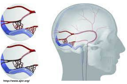
Improper conjunction of blood vessels in your spinal cord or brain where more than one artery is directly linked to other veins is known as a brain arteriovenous fistula.
The most critical issue with DAVFs (Dural arteriovenous fistulas) is they allow blood from the high-pressure artery to flow into veins that have low pressure of blood. Then drain blood from the brain and spinal cord.
It increases venous pressure around the spinal cord and brain, which can cause a variety of complications.
If you are searching for a treatment for arteriovenous brain fistula, visit Dr. Rao’s hospital in Guntur for the best treatment.
Now let us discuss the signs of arteriovenous brain fistula?
Patients in Guntur who have brain fistula or dural AVFs therapy complain of a rumbling sensation (tinnitus) in one of their ears that accompanies the heartbeat.
Tinnitus is a condition in which a patient may listen to blood flow right behind the ear like a distinct whooshing sound in sync with their heartbeat.
Fistulas can occur in the cavernous sinus that serves your eye. The eye might become swollen, painful, and red as a result of this.
The worst sort of fistula causes no symptoms in the beginning. It hampers the brain’s natural venous drainage, resulting in various brain dysfunctions such as edema, strokes,and seizures.

What causes arteriovenous fistulas in the brain?
It’s still unknown why dural brain fistulas occur. Your doctor can identify the disease through the symptoms, but it is tough to explain the reason. A patient may have a history of major trauma, surgery, or infection in the location where the fistula develops.
So, how does your doctor diagnose the complication?
Your doctor may offer diagnostic testing if you have signs or symptoms of the complication, such as:
Imaging:
Cross-sectional images from noncontract head computed tomography (CT) and magnetic resonance imaging (MRI) are commonly used in the first examination.
CT head scans can reveal both fluid accumulation and actual bleeding, which a DAVF may trigger but happen in some other part of your venous system in your brain.
MRIs are used to assess the form and size of a DAVF. It helps to identify any micro-hemorrhages (extremely minute bleed spots) and the impact of any unnatural blood vessel associated with the fistula.
So, what are the treatment alternatives available?
Arteriovenous fistulas can be treated by
- endovascular embolization
- stereotactic radiosurgery
- microsurgery.
Your doctor and team at Dr. Rao’s hospital assess each challenging situation and identify the best course of action for you. They provide you highly effective treatment strategy for your complication.
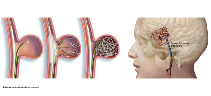
Endovascular embolization: The most frequent treatment procedure is endovascular embolization. A catheter is inserted into one of the arteries to accomplish this treatment. Then, using fluoroscopic or X-ray imaging as a guide, your doctor inserts it into the fistula’s position.
Then, to block the blood flow, he injects the device into the place where your artery and vein connect. The devices your doctor uses here are-
- Coils
- Detachable balloons
- Embolization particles
- Vascular plugs
- Embolization glue
- Embolization material
A covered stent works most effectively when a fistula occurs between a vein and a vital artery next to it. It is inserted into the artery or the vein. The procedure usually heals AVF while leaving the vein and artery unharmed. Surgical closure of the fistula is a third option.

Microsurgery: It can be done separately or along with the endovascular embolization. If your doctor believes there is a need for microsurgery, your doctor will inform you of all the necessary information about the same.
Microsurgery is used to examine complications under the microscope. It implants a titanium clip across the faulty connection. It helps to stop blood flowing irregularly from your artery to your vein. We usually repeat the angiography right after the treatment to ensure correctness.
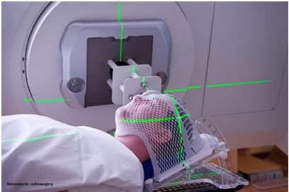
Stereotactic radiosurgery: In critical situations, your doctor will suggest you go for stereotactic radiosurgery. It is an outpatient and painless operation. Your doctor places a stereotactic frame on your head. Music therapy helps to make the procedure as easy as possible.
After the frame for the head is placed, your doctor will perform a CT scan of your head. The procedure takes approximately 30 minutes.

Frequently Asked Questions:
What causes brain arteriovenous fistula?
The causes of arteriovenous brain fistula are still now unknown. But treatment options are available, and they are effective.
Can brain arteriovenous fistula be cured?
Yes, It can be cured through various treatment options like –
- endovascular embolization
- microsurgery
- stereotactic radiosurgery.
Is it a hereditary disease?
No, it is not a hereditary disease. It can occur from any trauma or infection, or anything else.
Enquiry Now

