Author Archives: Dr RAO

meningitis-the-best-treatment-is-at-dr-raos-guntur

Meningitis – the best treatment is at Dr Raos, Guntur

 Guntur

Meningitis – the best treatment is at Dr Raos, Guntur

Introduction

Meningitis is a serious infection of the meninges, the thin membranes that surround and protect your brain and spinal cord. It’s usually caused by a virus, but can also be caused by bacteria, fungi, or parasites. Meningitis can occur in people of any age, but is most common in infants and young children. Meningitis is a medical emergency. It can cause death or permanent disability if not treated promptly. Early diagnosis and treatment are critical. Looking for the best neurology and neurosurgery treatment in Guntur look no further than Dr Raos hospital founded by Dr Rao.

causes

Meningitis is most often caused by a viral infection, but can also be caused by bacteria, fungi, or parasites. The most common cause of meningitis in the United States is the virus that causes the common cold. Other viruses that can cause meningitis include the flu, mumps, and measles. Bacterial meningitis is much less common than viral meningitis but is much more serious. The bacteria that most often cause meningitis are Streptococcus pneumoniae (pneumococcus), Haemophilus influenzae type b (Hib), Neisseria meningitidis (meningococcus), and Listeria monocytogenes. Fungal meningitis is very rare but can occur in people with weakened immune systems.
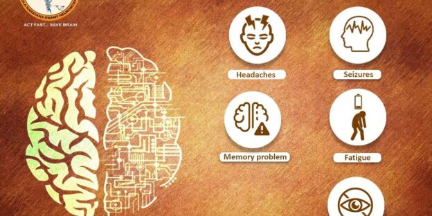
symptoms

Symptoms of meningitis can vary depending on the person, but there are some common symptoms that are seen in most cases. These include a high fever, severe headache, and a stiff neck. In some cases, people may also experience nausea, vomiting, and increased sensitivity to light. If you or someone you know is experiencing these symptoms, it is important to seek medical attention immediately as meningitis can be a very serious condition.

Diagnosis

A lumbar puncture, also called a spinal tap, is the most common test used to diagnose meningitis. This procedure involves removing a small amount of cerebrospinal fluid from the lower back for testing. The fluid is examined for bacteria, viruses, or other organisms that may be causing meningitis. A lumbar puncture can also be used to determine the type of meningitis you have.

treatment

There are two types of meningitis, viral and bacterial. Viral meningitis is less serious and usually goes away on its own. Bacterial meningitis is more serious and can be deadly. Bacterial meningitis is treated with antibiotics. Antibiotics kill the bacteria that are causing the infection. It is important to start treatment as soon as possible. The earlier you start, the better your chances are of surviving. If you have meningitis, you will be hospitalized so that you can be closely monitored. You will likely be given intravenous (IV) antibiotics. You may also need other treatments, such as: – Fluids through an IV to prevent dehydration – Pain relief medication – Oxygen therapy – Corticosteroids to reduce inflammation – seizure medication

antibiotic resistance

Antibiotic resistance is a major problem with treating meningitis. The bacteria that cause meningitis are constantly changing and becoming more resistant to antibiotics. This means that the antibiotics that were once effective against meningitis may no longer work. There are a few reasons why antibiotic resistance is such a problem with meningitis. First, meningitis is a very serious disease and even a small delay in treatment can be deadly. Second, the bacteria that cause meningitis are very good at surviving in the body and are difficult to kill. Finally, there are not many different types of antibiotics that are effective against meningitis. The best way to avoid antibiotic resistance is to prevent meningitis in the first place. Vaccines are available that can protect against some of the most common types of bacteria that cause meningitis. It is also important to finish all of the antibiotics prescribed for meningitis even if you start to feel better. This will help to make sure all of the bacteria are killed and prevent them from becoming resistant.

Conclusion

Meningitis is a serious infection of the meninges, the protective membranes that surround the brain and spinal cord. It can be caused by bacteria, viruses, or fungi, and can lead to death or permanent disability if not treated promptly and properly. Early diagnosis and treatment are critical to a good outcome. While most people recover from meningitis with no lasting effects, some people experience long-term problems such as hearing loss, seizures, or learning disabilities. Antibiotic resistance is a growing problem, making it more difficult to treat meningitis effectively. Meningitis is a serious disease that can have devastating consequences. prompt diagnosis and treatment are essential for the best possible outcome. Looking for the best neurology and neurosurgery treatment in Guntur look no further than Dr Raos hospital founded by Dr Rao.

brain-cancer-the-best-treatment-is-at-dr-raos-guntur

Brain Cancer – The best treatment is at Dr Raos, Guntur

Brain Cancer – The best treatment is at Dr Raos, Guntur

Introduction

Cancer is a class of diseases in which a group of cells display uncontrolled growth (division beyond the normal limits), invasion (intrusion into adjacent tissues), and sometimes metastasis (spread to other locations in the body via lymph or blood). Brain cancer, specifically, is an abnormal growth of cells within the brain. It can occur in both children and adults, though brain tumors are most common in people over the age of 60. There are many different types of brain cancer, each with its own set of symptoms, treatment options, and prognosis. Brain cancer is relatively rare, accounting for only about 2% of all cancers diagnosed each year. However, it is one of the most deadly types of cancer. The five-year survival rate for people with brain cancer is just 34%. This is largely due to the fact that brain tumors are very difficult to treat. They are located deep within the brain, making them difficult to reach with surgery or radiation. In addition, because the brain is such a complex organ, it is difficult to target cancer cells with chemotherapy without damaging healthy cells. As a result, most people with brain cancer will eventually die from their disease. There are many different types of brain cancer, each with its own set of symptoms, treatment options, and prognosis. The most common type of brain cancer is glioma, which accounts for about 80% of all cases. Gliomas can be further divided into astrocytomas and oligodendrogliomas, depending on the type of cell that is affected. Other types of brain cancer include meningiomas, medulloblastomas, and pituitary adenomas. Looking for the best treatment for your brain tumor or brain cancer, look no further than Dr Raos hospital, Guntur, the best neurosurgery and spine surgery hospital. Dr Rao is the fellowship trained neurooncologist and stereotactic radiosurgeon.

causes

There is no one definitive answer to the question of what causes brain cancer. However, there are a number of possible contributing factors that doctors and researchers believe may play a role in its development. These include exposure to certain toxins and chemicals, previous radiation therapy to the head or neck, and certain genetic conditions. Additionally, brain tumors are more common in people over the age of 60. While the exact cause of brain cancer remains unknown, understanding the possible risk factors can help doctors better identify those who may be at a higher risk for developing the disease.

symptoms

The most common symptom of brain cancer is a headache. Other symptoms can include nausea, vomiting, seizures, drowsiness, and changes in mood or personality.

Diagnosis

There are a few different ways that doctors can diagnose brain cancer. The first is through a medical history and physical examination. Your doctor will ask you about your symptoms and health history. They will also do a physical exam. This can help them rule out other conditions that may be causing your symptoms. The next step is usually an imaging test. This can help your doctor see if there is a mass or tumor in your brain. The most common imaging tests for brain cancer are computed tomography (CT) scans and magnetic resonance imaging (MRI) scans. If an imaging test shows that you have a mass in your brain, the next step is usually a biopsy. During a biopsy, your doctor removes a small sample of tissue from the mass so it can be tested for cancer cells. There are a few different ways to do this, but the most common is to insert a needle into your brain to remove the tissue sample. Once the tissue sample is removed, it will be sent to a lab where it will be examined under a microscope. If cancer cells are found, your doctor will then work to determine what type of brain cancer you have.

treatment

There are a number of different treatment options available for brain cancer patients, and the best course of action will be determined by a number of factors, including the type and stage of the cancer, as well as the overall health of the patient. Surgery is often the first line of treatment for brain cancer, and it may be used to remove as much of the tumor as possible. In some cases, radiation therapy may be used before or after surgery to help kill any remaining cancer cells. Chemotherapy is another common treatment option for brain cancer, and it uses drugs to kill cancer cells. This treatment can be given intravenously (through an IV) or orally (in pill form). Targeted therapy is a newer type of treatment that uses drugs or other substances to specifically target and kill cancer cells while sparing healthy cells. This type of therapy is often used in combination with other treatments, such as surgery, radiation therapy, or chemotherapy. The decision about which treatment to pursue will be made by the patient and their medical team after careful consideration of all of the available options.

Chemotherapy and Radiotherapy

Chemotherapy and radiotherapy are two of the most common treatments for brain cancer. Chemotherapy uses drugs to kill cancer cells, while radiotherapy uses high-energy beams of radiation to kill cancer cells. Both chemotherapy and radiotherapy can be used as standalone treatments or in combination with each other.

Conclusion

In conclusion, brain cancer is a serious illness that can be difficult to treat. However, with the right medical care and treatment, many people with brain cancer can lead long and healthy lives. Chemotherapy and radiotherapy are often effective treatments for brain cancer, and new treatments are being developed all the time. If you or someone you know has been diagnosed with brain cancer, don’t hesitate to seek out the best possible medical care and treatment. Looking for the best treatment for your brain tumor or brain cancer, look no further than Dr Raos hospital, Guntur, the best neurosurgery and spine surgery hospital. Dr Rao is the fellowship trained neurooncologist and stereotactic radiosurgeon.

bipolar-disorder-the-best-treatment-is-at-dr-raos-guntur

Bipolar disorder – the best treatment is at Dr Raos, Guntur

Bipolar disorder – the best treatment is at Dr Raos, Guntur

Introduction

Bipolar disorder is a mental illness that causes extreme mood swings. These swings can include periods of depression, where a person feels hopeless and down, and periods of mania, where a person feels excessively happy and energetic. Bipolar disorder can be very disruptive to a person’s life, making it difficult to maintain relationships, hold down a job, or even take care of oneself. There is no single cause of bipolar disorder, but it is thought to be caused by a combination of genetic and environmental factors. Treatment for bipolar disorder usually includes medication and therapy. With treatment, most people with bipolar disorder are able to live normal, productive lives. Looking for the best neurology or psychiatry services look no further than Dr Raos hospital, Guntur with best neurosurgeon Dr. Rao.

risk factors

There are several risk factors associated with bipolar disorder, and it is important to be aware of them. Genetics plays a role in bipolar disorder, as it does in many mental illnesses. If you have a family member with bipolar disorder, you are more likely to develop the condition yourself. Other risk factors include stress, trauma, and substance abuse. Bipolar disorder can be difficult to manage, but it is important to seek help if you think you may be suffering from the condition. If left untreated, bipolar disorder can lead to serious problems such as job loss, financial difficulties, and relationship problems. If you think you may be at risk for bipolar disorder, talk to your doctor about your concerns.

causes

The causes of bipolar disorder are not fully understood, but it is thought to be a combination of genetic and environmental factors. Bipolar disorder tends to run in families, so it is thought that genetic factors may play a role. However, not everyone with a family history of bipolar disorder will develop the condition, and other factors must also be involved. It is also thought that environmental factors, such as stress or trauma, may trigger the development of bipolar disorder in people who are genetically predisposed to the condition.

symptoms

The symptoms of bipolar disorder can be divided into two categories: manic symptoms and depressive symptoms. Manic symptoms include: – feeling excessively happy or “high” – having lots of energy – feeling like you can do anything – talking very fast – feeling like your thoughts are racing – being easily distracted – being impulsive or reckless – sleeping less than usual Depressive symptoms include: – feeling sad, hopeless, or empty – losing interest in activities you used to enjoy – having trouble sleeping or sleeping too much – feeling tired all the time – having difficulty concentrating or making decisions – experiencing changes in appetite or weight – feeling worthless or guilty

diagnosis

The diagnosis of bipolar disorder is made by a mental health professional based on a thorough clinical assessment. The assessment includes taking into account the person’s symptoms, medical history, family history, and any other relevant information. There is no single test that can diagnose bipolar disorder. However, there are certain tools that mental health professionals can use to help make a diagnosis, such as the Mood Disorder Questionnaire or the Hamilton Depression Rating Scale. In order to be diagnosed with bipolar disorder, a person must have had at least one episode of mania or hypomania. A manic episode is characterized by an abnormally elevated mood, energy levels, and activity levels. A hypomanic episode is similar to a manic episode but is less severe and does not impair functioning to the same degree. Bipolar disorder can be difficult to diagnose because it can resemble other mental disorders, such as depression, anxiety disorders, or substance abuse disorders. For this reason, it is important to seek professional help if you are experiencing any symptoms that are causing you distress or interfering with your ability to function in your everyday life.

treatment and prevention

There is no one-size-fits-all approach to treating bipolar disorder, as the condition can vary greatly from person to person. However, there are a number of effective treatments and strategies that can help manage the symptoms and improve quality of life. Medication is often the first line of treatment for bipolar disorder, and there are a number of different types of medication that can be effective. Antidepressants, mood stabilizers, antipsychotics, and anticonvulsants are all commonly prescribed medications for bipolar disorder. In some cases, a combination of medication may be necessary to effectively manage symptoms. In addition to medication, psychotherapy can be an important part of treatment for bipolar disorder. Cognitive behavioral therapy (CBT) is one type of therapy that has been shown to be particularly effective in treating bipolar disorder. CBT can help people learn how to identify and manage their symptoms, cope with stressors, and make positive lifestyle changes. Self-care is also an important part of managing bipolar disorder. Getting regular exercise, eating a healthy diet, getting enough sleep, and avoiding alcohol and drugs can all help improve symptoms and prevent relapse.

Conclusion

In conclusion, bipolar disorder is a serious mental illness that can have a profound impact on an individual’s life. While there is no cure for the condition, it is possible to manage the symptoms with medication and therapy. With proper treatment, people with bipolar disorder can lead full and productive lives. Looking for the best neurology or psychiatry services look no further than Dr Raos hospital, Guntur with best neurosurgeon Dr. Rao.

smoking-and-neurosurgery-prevention-of-smoking-is-the-best

Smoking and Neurosurgery – Prevention of smoking is the best

Smoking and Neurosurgery – Prevention of smoking is the best

Introduction

Smoking cigarettes has been proven to have numerous negative effects on the human body, including the brain, bones, and spine. In this article, we will explore the implications of smoking on brain function, bone growth, and spine health. We will also discuss some of the diseases associated with smoking, and how to prevent them. Looking for the best smoking cessation program please contact Dr Raos hospital. Dr Rao is the best neurosurgeon in India.

implications on brain

Smoking cigarettes has been shown to have implications on the brain. Studies have shown that smokers are more likely to suffer from memory problems and cognitive decline than non-smokers. Additionally, smokers are also at a higher risk for developing Alzheimer’s disease and other forms of dementia. The chemicals in cigarettes can damage the brain and lead to these problems.

disease associated with brain

Diseases associated with brain: There are many diseases that have been linked to smoking, and many of them affect the brain. These include stroke, dementia, and Alzheimer’s disease. Smoking also increases the risk of developing Parkinson’s disease.

implications on bone growth and spine

Smoking cigarettes has been shown to have implications on bone growth and the spine. Studies have shown that smokers are more likely to have a lower bone density than nonsmokers, which can lead to an increased risk of fractures. In addition, smoking has also been linked to an increased risk of developing degenerative diseases of the spine, such as osteoporosis and disk disease.

diseases associated with spine

Diseases associated with spine can include:

– Spinal stenosis: This is a condition in which the spinal canal narrows, putting pressure on the spinal cord. This can lead to pain, numbness, and weakness in the legs.

– Spondylolisthesis: This is a condition in which one of the vertebrae slips out of place, putting pressure on the spinal cord or nerves. This can lead to pain and numbness in the legs.

– Degenerative disc disease: This is a condition in which the discs between the vertebrae break down, leading to pain, stiffness, and loss of mobility.

– Osteoarthritis: This is a condition in which the joints degenerate, leading to pain, stiffness, and loss of mobility.

prevention

There are many ways to prevent smoking-related diseases. The best way to prevent smoking-related diseases is to never start smoking. If you currently smoke, quitting smoking is the best way to prevent smoking-related diseases. There are many resources available to help you quit smoking, including:

• nicotine replacement therapy (NRT), which can help reduce withdrawal symptoms and cravings

• prescription medications, such as bupropion or varenicline, which can also help reduce withdrawal symptoms and cravings

• counseling and support groups, which can provide motivation and encouragement If you’re struggling to quit smoking on your own, don’t hesitate to ask for help.

There are many people and organizations who want to see you succeed in quitting smoking.

Conclusion

In conclusion, smoking has a variety of implications on both the brain and the spine. These implications can lead to a number of different diseases, some of which are potentially fatal. However, there are ways to prevent these diseases from developing, and it is important to be aware of them.

State-of-the-art keyhole neurosurgery at Dr. Rao's Hospital, Guntur, providing advanced minimally invasive treatments for brain and spine conditions.

Biopsy and Neurosurgery – the best at Dr Raos, Guntur

Biopsy and Neurosurgery – the best at Dr Raos, Guntur

Introduction

A biopsy is a medical procedure in which a small sample of tissue is removed from the body for examination. Biopsies are performed for many reasons, including to diagnose cancer, to determine the cause of a suspicious lump or mass, or to assess whether an organ is functioning properly. There are many different types of biopsies, and the specific procedure used will depend on the location and nature of the tissue being sampled. Common types of biopsies include needle biopsies, endoscopic biopsies, and open surgical biopsies. Needle biopsies are the most common type of biopsy and can be performed using either fine-needle aspiration (FNA) or core needle biopsy (CNB). FNA involves inserting a thin needle into the suspicious area and withdrawing a small amount of cells or fluid. CNB involves removing a small cylinder of tissue (a core) using a larger-gauge needle. Endoscopic biopsies are performed using an endoscope, a long, thin tube equipped with a light and camera that is inserted through the mouth or rectum. Open surgical biopsies involve making an incision in the skin to access the tissue in question. The risks associated with biopsy procedures vary depending on the type of procedure being performed. In general, however, complications from biopsies are rare and typically minor. The most common complication is bleeding, which can usually be controlled with pressure or medication. Infection is another potential complication, although this is also rare. If you have been asked to undergo a biopsy, it is important to ask your doctor any questions you may have about the procedure. You should also be sure

Brian biopsy

A brain biopsy is a medical procedure in which a small sample of tissue is taken from the brain for diagnostic purposes. It is usually performed under general anesthesia, and involves making an incision in the skull in order to access the brain. A brain biopsy can be used to diagnose a variety of conditions, including tumors, infections, and inflammatory diseases. It can also be used to determine the cause of certain types of seizures.

muscle biopsy

A muscle biopsy is a diagnostic procedure during which a small sample of muscle tissue is removed from the body for examination. The purpose of a muscle biopsy is to help doctors diagnose various muscle disorders, such as muscular dystrophy and inflammatory myopathies. There are two main types of muscle biopsies: needle biopsies and open biopsies. Needle biopsies are less invasive than open biopsies and can be performed on an outpatient basis. During a needle biopsy, a needle is inserted into the muscle through a small incision in the skin. A small amount of muscle tissue is then removed and examined under a microscope. Open biopsies are more invasive than needle biopsies and usually require a hospital stay. During an open biopsy, a surgeon makes an incision in the skin and removes a larger piece of muscle tissue. This tissue is then examined under a microscope. Both types of muscle biopsies carry some risks, such as bleeding, infection, and pain at the site of the incision. However, these risks are generally low and most people tolerate the procedure well.

nerve biopsy

Nerve biopsy is a diagnostic procedure used to obtain a small sample of nerve tissue for examination. The tissue sample can be obtained from any number of nerves, but is most commonly taken from the sural nerve in the leg. Nerve biopsy is performed using local anesthesia to numb the area where the biopsy will be performed. A small incision is made and a needle is inserted into the nerve. Using special instruments, a small portion of the nerve is removed and sent to a laboratory for analysis. The results of a nerve biopsy can provide important information about the cause of a person’s nerve damage. Nerve biopsy is generally safe and complications are rare.

procedure

A biopsy is a medical procedure in which a small sample of tissue is removed from the body for examination. The tissue may be removed from any part of the body, but is most commonly taken from the skin, liver, or kidney. There are several different types of biopsy procedures, but the most common is the needle biopsy. In this procedure, a needle is inserted into the body through the skin and into the area where the tissue sample will be taken. A small amount of tissue is then removed and sent to a laboratory for analysis. The results of a biopsy can help to diagnose or rule out certain diseases and conditions. It can also be used to determine how well a treatment is working.

prevention of complications

There are a few complications that can occur during or after a biopsy. The most common complication is bleeding. To help prevent this, your doctor may prescribe a blood thinner. Other potential complications include infection, pain, and nerve damage.

Conclusion

The biopsy is a vital medical procedure that can provide invaluable information about the health of a patient. When performed correctly, it is a safe and effective way to obtain tissue samples for diagnostic purposes. However, as with any medical procedure, there are certain risks and complications associated with biopsy procedures. It is important for patients to be aware of these risks and to work closely with their healthcare team to ensure that the biopsy is performed correctly and safely. Looking for the biopsy in neurosurgery either brain tumor biopsy or nerve biopsy or muscle biopsy look no further than Dr Raos hospital, Guntur. Dr Rao is the best neurosurgeon in Guntur, Andhra Pradesh.

pregnancy-and-back-pain-the-best-treatment-is-at-dr-raos-guntur

Pregnancy and Back Pain – the best treatment is at Dr Raos, Guntur

Pregnancy and Back Pain – the best treatment is at Dr Raos, Guntur

Introduction

Back pain is a common complaint during pregnancy. In fact, it is estimated that 50-70% of pregnant women will experience some degree of back pain at some point during their pregnancy (1). The good news is that there are things you can do to help ease the discomfort and prevent further pain. The causes of back pain during pregnancy are often related to the physical changes that occur in your body as it prepares for childbirth. Looking for the best treatment for back pain in pregnancy look no further than Dr. Raos hospital in Guntur; Dr. Rao is the best neurosurgeon for the surgery.

These changes can put extra strain on your back and cause pain. Some other common causes of back pain during pregnancy include:

• Weight gain: As your baby grows, your body will gain weight. This extra weight can put strain on your back and cause pain.

• Posture: As your belly grows, your posture will change. This can cause strain on your back and lead to pain.

• Hormones: The hormones released during pregnancy can loosen the joints in your pelvis and back, which can lead to pain.

causes

There are many different causes of back pain during pregnancy. Some women experience it because of the extra weight they are carrying, while others may have back pain due to changes in their posture or hormones. Additionally, some pregnant women may have underlying health conditions that can contribute to back pain. Some of the most common causes of back pain during pregnancy include:

• Carrying extra weight: As your baby grows, you will gain weight. This extra weight can put strain on your back and cause pain.

• Changes in posture: As your belly grows, your posture will change. This can cause strain on your back and lead to pain.

• Hormonal changes: During pregnancy, your body produces a hormone called relaxin. This hormone relaxes the ligaments in your pelvis, which can lead to instability and pain in the lower back.

• Underlying health conditions: If you have a history of back pain or other health conditions such as arthritis, you may be more likely to experience back pain during pregnancy.

risk factors

There are several risk factors that can contribute to back pain during pregnancy. These include:

• Weight gain – as your baby and your body grow, the extra weight can put strain on your back and cause pain.

• Poor posture – as your belly grows, it can pull your spine out of alignment and lead to pain.

• Inactivity – not getting enough exercise can make your muscles weak and increase your risk of pain.

• An existing back condition – if you have a history of back pain or other problems, you may be more likely to experience pain during pregnancy.

diagnosis

The diagnosis of back pain during pregnancy is based on the symptoms and medical history of the woman. Physical examination can help to rule out other causes of back pain, such as a herniated disc or spinal stenosis. Imaging tests, such as X-rays, CT scans, or MRIs, are not usually needed to diagnose back pain during pregnancy.

treatment

There are many ways to treat back pain during pregnancy. Some women find relief with over-the-counter (OTC) medications, such as acetaminophen (Tylenol) or ibuprofen (Advil, Motrin). Others find relief with home remedies, such as heat or ice packs, massage, and stretching. If OTC medications don’t relieve your pain, your doctor may prescribe stronger medication. In some cases, epidural steroid injections or nerve blocks may be necessary. If you have severe back pain, your doctor may recommend surgery. Most women find that their back pain improves significantly after they give birth. However, some women continue to have back pain after delivery. If your back pain does not improve after delivery or gets worse, be sure to talk to your doctor about other treatment options.

prevention and exercises

There are a few things you can do to help prevent back pain during pregnancy. First, avoid high-heeled shoes and wear comfortable, supportive shoes. Second, when sitting, use a chair that supports your back and avoid sitting for long periods of time. Third, when lifting, bend at your knees and not at your waist. Finally, exercise regularly to maintain good muscle tone and flexibility in your back and abdominal muscles. There are also specific exercises you can do to help prevent or relieve back pain during pregnancy. First, start by doing pelvic tilts. To do this, stand with your feet hip-width apart and place your hands on your hips. As you exhale, tilt your pelvis forward and tuck in your buttocks. As you inhale, return to the starting position. Second, do hamstring stretches by lying on your back with a pillow under your knees. Bend one knee and bring it toward your chest while keeping the other leg straight. Hold for 30 seconds and then switch legs. Third, do modified planks by lying on your forearms and knees with your feet hip-width apart. Slowly lift up your hips so that your body forms a straight line from shoulders to knees. Hold for 30 seconds and then lower down. Repeat 3 times. These exercises can help prevent or relieve back pain during pregnancy. However, if you experience severe back pain, please consult with your doctor or healthcare provider.

Conclusion

In conclusion, pregnancy and back pain can be a difficult experience for many women. However, by understanding the causes and risk factors, as well as the available treatments and preventative measures, it is possible to manage the condition and improve quality of life. With the proper care, pregnant women can enjoy a healthy pregnancy and delivery. Looking for the best treatment for back pain in pregnancy look no further than Dr. Raos hospital in Guntur; Dr. Rao is the best neurosurgeon for the surgery.

acute-low-back-pain-the-best-treatment-is-at-dr-raos-guntur

Acute Low back pain – the best treatment is at Dr Raos, Guntur

Acute Low back pain – the best treatment is at Dr Raos, Guntur

Introduction

Acute low back pain is a common condition that affects millions of people each year. The vast majority of cases will resolve on their own with self-care and home treatment, but some may require medical intervention. This article will provide an overview of the anatomy and physiology of the lower back, as well as common causes and treatments for acute low back pain.

Muscles of lower back

There are several muscles in the lower back that can contribute to pain and dysfunction. The erector spinae is a large muscle group that extends along the length of the spine. This muscle is responsible for maintaining upright posture and can become tight and painful with prolonged sitting or standing. The quadratus lumborum is a large muscle that runs from the pelvis to the ribs and helps to stabilize the spine. This muscle can also become tight and painful with prolonged sitting or standing. The gluteus medius and minimus are muscles in the buttocks that help to stabilize the hip and pelvis. These muscles can become weak and painful with overuse.

Vertebrae and discs of lower back

The vertebrae are the bones that make up the spine and protect the spinal cord. The discs are the cushions between the vertebrae. They help to absorb shock and keep the spine flexible. There are five vertebrae in the lower back: • L1–L5

The discs between these vertebrae are: • L4/5 • L5/S1

The L4/5 disc is located between the L4 and L5 vertebrae.

The L5/S1 disc is located between the L5 and S1 vertebrae.

diagnosis

There are a few different ways to diagnose acute low back pain. The first is through a physical examination. Your doctor will likely ask you to describe your pain, how long you’ve been experiencing it, and what activities make it worse or better. They will also ask about your medical history and whether you’ve had any previous injuries to your back. Your doctor will then do a physical examination of your back. They may check your range of motion, reflexes, and muscle strength. They may also order imaging tests, such as an X-ray, MRI, or CT scan, to further assess the cause of your pain. Once your doctor has made a diagnosis, they can develop a treatment plan to help relieve your pain.

Acute management

Acute management of low back pain generally focuses on the short-term relief of symptoms. In most cases, this will involve a combination of medication and physical therapy. Medication can help to reduce inflammation and pain, while physical therapy can help to stretch and strengthen the muscles in the back. In some cases, surgery may be necessary to correct any underlying problems.

Exercises for acute back pain

There are a few key exercises that can help alleviate acute low back pain. First, start by lying on your back with both knees bent and your feet flat on the ground. Place a small, rolled up towel under your lower back for added support. Then, slowly raise your right leg up into the air, keeping your knee bent. Hold this position for a few seconds, then lower your leg back down to the starting position. Repeat this exercise 10 times on each side. Next, still lying on your back with both knees bent and feet flat on the ground, place your hands behind your head. Slowly lift your shoulders off the ground, contracting your abdominal muscles as you do so. Hold this position for a few seconds before slowly lowering yourself back down to the starting position. Repeat this exercise 10-15 times. Finally, sit on the ground with your legs straight out in front of you and your feet together. Reach forward with both arms, keeping your back straight, and try to touch your toes. Hold this stretch for 30 seconds before releasing and repeating 2-3 times. These exercises are just a few of the many that can help to relieve acute low back pain. Be sure to speak with your doctor or physical therapist before beginning any new exercise routine to ensure that it is safe for you to do so.

Conclusion

In conclusion, acute low back pain is a common condition that can be managed effectively with a combination of self-care, medication, and physical therapy. With proper treatment, most people with acute low back pain will recover fully and be able to return to their normal activities. Looking for the best neurosurgery or back surgery or spine surgery look no further than Dr Raos hospital, Guntur. Dr Rao is the best spine surgeon in India.

First biplane cathlab in Andhra Pradesh and Telangana diagnostic-tests-in-neurosurgery-the-best-at-dr-raos-guntur

Diagnostic tests in Neurosurgery – the best at Dr Raos, Guntur

Diagnostic tests in Neurosurgery – the best at Dr Raos, Guntur

Introduction

Neurosurgery is a branch of medicine that deals with the diagnosis and treatment of disorders of the nervous system. The nervous system includes the brain, spinal cord, and nerves. Neurosurgeons are medical doctors who have completed training in neurosurgery. A variety of diagnostic tests are used in neurosurgery to help identify problems with the nervous system. Blood tests, cerebrospinal fluid (CSF) analysis, imaging, and special tests may be used. Prevention of complications is also important in neurosurgery. The various diagnostic tests used in neurosurgery will be discussed in this article. Looking for the best neuro treatment in Neurology, neurosurgery or spine surgery, look no further than Dr Raos hospital, Dr Rao the best neurosurgeon in India.

Blood tests

Blood tests are an important part of the diagnostic process for neurosurgery. They can help to rule out other causes of symptoms, and can provide information about the level of inflammation present. There are a number of different blood tests that can be used in neurosurgery. The most common are the white blood cell count (WBC) and the erythrocyte sedimentation rate (ESR). The WBC is a measure of the level of inflammation present, and the ESR is a measure of the rate at which red blood cells settle in a tube of blood. Other blood tests that may be used include the C-reactive protein (CRP) test, which measures the level of CRP in the blood. CRP is a protein that is produced by the liver in response to inflammation. The CRP test can be used to monitor the level of inflammation present, and can also be used to monitor the response to treatment. The blood tests that are used in neurosurgery will vary depending on the individual case. However, they all play an important role in helping to diagnose and treat conditions affecting the nervous system.

CSF analysis

A lumbar puncture, also called a spinal tap, is a diagnostic procedure used to collect cerebrospinal fluid (CSF) for analysis. CSF is the clear fluid that surrounds and protects the brain and spinal cord. A lumbar puncture is performed by inserting a needle into the lower back to collect a small amount of CSF. CSF analysis can help diagnose various conditions, such as infections, inflammation, tumors, and bleeding in or around the brain. CSF can also be used to measure the pressure inside the skull. A lumbar puncture is generally safe and complications are rare.

Imaging

Imaging is critical in the evaluation of patients with suspected neurosurgical disorders. A variety of imaging modalities are available, each with its own advantages and disadvantages.

CT is the imaging modality of choice for many neurosurgical disorders, as it provides excellent anatomical detail.

MRI is often used for disorders that require more detailed anatomical information, such as tumors or vascular abnormalities.

PET and SPECT are useful for functional imaging, and can be used to evaluate cerebral metabolism or blood flow.

Angiography is occasionally used to evaluate vascular abnormalities.

Myelography is used to evaluate disorders of the spinal cord, and can be performed with CT or MRI.

Special tests

There are a number of special tests that may be used to diagnose neurological conditions. These include:

-Electroencephalography (EEG): This test measures electrical activity in the brain and can be used to diagnose conditions such as epilepsy.

-Evoked potentials: These tests measure the brain’s response to stimuli and can be used to diagnose conditions such as multiple sclerosis.

-Magnetic resonance imaging (MRI): This test uses magnetic fields and radio waves to create images of the brain and can be used to diagnose conditions such as brain tumors.

-Positron emission tomography (PET): This test uses radioactive tracers to create images of the brain and can be used to diagnose conditions such as Alzheimer’s disease.

prevention of complications

The best way to prevent complications is to have a thorough understanding of the risks involved with the procedure. There are many potential risks associated with any surgery, but some are more common than others. It is important to discuss all of the possible complications with your surgeon prior to having the procedure. Additionally, it is important to follow all of the post-operative instructions given to you by your surgeon in order to minimize the risk of complications.

Conclusion

Neurosurgery is a branch of medicine that deals with the diagnosis and treatment of disorders of the nervous system. The nervous system includes the brain, spinal cord, and peripheral nerves. Neurosurgery is a very delicate and complex field of medicine. There are many different diagnostic tests that are used in neurosurgery in order to make an accurate diagnosis and to determine the best course of treatment. The various diagnostic tests that are used in neurosurgery include blood tests, CSF analysis, imaging, and special tests. Each of these tests has its own advantages and disadvantages. Blood tests can be used to detect infections or inflammation. CSF analysis can be used to detect abnormalities in the brain or spinal cord. Imaging can be used to detect tumors or other abnormalities. Special tests can be used to detect problems with the blood vessels or nerves. The decision on which diagnostic test to use depends on the individual patient’s symptoms and medical history. The goal of all of these tests is to make an accurate diagnosis so that the best possible treatment can be provided. Looking for the best neuro treatment in Neurology, neurosurgery or spine surgery, look no further than Dr Raos hospital, Dr Rao the best neurosurgeon in India. Deccanherald say about us.

Biplane Cath Lab - cerebral-angiography-the-best-at-dr-raos-guntur

Cerebral Angiography – the best at Dr Raos, Guntur

Cerebral Angiography – the best at Dr Raos, Guntur

Introduction

Cerebral angiography is a diagnostic procedure used to visualize the blood vessels in the brain. It is often used to evaluate patients with stroke or transient ischemic attack (TIA). The procedure involves injecting a contrast agent into the carotid artery and then performing an X-ray study of the brain. Cerebral angiography is a minimally invasive procedure that can be performed on an outpatient basis. The risks of the procedure are low and complications are rare.

Indication

There are many indications for cerebral angiography. The most common indication is to evaluate for stenosis or occlusion of the cerebral arteries. This can be done to evaluate for stroke or transient ischemic attack (TIA). Other indications include aneurysms, arteriovenous malformations (AVMs), and dural arteriovenous fistulas (DAVFs). Cerebral angiography can also be done to evaluate for vasculitis, such as Takayasu’s arteritis. It can also be done to evaluate for congenital abnormalities, such as moyamoya disease. Finally, it can be done to help guide interventional procedures, such as coil embolization of an aneurysm or stenting of a stenotic artery.

preparation

Preparation for cerebral angiography generally begins the night before the procedure is scheduled. The patient will be asked to fast for at least six hours prior to the procedure, and to avoid drinking any caffeinated beverages. It is important that the patient arrive on time and relaxed, as the procedure can be quite stressful.

procedure

The procedure for cerebral angiography is as follows: 1. The patient is placed in a supine position on the angiography table. 2. A local anesthetic is injected into the skin over the femoral artery in the groin. 3. A catheter is inserted into the femoral artery and threaded through the aorta and into the carotid artery. 4. contrast material is injected through the catheter and X-rays are taken to visualize the arteries of the brain.

complications

There are several potential complications that can occur during or after a cerebral angiography procedure. These include: -Allergic reaction to the contrast dye used during the procedure -Brain hemorrhage -Stroke -Seizures -Infection Fortunately, most of these complications are rare. However, it is important to be aware of them and to seek immediate medical attention if any occur.

prevention of complications

There are several ways to prevent complications during cerebral angiography. First, the patient should be well hydrated before the procedure. Second, the operator should use a small-gauge catheter to minimize the risk of damage to the blood vessels. Third, care should be taken to avoid injecting contrast material into the brain tissue. Fourth, the operator should be experienced in performing the procedure and be familiar with the anatomy of the blood vessels in the brain. Finally, if any complications do occur, they should be treated immediately.

Conclusion

The conclusion of this article is that cerebral angiography is a safe and effective procedure when performed by a skilled physician. There are potential complications associated with the procedure, but these can be effectively prevented with proper planning and execution. Looking for the best neurosurgery or neurology or spine surgery or interventional neurology services look no further than Dr Raos hospital, the best neurosurgery hospital in Guntur, Andhra Pradesh and in India. Dr Rao is the best spine surgeon, best neurosurgeon and the best endovascular neurosurgeon in India.

Cobra Pose, Bhudjangasana

Back Pain – The best physiotherapy exercises at Dr Raos

Back Pain – The best physiotherapy exercises at Dr Raos

Introduction

Back pain is a common issue that can affect people of all ages. It can be caused by a variety of factors, including muscle strain, poor posture, and injury. There are several things you can do to help relieve back pain, including strengthening exercises and using proper backpacking techniques. Looking for the best chronic pain exercises or physiotherapy look no further than Dr Rao for the guidance at Dr Raos hospital.

Muscles of back

There are many muscles in the back that work together to provide support and movement. The largest muscle group is the erector spinae, which runs along either side of the spine. This muscle group is responsible for keeping the spine upright and also aids in bending and twisting movements. The latissimus dorsi, or “lats,” are the large muscles on either side of the back that give the body its width. These muscles are important for pulling movements, such as when you row a boat. The trapezius is a triangular muscle that extends from the base of the skull to the middle of the back. This muscle is responsible for shrugging the shoulders and moving the head from side to side. The rhomboids are a small muscle group that runs between the shoulder blades. These muscles help to pull the shoulder blades together and also aid in stabilizing the shoulder joint.

Vertebrae and discs

The spine is made up of 33 vertebrae, which are separated by discs. The discs act as shock absorbers and allow the spine to move. There are four main types of vertebrae: cervical (neck), thoracic (upper back), lumbar (lower back), and sacral (tailbone). The discs are made up of a tough outer layer (the annulus) and a soft inner layer (the nucleus). The discs allow the spine to move and act as shock absorbers. When a disc is damaged, it can bulge or rupture. This can put pressure on the nerves, causing pain.

strengthening exercises

There are many different exercises that can help to strengthen the muscles in your back and help to prevent back pain. Some of these exercises may include:

1. Pilates: Pilates is a great way to strengthen the muscles in your back and help to improve your posture. 2. Yoga: Yoga is another great way to help improve your posture and also helps to stretch and lengthen the muscles in your back.

3. Strength Training: Strength training is a great way to build up the muscles in your back and help to prevent back pain.

4. Cardio Exercise: Cardio exercise is also important for overall health and can help to improve blood circulation and reduce inflammation throughout the body, which can lead to less back pain.

Back pack safety

One of the best ways to avoid back pain is to be mindful of your posture and how you carry yourself throughout the day. This is especially important when carrying a heavy backpack, as improper posture and lifting techniques can lead to serious strain on your back muscles. When carrying a backpack, always make sure that it is evenly balanced and not too heavy. If possible, distribute the weight evenly between both shoulders. If the backpack is too heavy or unbalanced, it will pull you backwards and cause you to lean forward in an attempt to compensate. This puts a lot of strain on your back and can lead to pain. In addition, be sure to stand up straight and avoid slouching when wearing a backpack. This will help keep your spine in alignment and prevent strain on your back muscles. Finally, when lifting a backpack, always use your legs to lift it up rather than your back. This will help prevent injury.

Exercises for acute and chronic back pain

There are a number of exercises that can help alleviate acute and chronic back pain. For those with acute back pain, it is important to focus on exercises that stretch the muscles and improve flexibility. For those with chronic back pain, it is important to focus on exercises that strengthen the muscles and improve posture. Some specific exercises that can help relieve acute and chronic back pain include:

– Cat/cow stretches: These stretches help to lengthen the spine and release tension in the back.

cat or cow pose


Cat or Cow stretches to relieve chronic back pain

– Child’s pose: This stretch helps to lengthen the spine and release tension in the back.

child pose


Child pose

– Cobra pose: This pose helps to strengthen the muscles in the back and improve posture.

Cobra pose


Sporty young woman practicing yoga, doing cobra asana, bhudjangasana

– Pelvic tilts: These tilts help to strengthen the muscles in the lower back and improve flexibility.

Pelvic tilt


Pelvic tilt

– Knee to chest stretches: These stretches help to lengthen the muscles in the back and release tension.

Knee to chest pose


Knee to chest pose

Conclusion

In conclusion, back pain is a common condition that can be caused by a variety of factors. However, there are many things that you can do to help prevent and treat back pain. By strengthening the muscles of your back and practicing good back pack safety, you can help to prevent back pain. If you already have back pain, there are many exercises that you can do to help relieve your pain.

Back Pain – The best physiotherapy exercises at Dr RaosBack Pain – The best physiotherapy exercises at Dr Raos