Severe Spasticity in Cerebral Palsy Should Not Simply Be Accepted: When Specialist Assessment Can Help
Cerebral palsy is a lifelong condition affecting movement and posture, but severe
muscle stiffness should not automatically be accepted as something a child simply
has to live with. When spasticity begins limiting movement, walking, sitting,
transfers, dressing, hygiene or rehabilitation, a detailed specialist assessment
can help determine whether there are treatable components.
What Is Cerebral Palsy?
Cerebral palsy is a lifelong disorder of movement and posture caused by disturbances
in the developing brain. Its clinical presentation can vary considerably from one
child to another.
Some children have predominantly spasticity, while others may have dystonia, weakness, impaired coordination, or a combination of
these problems.
This difference is important because the same treatment is not appropriate for every
child with cerebral palsy.
The first step should therefore be to understand what is actually limiting the
child’s function and whether any part of that limitation may be modifiable.
What Is Spasticity in Cerebral Palsy?
Spasticity refers to abnormal muscle tone that can make movement stiff or difficult.
In a child with cerebral palsy, significant spasticity may restrict movement that
the child otherwise has the potential to perform.
The important clinical question is not simply whether a child has cerebral palsy.
It is whether abnormal muscle tone is preventing the child from using voluntary
movement that is already present.
The key question is whether spasticity is limiting movement already present and,
if so, whether reducing that spasticity could improve movement, daily activities
and rehabilitation.
Severe Spasticity Should Not Automatically Be Considered “Just Part of CP”
A diagnosis of cerebral palsy does not mean that every functional limitation should
automatically be attributed to the underlying condition.
When increasing stiffness begins interfering with a child’s function, specialist
assessment may help determine whether spasticity, dystonia, weakness or another
neurological problem is the dominant limitation.
The goal is not to promise that surgery will make a child “normal.” The goal is to
identify potentially modifiable problems, reduce barriers to movement where
appropriate, and give the child the best possible opportunity to use the function
they have.
When Should a Child With Cerebral Palsy Be Reassessed?
Earlier specialist assessment does not necessarily mean earlier surgery.
It means identifying potentially treatable problems before they become accepted as
an unavoidable explanation for every functional limitation.
A specialist assessment may be particularly relevant when:
Increasing stiffness interferes with walking or mobility
Sitting becomes increasingly difficult
Transfers become more difficult
Dressing or personal hygiene becomes difficult because of abnormal tone
Functional progress has plateaued despite appropriate rehabilitation
Repeated focal treatments provide only temporary benefit
It is unclear whether spasticity, dystonia, weakness or another neurological
problem is the main functional limitation
The objective is to identify the child’s modifiable problems and establish
realistic functional goals.
Cerebral Palsy Treatment Is Multidisciplinary
Cerebral palsy management should not be viewed as a choice between rehabilitation
and neurosurgery.
Depending on the individual child, treatment may involve a combination of:
Physiotherapy
Occupational therapy
Antispastic medications
Botulinum toxin injections
Intrathecal baclofen
Orthopaedic procedures
Selected neurosurgical procedures
Neurosurgery, when appropriate, should be considered as one component of a much
longer rehabilitation pathway rather than as the end of treatment.
Reducing abnormal muscle tone may, in carefully selected children, create a better
environment in which rehabilitation can help the child make use of the movement
that remains.
What Is Selective Dorsal Rhizotomy (SDR)?
Selective dorsal rhizotomy (SDR) is a neurosurgical procedure
used in carefully selected children with significant lower-limb spasticity.
During SDR, selected sensory nerve rootlets contributing to lower-limb spasticity
are divided with the aim of reducing abnormal muscle tone.
Because SDR is an irreversible procedure, the decision to perform
it should never be based on the diagnosis of cerebral palsy alone.
Appropriate patient selection, detailed neurological assessment, functional goals,
multidisciplinary evaluation and discussion of potential complications and
uncertainties are essential.
Who May Be Considered for Selective Dorsal Rhizotomy?
Not every child with cerebral palsy and spasticity is a candidate for SDR.
The press release cites NICE guidance that recommends consideration of selective
dorsal rhizotomy for appropriately selected children and young people with
spasticity affecting the lower limbs, particularly those at GMFCS levels II or III,
following multidisciplinary assessment.
The decision should also take into account the irreversible nature of the procedure,
known complications, uncertainty surrounding some long-term outcomes, and the
availability of intensive rehabilitation after treatment.
What Does the Evidence Say About Selective Dorsal Rhizotomy?
The evidence surrounding SDR supports a careful and selective approach rather than
offering the procedure to every child with cerebral palsy.
A 2025 systematic review and meta-analysis cited in the release found significant
improvements in measures of lower-limb spasticity and gross motor function following
SDR in children with cerebral palsy, while also noting limitations in the available
evidence and follow-up.
Longer-term reviews have highlighted uncertainties regarding functional outcomes
and the quality and consistency of the available evidence.
Potential complications also need to be considered. Reviews of SDR complications
have reported structural and neurological complications, reinforcing the importance
of careful patient selection and long-term surveillance during growth.
Therefore, the evidence does not support treating SDR as a procedure that should be
automatically offered to every child with cerebral palsy.
Why Specialist Assessment Matters More Than Simply Choosing Surgery
The important question for a child with cerebral palsy is not simply:
“Does my child need surgery?”
A more useful starting question is:
“What is actually limiting my child’s function, and is any part of it treatable?”
This approach helps distinguish between spasticity, dystonia, weakness and other
factors that may affect function.
It also allows the treatment plan to be built around the child’s actual functional
goals rather than around a procedure.
Dr. Rao’s Hospital Reports Surgical Treatment of 104 Patients With Cerebral Palsy
Dr. Rao’s Hospital – International Institute of Neurosciences (IIN), Guntur,
reports surgical treatment of 104 patients with cerebral palsy.
According to the institutional experience described in the World Cerebral Palsy Day
release, this experience has reinforced the importance of individualized treatment
rather than applying one procedure to every child.
The hospital evaluates and provides a range of approaches for clinically significant
spasticity, including medical management, botulinum toxin therapy, intrathecal
baclofen and selected neurosurgical procedures.
Decisions regarding surgery are based on the individual child’s neurological
examination, functional limitations, pattern of tone, treatment goals,
rehabilitation potential and multidisciplinary assessment.
Three Questions Every Family Should Ask During a Specialist Assessment
Families considering treatment for significant spasticity may find it useful to
discuss these three questions with the treating team:
Is the main problem spasticity, dystonia, weakness or a combination of these?
What voluntary movement does my child already have that may be hidden or
restricted by abnormal muscle tone?
If treatment reduces the abnormal tone, what specific rehabilitation programme
will help my child use the movement that remains?
These questions shift the conversation away from simply asking how to reduce muscle
stiffness. They bring the discussion back to the real goal: improving the child’s
function and independence.
What Is the Goal of Cerebral Palsy Surgery?
The purpose of cerebral palsy surgery is not to “cure” cerebral palsy or to make a
child normal.
The purpose is to identify what may be modifiable, reduce barriers to movement where
appropriate, and provide the child with the best possible opportunity to use their
existing functional potential.
For appropriately selected children, treatment of significant spasticity may become
one component of a broader rehabilitation programme.
This is why postoperative rehabilitation is not an optional afterthought. It is an
important part of the overall treatment pathway.
World Cerebral Palsy Day: A Different Conversation About Spasticity
World Cerebral Palsy Day provides an opportunity to recognize people living with
cerebral palsy and the families and professionals supporting them.
It is also an opportunity to look beyond the diagnosis itself and consider potentially
modifiable factors that influence function throughout childhood.
Severe, function-limiting spasticity should not automatically be accepted as an
unavoidable part of cerebral palsy.
At the same time, this does not mean that every child with cerebral palsy needs
neurosurgery. The appropriate approach is individualized assessment followed by
treatment that matches the child’s neurological findings, functional goals,
rehabilitation potential and overall needs.
When Should Parents Seek a Specialist Opinion?
Parents may consider specialist assessment when their child’s increasing stiffness
begins to interfere with function or when progress has stopped despite appropriate
rehabilitation.
A specialist assessment can help determine:
What type of abnormal muscle tone is present
Which movements are being restricted
Whether spasticity is the principal functional limitation
What treatment options are available
Whether rehabilitation alone may be appropriate
Whether medication, injections, intrathecal therapy, orthopaedic treatment or neurosurgery should be considered
What functional goals should guide treatment
Cerebral Palsy Treatment Should Be Built Around the Child
Every child with cerebral palsy is different. The pattern of spasticity, dystonia,
weakness, coordination problems, existing voluntary movement, functional goals and
rehabilitation potential can vary substantially.
Therefore, treatment should not begin with a procedure. It should begin with a
careful assessment of the child and a clear understanding of what is limiting
function.
For selected children, reducing severe spasticity may create an opportunity for
more effective rehabilitation and improved use of existing movement. For others,
non-surgical management may remain the most appropriate strategy.
The goal is always individualized care rather than a one-size-fits-all approach.
Dr. Mohana Rao Patibandla – Pediatric Neurosurgeon in Guntur
Dr. Mohana Rao Patibandla is the Founder and Chief Neurosurgeon of Dr. Rao’s Hospital – International Institute of Neurosciences (IIN)
in Guntur, Andhra Pradesh.
His clinical training and practice include pediatric neurosurgery, minimally invasive
neurosurgery, skull base surgery, cerebrovascular and endovascular neurosurgery,
neuro-oncology and stereotactic radiosurgery.
Dr. Rao’s Hospital is a dedicated neurosciences centre providing neurological and
neurosurgical care, including pediatric neurosurgery and management of complex
neurological conditions.
Cerebral Palsy and Spasticity Evaluation in Guntur
If your child has cerebral palsy and severe or increasing spasticity is affecting
walking, sitting, transfers, dressing, hygiene, rehabilitation or other daily
activities, a specialist assessment can help identify the factors limiting function
and discuss appropriate treatment options.
Dr. Rao’s Hospital – International Institute of Neurosciences (IIN)
Frequently Asked Questions About Cerebral Palsy and Spasticity
Can cerebral palsy be cured?
Cerebral palsy is a lifelong disorder related to disturbances in the developing brain.
The purpose of treatment is not to cure cerebral palsy, but to manage symptoms,
reduce functional barriers where possible and help the child achieve the best
possible function and independence.
Is severe spasticity always part of cerebral palsy?
Spasticity is common in cerebral palsy, but not every child has the same pattern of
abnormal tone. Some children may have dystonia, weakness, impaired coordination or
combinations of these problems. A specialist assessment can help determine what is
actually limiting function.
Does every child with cerebral palsy need neurosurgery?
No. The appropriate treatment depends on the individual child. Management may include
rehabilitation, medications, botulinum toxin, intrathecal baclofen, orthopaedic
treatment or selected neurosurgical procedures.
What is selective dorsal rhizotomy?
Selective dorsal rhizotomy, or SDR, is a neurosurgical procedure in which selected
sensory nerve rootlets contributing to lower-limb spasticity are divided to reduce
abnormal muscle tone. Because the procedure is irreversible, careful patient
selection and multidisciplinary assessment are essential.
Is selective dorsal rhizotomy suitable for every child with cerebral palsy?
No. SDR is considered only for appropriately selected children. The child’s pattern
of spasticity, neurological findings, functional goals, rehabilitation potential,
potential complications and the uncertainty surrounding some long-term outcomes
should all be discussed before treatment.
Why is rehabilitation important after treatment for spasticity?
Reducing abnormal muscle tone does not by itself guarantee functional improvement.
Rehabilitation helps the child learn to use the movement that remains and work toward
specific functional goals.
When should parents seek a specialist opinion for spasticity?
Specialist assessment may be appropriate when increasing stiffness interferes with
walking, sitting, transfers, dressing, hygiene or rehabilitation, when functional
progress plateaus, or when it is unclear whether spasticity, dystonia, weakness or
another neurological problem is the main limitation.
Clinical Evidence and References
The educational information in this article is based on the World Cerebral Palsy Day
discussion published by Dr. Rao’s Hospital and the clinical evidence cited in that
release, including NICE guidance and systematic reviews concerning selective dorsal
rhizotomy.
National Institute for Health and Care Excellence (NICE).
Cerebral palsy in under 25s: assessment and management. NICE Guideline NG62.
National Institute for Health and Care Excellence (NICE).
Spasticity in under 19s: management. NICE Guideline CG145.
Otero-Luis I, et al. Efficacy of selective dorsal rhizotomy in the treatment of
spasticity in children with cerebral palsy: a systematic review and meta-analysis.
Journal of Neurosurgery: Pediatrics. 2025;35(6):571–580.
Himmelmann K, et al. Long-term effects of selective dorsal rhizotomy in children
with cerebral palsy: a systematic review. Developmental Medicine & Child Neurology.
2019.
Systematic review of complications following selective dorsal rhizotomy in cerebral
palsy, as cited in the World Cerebral Palsy Day release.
Medical Disclaimer
This article is intended for general educational and informational purposes only.
It does not replace an individual medical consultation, diagnosis or treatment plan.
Not every child with cerebral palsy or spasticity is a candidate for surgery.
Treatment decisions should be made following appropriate neurological, functional
and multidisciplinary assessment and discussion of the potential benefits, risks,
alternatives and uncertainties with the child’s treating specialists.
Trigeminal Neuralgia Treatment: Why Finding the Cause of Facial Pain Matters
Trigeminal neuralgia can cause sudden, severe facial pain that feels like an electric shock.
But for some patients, the most important question is not simply how to stop the pain—it is
whether an underlying cause can be identified and treated.
What Is Trigeminal Neuralgia?
Trigeminal neuralgia is a neurological condition characterized by recurrent attacks of
severe facial pain. Patients may describe the pain as electric shocks, stabbing sensations,
or lightning-like pain.
Everyday activities such as talking, chewing, brushing the teeth, touching the face, or even
exposure to a light breeze may trigger an attack.
Because the pain may occur around the jaw, cheek, or teeth, some patients initially consult
a dentist. When dental disease does not adequately explain the symptoms, a neurological
cause should be considered.
Trigeminal Neuralgia Does Not Have One Best Treatment for Everyone
One of the most important principles in treating trigeminal neuralgia is that the same
procedure is not necessarily appropriate for every patient.
Treatment should take into account the patient’s symptoms, neurological examination,
MRI findings, age, medical fitness, previous treatments, and willingness to accept the
risks associated with a particular procedure.
The goal is not simply to choose the procedure with the highest reported success rate.
The more useful question is:
Which treatment provides the best balance of durable pain control, preservation
of facial sensation, procedural risk, and the patient’s individual preferences?
Why MRI Matters in Trigeminal Neuralgia
Appropriate brain MRI is an important part of evaluating suspected trigeminal neuralgia.
Imaging can help exclude other causes and can provide information relevant to treatment
planning and surgical candidacy.
However, an MRI should not be interpreted in isolation. Simply seeing a blood vessel close
to the trigeminal nerve does not automatically establish that the vessel is responsible
for the patient’s pain.
The clinical symptoms and imaging findings need to tell a consistent story.
When a patient has clinical features consistent with classical trigeminal neuralgia and
MRI demonstrates a convincing neurovascular relationship, the treatment discussion may
change significantly.
What Is Neurovascular Compression?
In classical trigeminal neuralgia, a blood vessel may compress or contact the trigeminal
nerve and contribute to the characteristic attacks of facial pain.
When the clinical diagnosis and imaging support this mechanism, treatment may be directed
at the underlying neurovascular conflict rather than only interrupting pain transmission.
This distinction is particularly important when long-term pain control and preservation
of facial sensation are important goals.
What Is Microvascular Decompression (MVD)?
Microvascular decompression, commonly called MVD, is a surgical procedure designed to
address the underlying neurovascular conflict in appropriately selected patients.
Unlike procedures that intentionally injure or interrupt the trigeminal nerve to control
pain, MVD involves identifying the offending vascular structure and separating it from
the nerve, typically by placing protective material between them.
This makes MVD fundamentally different from procedures that intentionally interrupt
trigeminal nerve transmission.
The European Academy of Neurology guideline cited in the recent discussion recommends
microvascular decompression as first-line surgery for classical trigeminal neuralgia
when surgery is indicated.
Why MVD May Be Considered for Long-Term Pain Relief
The objective of treatment is not always simply immediate pain relief. For appropriately
selected patients, durable pain control while preserving facial sensation may be an
important consideration.
Comparative evidence cited in the EIN Presswire release reports higher short- and
long-term pain freedom with MVD compared with stereotactic radiosurgery in selected
patients, together with lower rates of facial numbness and dysesthesia, although MVD
carries greater postoperative procedural risk.
One prospective long-term comparison cited in the release reported pain-free rates
after MVD of 83% at five years compared with 47% after stereotactic radiosurgery,
with a longer median time to pain recurrence following MVD.
These findings do not mean that MVD is the right procedure for every patient. Instead,
they reinforce the importance of matching the treatment to the patient’s diagnosis,
anatomy, health, and treatment goals.
Is Microvascular Decompression Suitable for Everyone?
No. Not every person with trigeminal neuralgia is an appropriate candidate for MVD.
An older patient with significant medical comorbidities, a patient who does not want
intracranial surgery, or a patient without convincing neurovascular compression may be
better served by a less invasive treatment approach.
The decision should therefore be individualized rather than based solely on the name
of the procedure.
Other Treatment Options for Trigeminal Neuralgia
Depending on the patient’s diagnosis and individual circumstances, treatment options
may include:
Medical treatment for patients who achieve adequate pain control
Nerve-block and injection-based treatment in selected circumstances
Radiofrequency ablation through the foramen ovale
Balloon compression or rhizotomy
Stereotactic radiosurgery
Microvascular decompression
Endoscopic-assisted or endoscopic microvascular decompression in appropriately selected cases
The European Academy of Neurology guideline cited in the release recommends surgery when
pain is inadequately controlled with medication or medication is poorly tolerated, with
the choice of procedure guided by the type of trigeminal neuralgia and individual
circumstances.
More Than 440 Trigeminal Neuralgia Patients: Experience Across the Treatment Spectrum
Dr. Rao’s Hospital – International Institute of Neurosciences (IIN) reports treatment
experience involving more than 440 patients with trigeminal neuralgia.
The institutional procedural experience reported in the EIN Presswire release includes
approximately:
213 microvascular decompression procedures
114 endoscopic MVD procedures
76 stereotactic radiosurgery procedures
35 balloon compression/rhizotomy procedures
120 radiofrequency ablation procedures
These figures represent institutional procedural experience rather than unique patient
counts. An individual patient may undergo more than one procedure during the course of
treatment.
This breadth of experience highlights an important principle: having multiple treatment
options is valuable only when those options are used selectively and appropriately.
When Recurrent Trigeminal Neuralgia Should Prompt Reassessment
Repeated procedures can provide meaningful pain relief and may remain appropriate for
patients who are not candidates for MVD or who prefer a less invasive approach.
However, when pain repeatedly returns, reassessment may be appropriate rather than
automatically repeating the same procedure.
Recurrent pain should prompt consideration of whether the diagnosis remains consistent
with classical trigeminal neuralgia, whether there is convincing neurovascular conflict,
what previous procedures have changed, and whether the treatment strategy should be
modified.
Five Questions to Ask Before Trigeminal Neuralgia Treatment
Patients considering a procedure for trigeminal neuralgia may benefit from discussing
the following questions with their treating specialist:
Does my clinical picture fit classical trigeminal neuralgia?
Does my MRI show convincing neurovascular compression of the trigeminal nerve?
If MVD is being considered, what is my expected chance of durable pain relief and what are the operative risks?
If a less invasive procedure is recommended, how quickly should I expect pain relief, how durable is it likely to be, and what is the risk of facial numbness?
If the pain returns, what will the next treatment option be?
Trigeminal Neuralgia: Treat the Cause When the Cause Can Be Identified
Trigeminal neuralgia is often described simply as severe facial pain. For some patients,
however, there may be an identifiable anatomical problem behind that pain.
When the clinical diagnosis, imaging, and patient profile point toward convincing
neurovascular compression, it may be appropriate to discuss whether the underlying
cause can be treated rather than repeatedly treating only the symptom.
This does not mean that every patient with trigeminal neuralgia requires surgery.
Rather, when classical trigeminal neuralgia, convincing neurovascular compression,
and appropriate surgical candidacy come together, microvascular decompression may
deserve serious consideration because it can address the underlying neurovascular
conflict while aiming to preserve facial sensation.
Why Treatment Should Be Individualized
There is no universal “best procedure” for every patient with trigeminal neuralgia.
The most appropriate treatment depends on the mechanism of pain, MRI findings,
overall health, previous treatment, age, procedural risks, and patient preferences.
The goal should be a carefully considered treatment strategy that balances pain relief,
durability, preservation of sensation, procedural risk, and the patient’s individual
circumstances.
If facial pain continues despite treatment, a specialist reassessment can help determine
whether the diagnosis, imaging interpretation, or treatment strategy needs to be revisited.
Dr. Mohana Rao Patibandla – Neurosurgeon in Guntur
Dr. Mohana Rao Patibandla is the Founder and Chief Neurosurgeon of
Dr. Rao’s Hospital – International Institute of Neurosciences (IIN), Guntur, Andhra Pradesh.
His clinical practice includes minimally invasive neurosurgery, skull base surgery,
pediatric neurosurgery, cerebrovascular and endovascular neurosurgery, neuro-oncology,
and stereotactic radiosurgery.
Dr. Rao’s Hospital is a dedicated neurosciences centre providing neurosurgical and
neurological care with infrastructure that includes intraoperative neurophysiological
monitoring, neuronavigation, a hybrid operating theatre, and a biplane catheterisation
laboratory.
Trigeminal Neuralgia Treatment in Guntur
If you or a family member is experiencing recurrent electric-shock-like facial pain,
pain triggered by chewing or touching the face, or persistent facial pain despite
previous treatment, specialist neurological evaluation may help determine the underlying
cause and appropriate treatment options.
Dr. Rao’s Hospital – International Institute of Neurosciences (IIN)
Old Bank Road, Kothapeta, Guntur, Andhra Pradesh, India
Phone: +91 90100 56444
Email: info@drraoshospitals.com
This article is intended for general educational and informational purposes only.
It does not replace an individual medical consultation, diagnosis, or treatment plan.
Treatment decisions for trigeminal neuralgia should be made after appropriate clinical
evaluation, imaging review, and discussion of the potential benefits and risks with a
qualified neurosurgeon or neurologist.
Dr. Mohana Rao Patibandla Featured in Forbes India’s “Dynamic Leaders Powering India’s Future”
Guntur, Andhra Pradesh — October 2026
The journey of building advanced neurosurgical care in India is increasingly being shaped by doctors who combine clinical expertise, international training, research and healthcare leadership. Dr. Mohana Rao Patibandla, Founder & CEO of Dr. Rao’s Hospital – International Institute of Neurosciences (IIN), Guntur, has been featured in the Forbes India Brand Connect feature “Dynamic leaders powering India’s future.”
The feature brings together leaders from different sectors who are contributing to India’s evolving economic and professional landscape. Within the healthcare sector, Dr. Mohana Rao Patibandla is profiled for his work in advancing specialized neurosurgical care from Guntur, Andhra Pradesh.
From Neurosurgical Training to Building an Institution
Dr. Mohana Rao Patibandla’s profile reflects a career built around advanced neurosurgical training and the development of specialized healthcare infrastructure.
According to the Forbes India profile, Dr. Rao completed his neurosurgery training at Nizam’s Institute of Medical Sciences (NIMS), Hyderabad, before pursuing fellowship training in the United States across multiple neurosurgical subspecialties. His international training included skull base surgery, pediatric neurosurgery, neuro-oncology, cerebrovascular and endovascular neurosurgery, functional stereotactic radiosurgery, and minimally invasive techniques.
This multidisciplinary exposure has been an important part of his approach to neurosurgery, where complex neurological conditions may require expertise extending across several subspecialties.
Today, he leads Dr. Rao’s Hospital – International Institute of Neurosciences (IIN), Guntur, which the profile describes as a standalone institute dedicated to advanced brain, spine, stroke and nerve care.
Bringing Internationally Benchmarked Neurosurgery to Guntur
One of the central themes of Dr. Rao’s professional journey has been the effort to make advanced neurosurgical expertise available closer to patients in India.
International training can provide exposure to different surgical techniques, technologies, multidisciplinary systems and approaches to complex neurological disorders. Translating that experience into an Indian healthcare environment, however, requires more than individual surgical expertise. It requires infrastructure, trained teams, technology, research and a sustained commitment to patient outcomes.
The Forbes India profile describes Dr. Rao’s continuing focus as bringing “globally benchmarked, evidence-based neurosurgical care to India” while advancing innovation, education and patient outcomes.
For a healthcare institution based in Guntur, this vision carries particular significance. It reflects a broader shift in Indian healthcare in which sophisticated specialty care is increasingly being developed outside the country’s traditional metropolitan medical centres.
Research as an Integral Part of Neurosurgical Practice
The Forbes India profile also highlights Dr. Rao’s research achievements.
His work has received recognition including the Brainlab Neurosurgery Best Paper Award at the Congress of Neurological Surgeons (CNS), USA, as well as Best Paper honours at MISSABCON and PASMISSCON. The profile also notes the COSSCON Gold Medal for spine surgery research.
These recognitions add another dimension to his professional journey.
Modern neurosurgery is not limited to performing operations. It also involves continually evaluating techniques, studying outcomes, presenting clinical experience, participating in academic meetings and contributing to the wider body of medical knowledge.
For Dr. Rao, research and clinical practice are therefore closely connected.
A Healthcare Entrepreneur With a Neurosurgical Focus
The Forbes India profile identifies Dr. Rao not only as a neurosurgeon but also as a healthcare entrepreneur.
This distinction is important.
Building a specialized neuroscience institution involves bringing together clinical expertise with healthcare infrastructure, technology, people, systems and long-term institutional planning.
The objective is not simply to create a hospital building. It is to create an environment in which patients with complex neurological and neurosurgical conditions can receive coordinated care.
This combination of medicine and institution-building forms an important part of Dr. Rao’s professional identity.
The profile places Dr. Rao among a wider group of professionals described under the theme “Dynamic leaders powering India’s future.” The feature discusses leaders across sectors including technology, architecture, pharmaceuticals, manufacturing, real estate and healthcare.
Within that broader context, Dr. Rao’s story represents the role that specialized healthcare can play in India’s development.
India’s healthcare needs are changing rapidly. Patients increasingly seek access to sophisticated diagnostic and treatment options, while advances in technology are expanding what can be achieved in neurosurgery.
The challenge is to translate those advances into safe, evidence-based and accessible care.
That is the direction reflected in Dr. Rao’s stated commitment to globally benchmarked neurosurgical care, innovation, education and patient outcomes.
Being included in a national leadership feature provides an opportunity to place a regional healthcare story within a larger national narrative.
For Dr. Mohana Rao Patibandla, the feature brings together several aspects of his professional journey:
International training.
Advanced fellowship exposure across multiple neurosurgical subspecialties in the United States.
Institution building.
Leadership of Dr. Rao’s Hospital – International Institute of Neurosciences in Guntur.
Research.
Recognition through international and national neurosurgical research awards.
Innovation.
A professional focus that includes minimally invasive and advanced neurosurgical techniques.
Evidence-based care.
A stated commitment to globally benchmarked, evidence-based neurosurgical practice and improved patient outcomes.
Together, these elements provide a picture of a neurosurgeon whose work extends beyond the operating room into research, education, innovation and healthcare institution building.
The future of neurosurgery in India will depend not only on technological advancement but also on the ability to develop skilled teams, research-driven clinical practice and specialized institutions capable of managing increasingly complex neurological disorders.
Dr. Mohana Rao Patibandla’s journey from advanced neurosurgical training to building a dedicated neuroscience institution in Guntur reflects this broader evolution.
His Forbes India profile highlights a simple but important idea: world-class healthcare does not have to be limited to the largest metropolitan centres.
With the right combination of expertise, infrastructure, research and commitment to patient outcomes, advanced neurosurgical care can continue to develop across India.
For Dr. Rao and Dr. Rao’s Hospital – International Institute of Neurosciences, the focus remains on advancing neurological and neurosurgical care while contributing to innovation, education and better patient outcomes.
Dr. Mohana Rao Patibandla is an internationally trained neurosurgeon and healthcare entrepreneur and the Founder & CEO of Dr. Rao’s Hospital – International Institute of Neurosciences (IIN), Guntur. His training includes neurosurgery at NIMS, Hyderabad, followed by fellowship exposure in skull base surgery, pediatric neurosurgery, neuro-oncology, cerebrovascular and endovascular neurosurgery, functional stereotactic radiosurgery and minimally invasive techniques in the United States. His research has received recognition including the Brainlab Neurosurgery Best Paper Award at CNS, USA, Best Paper honours at MISSABCON and PASMISSCON, and the COSSCON Gold Medal for spine surgery research.
About the Forbes India Feature
The profile “Dynamic leaders powering India’s future” was published under Forbes India Brand Connect and includes profiles of leaders from multiple sectors. Forbes India explicitly notes that Brand Connect pages are equivalent to advertisements and are not written or produced by Forbes India journalists.
Best Spine Specialist Doctor in India – Dr. Rao at Dr. Rao’s Hospital
World Heart Day 2026: The Brain–Heart Connection
Why Every Heartbeat Can Protect — or Permanently Damage — Your Brain
GUNTUR, Andhra Pradesh, India — September 29, 2026
Every heartbeat tells a story.
For most people, it is the rhythm of life. For a neurosurgeon, it is also the rhythm that determines whether millions of brain cells live or die.
On World Heart Day 2026, the global campaign encourages people to “Don’t Miss a Beat“—a reminder that heart disease often develops silently before causing devastating consequences. Yet there is another organ whose health depends on every heartbeat: the brain. Cardiovascular disease remains the world’s leading cause of death, and stroke shares many of the same vascular risk factors. This year’s campaign highlights the importance of recognizing hidden cardiovascular disease before it becomes a life-changing emergency.
Most awareness campaigns discuss heart attacks, cholesterol, and blood pressure. Far fewer explain one of medicine’s most important relationships: the Brain–Heart Connection.
The brain receives approximately 20% of the body’s blood flow, despite accounting for only about 2% of body weight. Every minute, uninterrupted circulation supplies oxygen and glucose to nearly 86 billion neurons that control memory, speech, movement, vision, emotions, breathing, and consciousness. When blood flow stops—even briefly—the brain begins to suffer irreversible injury.
A diseased heart can trigger stroke.
A damaged brain can disrupt the heart.
High blood pressure can silently injure the brain years before symptoms appear.
Irregular heart rhythms can send clots directly into cerebral arteries.
Brain hemorrhage can temporarily weaken the heart muscle itself.
This is the science of neurocardiology, and it is becoming increasingly important in modern stroke prevention and cerebrovascular neurosurgery.
As a cerebrovascular and endovascular neurosurgeon, I believe World Heart Day is also an opportunity to educate families about protecting the brain through cardiovascular health. Preventing stroke, brain hemorrhage, aneurysm rupture, and silent vascular brain injury begins long before an emergency operation—it begins with understanding the connection between the heart and the brain.
This editorial explains that connection through the lens of neuroscience, evidence-based medicine, and modern neurovascular care.
Executive Summary
KEY TAKEAWAYS
The Brain–Heart Connection describes the two-way relationship between cardiovascular health and brain health. Diseases affecting the heart—including hypertension, atrial fibrillation, diabetes, high cholesterol, obesity, smoking, and vascular disease—significantly increase the risk of stroke, brain hemorrhage, dementia, and silent brain injury. Likewise, severe brain injuries such as aneurysm rupture and subarachnoid hemorrhage can cause dangerous cardiac dysfunction through autonomic nervous system disturbances.
What every family should know on World Heart Day 2026
Stroke and heart disease are closely connected.
High blood pressure is the leading preventable cause of brain hemorrhage.
Atrial fibrillation is a major cause of ischemic stroke.
Silent brain infarcts may occur years before symptoms develop.
Stroke is a time-critical medical emergency.
Protecting your heart also protects your memory, speech, movement, and independence.
The Brain–Heart Connection in One Minute
The heart and brain are connected through an intricate network of arteries, veins, nerves, hormones, and electrical signaling pathways.
Heart → Brain
The heart supplies oxygen-rich blood to the brain through the carotid and vertebral arteries. Reduced blood flow, blood clots, or vascular disease can cause ischemic stroke, transient ischemic attacks (TIAs), silent brain infarcts, or cognitive decline.
Brain → Heart
The brain regulates heart rate, blood pressure, and autonomic function. Brain hemorrhage, aneurysm rupture, traumatic brain injury, and severe stroke can trigger arrhythmias, ECG changes, elevated cardiac enzymes, and stress-induced cardiomyopathy.
Shared Risk Factors
Hypertension, diabetes, smoking, obesity, sedentary lifestyle, sleep apnea, high cholesterol, and atrial fibrillation simultaneously increase the risk of cardiovascular disease and neurological disease.
A healthy heart delivers uninterrupted blood flow to the brain. A damaged cardiovascular system silently damages the brain long before a person experiences paralysis, speech loss, or memory decline.
World Heart Day 2026 — Why This Matters More Than Ever
The message of World Heart Day has evolved from preventing heart attacks alone to recognizing cardiovascular disease before symptoms appear.
That message has enormous implications for neurosurgery.
Many patients arrive at emergency departments with sudden paralysis or unconsciousness without realizing that the underlying disease developed silently over years.
Irregular heart rhythm allows blood clots to form inside the heart and travel directly to the brain.
Statistics summarized from the World Heart Federation, WHO cardiovascular disease updates, World Stroke Organization, and peer-reviewed stroke literature.
India Snapshot — Why the Brain–Heart Connection Is a Public Health Priority
India faces a growing burden of cardiovascular disease and stroke because of rapid urbanization, hypertension, diabetes, obesity, tobacco use, and sedentary lifestyles.
Why this matters in India
Challenge
Brain Health Impact
Earlier onset of hypertension
Higher lifetime risk of stroke and brain hemorrhage.
Increasing diabetes prevalence
Accelerated small vessel disease and cognitive decline.
Undiagnosed atrial fibrillation
Preventable cardioembolic strokes.
Smoking and tobacco exposure
Higher risk of aneurysm formation and vascular disease.
Delayed hospital arrival after stroke symptoms
Reduced opportunity for thrombectomy and time-sensitive stroke treatment.
Many strokes in India occur in working-age adults, making prevention especially important for families and communities.
Why This Editorial Is Different from Traditional World Heart Day Articles
Most World Heart Day articles focus on:
Heart attacks.
Cholesterol.
Diet.
Exercise.
This editorial goes further by explaining the neuroscience behind cardiovascular health.
It answers questions patients rarely hear:
How does high blood pressure silently damage the brain?
Can heart rhythm disorders cause paralysis?
Why can a brain hemorrhage affect the heart?
What are silent brain infarcts?
What can families do before an emergency occurs?
This perspective bridges cardiology, neurology, neurosurgery, and preventive medicine.
Key Message of World Heart Day 2026
WORLD HEART DAY 2026 MESSAGE
Don’t Miss a Beat. Don’t Miss the Brain.
Every heartbeat nourishes the brain.
Every uncontrolled blood pressure reading can silently damage brain blood vessels.
Every irregular heartbeat has the potential to send a clot toward the brain.
Every minute during a stroke matters because brain cells cannot survive prolonged interruption of blood flow.
Protecting cardiovascular health is one of the most effective ways to protect memory, speech, movement, independence, and quality of life.
Editorial Theme:The heart keeps the brain alive. The brain keeps the heart beating.
Table of Contents
IN THIS SECTION
Section
Topic
2.1
What Is the Brain–Heart Connection?
2.2
The Science of Neurocardiology
2.3
Silent Brain Injury — The Hidden Epidemic
2.4
Hypertension — India’s Silent Brain Killer
2.5
Atrial Fibrillation — When an Irregular Heartbeat Causes Stroke
2.6
Carotid Artery Disease — The Highway to the Brain
2.7
Diabetes and Small Vessel Brain Disease
2.8
Obesity, Metabolic Syndrome & Brain Health
2.9
Sleep Apnea — The Night-Time Stroke Risk Nobody Talks About
2.1 What Is the Brain–Heart Connection?
The Brain–Heart Connection refers to the continuous two-way communication between the cardiovascular system and the nervous system. Every heartbeat delivers oxygen, glucose, hormones, and nutrients through an intricate network of arteries that sustain brain function. Simultaneously, the brain regulates heart rate, blood pressure, breathing, and vascular tone through the autonomic nervous system.
This relationship is so tightly integrated that dysfunction in one organ frequently produces disease in the other.
Why this connection matters
Heart → Brain
Reduced blood flow, blood clots, vascular disease, and cardiac rhythm disorders can cause stroke, cognitive decline, silent brain injury, and vascular dementia.
Brain → Heart
Brain hemorrhage, stroke, traumatic brain injury, epilepsy, and autonomic dysfunction can trigger dangerous cardiac rhythm abnormalities and heart muscle dysfunction.
Why neurosurgeons care about the heart
For a cerebrovascular neurosurgeon, cardiovascular health determines:
Stroke risk.
Brain hemorrhage risk.
Cerebral aneurysm rupture risk.
Outcomes after neurosurgery.
Recovery after traumatic brain injury.
Long-term cognitive function.
The healthiest brain begins with healthy circulation.
2.2 Neurocardiology — Where Neuroscience Meets Cardiology
What is Neurocardiology?
Neurocardiology is the science that studies interactions between the brain, heart, blood vessels, and autonomic nervous system.
Rather than viewing heart disease and brain disease separately, neurocardiology explains how both organs function as one integrated system.
The Brain Controls the Heart
Several brain regions regulate cardiovascular function.
Brain Region
Cardiovascular Function
Brainstem (Medulla)
Heart rate, breathing, blood pressure.
Hypothalamus
Hormonal regulation and autonomic balance.
Insular Cortex
Sympathetic and parasympathetic cardiovascular control.
Amygdala
Stress response affecting blood pressure and heart rhythm.
The Heart Sends Information Back to the Brain
The heart communicates with the brain through:
Blood pressure receptors.
Stretch receptors.
Hormonal signaling.
Vagus nerve pathways.
Heart rate variability.
This constant feedback helps maintain stable cerebral blood flow.
Quick Answer
What is neurocardiology?
Neurocardiology is the medical field that studies the two-way relationship between the brain and the heart, including how brain disorders affect heart function and how cardiovascular disease affects the brain.
2.3 Silent Brain Injury — The Hidden Epidemic
Millions Have Brain Damage Without Knowing It
One of the most important concepts in modern neuroscience is silent brain injury.
Unlike a major stroke, silent brain injury occurs without obvious paralysis, speech loss, or facial weakness.
MRI scans may reveal:
Silent cerebral infarcts.
White matter disease.
Microvascular ischemia.
Cerebral microbleeds.
Patients often feel perfectly normal.
What Are Silent Brain Infarcts?
A silent brain infarct is a small area of permanent brain tissue injury caused by interrupted blood supply.
Although symptoms are absent, these lesions are associated with:
Increased future stroke risk.
Memory decline.
Difficulty walking.
Depression.
Vascular dementia.
Why They Matter
Silent Brain Injury
Long-Term Consequence
Silent infarcts
Higher stroke risk.
White matter disease
Slow thinking and gait problems.
Microbleeds
Future hemorrhage risk.
Small vessel disease
Cognitive decline.
Did You Know?
A person can accumulate silent brain injuries over many years before experiencing their first noticeable stroke.
2.4 Hypertension — India’s Silent Brain Killer
High Blood Pressure Damages the Brain Every Day
Hypertension is one of the strongest modifiable risk factors for:
Brain hemorrhage.
Ischemic stroke.
Dementia.
Small vessel disease.
Aneurysm rupture.
Because it often causes no symptoms, hypertension is frequently called the silent killer.
For neurosurgeons, it is also the silent brain killer.
How Hypertension Damages Brain Blood Vessels
Chronic high blood pressure causes:
Thickening of arterial walls.
Narrowing of small penetrating arteries.
Reduced oxygen delivery.
Vessel rupture.
Chronic inflammation.
Eventually this results in:
Lacunar strokes.
Brain hemorrhage.
White matter disease.
Cognitive impairment.
Hypertension and Brain Hemorrhage
Hypertension is responsible for many spontaneous intracerebral hemorrhages.
Common locations include:
Brain Region
Symptoms
Basal Ganglia
Sudden paralysis.
Thalamus
Sensory loss.
Pons
Coma and breathing failure.
Cerebellum
Severe dizziness and imbalance.
Warning Signs That Require Emergency Care
Sudden severe headache.
Weakness of one side.
Difficulty speaking.
Loss of consciousness.
Double vision.
Persistent vomiting.
These symptoms require immediate emergency evaluation.
Quick Answer
Can high blood pressure damage the brain without symptoms?
Yes. Chronic hypertension silently damages small blood vessels in the brain for years before causing stroke, brain hemorrhage, memory decline, or walking difficulties.
2.5 Atrial Fibrillation — When an Irregular Heartbeat Causes Stroke
What Is Atrial Fibrillation?
Atrial fibrillation (AF) is the most common sustained cardiac rhythm disorder.
Instead of contracting normally, the atria quiver irregularly.
Blood stagnates inside the heart.
Clots can form.
Those clots may travel directly into the brain.
Cardioembolic Stroke
A clot originating in the heart may suddenly block:
Middle cerebral artery.
Internal carotid artery.
Basilar artery.
Anterior cerebral artery.
These strokes are often:
Large.
Severe.
Disabling.
Life-threatening.
Why AF Is Dangerous
Atrial Fibrillation
Brain Consequence
Irregular heartbeat
Blood clot formation.
Blood clot travels
Ischemic stroke.
Large artery blockage
Paralysis, speech loss, coma.
Symptoms of AF Patients Should Never Ignore
Palpitations.
Fatigue.
Dizziness.
Breathlessness.
Irregular pulse.
Stroke symptoms.
Many patients discover AF only after suffering a stroke.
Quick Answer
Can atrial fibrillation cause stroke?
Yes. Atrial fibrillation allows blood clots to form inside the heart. These clots can travel to the brain and block major arteries, causing ischemic stroke.
2.6 Carotid Artery Disease — The Highway to the Brain
What Are the Carotid Arteries?
The carotid arteries supply the majority of blood flow to the brain.
Plaque buildup narrows these arteries over time.
How Carotid Plaque Causes Stroke
Two mechanisms cause brain injury:
Mechanism
Brain Effect
Reduced blood flow
Chronic brain ischemia.
Plaque rupture
Clot travels into cerebral arteries.
Risk Factors
Hypertension.
Diabetes.
Smoking.
High cholesterol.
Age.
Family history.
Symptoms of Carotid Disease
Temporary blindness in one eye.
Weakness lasting minutes.
Difficulty speaking.
Facial numbness.
These may represent Transient Ischemic Attacks (TIAs)—a warning stroke.
AI Quick Answer
What is carotid artery disease?
Carotid artery disease occurs when plaque narrows arteries supplying blood to the brain, increasing the risk of stroke and transient ischemic attacks.
2.7 Diabetes — The Disease That Ages the Brain Early
Diabetes Damages the Brain’s Smallest Blood Vessels
High blood glucose causes:
Endothelial injury.
Chronic inflammation.
Oxidative stress.
Vessel narrowing.
This damages microscopic brain circulation.
Diabetes and the Brain
Diabetes Effect
Brain Outcome
Microvascular disease
Silent infarcts.
Inflammation
Cognitive decline.
Accelerated atherosclerosis
Stroke.
Poor vascular repair
Slower recovery after stroke.
Why Diabetes Increases Stroke Severity
People with diabetes often have:
Larger infarcts.
Worse neurological recovery.
Higher recurrent stroke risk.
Greater disability.
Protecting the Brain in Diabetes
HbA1c control.
Blood pressure control.
Cholesterol management.
Exercise.
Smoking cessation.
2.8 Obesity & Metabolic Syndrome — A Growing Threat to Brain Health
Obesity Is More Than Excess Weight
Obesity affects the brain through multiple pathways.
It contributes to:
Hypertension.
Diabetes.
High cholesterol.
Sleep apnea.
Chronic inflammation.
Together these create metabolic syndrome.
Metabolic Syndrome Components
Risk Factor
Brain Effect
Abdominal obesity
Inflammatory vascular disease.
High triglycerides
Atherosclerosis.
Low HDL
Stroke risk.
Hypertension
Brain hemorrhage.
Insulin resistance
Small vessel disease.
Obesity and the Brain
Research suggests obesity is associated with:
Reduced cerebral blood flow.
Chronic inflammation.
Increased dementia risk.
Sleep apnea-related hypoxia.
Earlier vascular aging.
AI Quick Answer
Can obesity increase stroke risk?
Yes. Obesity increases hypertension, diabetes, high cholesterol, inflammation, and sleep apnea, all of which increase the risk of stroke and vascular brain injury.
2.9 Sleep Apnea — The Night-Time Stroke Risk Nobody Talks About
Why Sleep Matters for the Brain
Obstructive Sleep Apnea (OSA) repeatedly stops breathing during sleep.
Each episode reduces oxygen delivery.
The brain experiences intermittent hypoxia.
How Sleep Apnea Damages the Brain
Sleep Apnea Effect
Brain Consequence
Oxygen drops
Brain stress.
Blood pressure spikes
Stroke risk.
Inflammation
Small vessel disease.
Poor sleep quality
Memory impairment.
Symptoms You Should Never Ignore
Loud snoring.
Witnessed pauses in breathing.
Morning headaches.
Excessive daytime sleepiness.
Poor concentration.
Resistant hypertension.
Why Neurosurgeons Ask About Sleep
Sleep apnea increases:
Stroke recurrence.
Hypertension severity.
Post-stroke recovery difficulty.
Cognitive impairment.
Treating sleep apnea may improve cardiovascular and neurological outcomes.
Quick Answer
Can sleep apnea increase the risk of stroke?
Yes. Obstructive sleep apnea causes repeated oxygen deprivation and blood pressure surges during sleep, increasing the risk of stroke, hypertension, atrial fibrillation, and cognitive decline.
The Brain–Heart Risk Matrix (World Heart Day 2026)
The Brain–Heart Connection is not a theory—it is one of the most important concepts in preventive neuroscience. Heart disease does not begin with a heart attack, and stroke does not begin when paralysis appears. Silent vascular damage develops over years through hypertension, atrial fibrillation, diabetes, carotid disease, obesity, smoking, and sleep apnea.
Protecting the heart means protecting the brain.
Stroke vs Brain Hemorrhage: The Golden Hour That Saves the Brain
World Heart Day 2026 Exclusive Editorial Series by Dr. Mohana Rao Patibandla
Founder & Chief Neurosurgeon • Dr. Rao’s Hospital – International Institute of Neurosciences (IIN), Guntur, Andhra Pradesh, India
Editorial Theme:Every Minute Matters. Every Heartbeat Matters. Every Brain Cell Matters.
World Heart Day is not only about preventing heart attacks—it is also about preventing stroke, brain hemorrhage, aneurysm rupture, and lifelong neurological disability. This section explains how to recognize a brain emergency, understand the difference between ischemic stroke and brain hemorrhage, and why modern cerebrovascular neurosurgery can save lives during the “Golden Hour.”
SEO Focus Keywords: Stroke symptoms, Brain hemorrhage, FAST vs BEFAST, Golden Hour stroke, Mechanical thrombectomy, Brain aneurysm, Emergency neurosurgery, Endovascular neurosurgery, World Heart Day 2026.
Table of Contents
IN THIS SECTION
Section
Topic
3.1
Stroke vs Brain Hemorrhage — Understanding the Difference
3.2
FAST vs BEFAST — Recognizing Stroke Early
3.3
The Golden Hour — Why Every Minute Saves Brain Cells
3.4
Mechanical Thrombectomy — The Revolution in Stroke Treatment
3.5
Brain Aneurysms — A Silent Time Bomb
3.6
Takotsubo Syndrome — When Brain Injury Breaks the Heart
3.7
Emergency Neurosurgery — When Seconds Decide Survival
3.1 Stroke vs Brain Hemorrhage — Two Different Brain Emergencies
6
Most People Use One Word for Two Completely Different Diseases
The word stroke is commonly used for any sudden brain attack. Medically, there are two major types, and the treatments are completely different.
ISCHEMIC STROKE
HEMORRHAGIC STROKE
Blood vessel is blocked.
Blood vessel ruptures and bleeds.
Reduced blood supply to brain tissue.
Blood leaks into brain tissue or surrounding spaces.
Accounts for about 85% of strokes.
Accounts for about 15% of strokes.
Often caused by atrial fibrillation, carotid disease, or blood clots.
Often caused by hypertension, aneurysms, AVMs, or bleeding disorders.
Treatment aims to restore blood flow.
Treatment aims to control bleeding and reduce brain pressure.
Key Message: Never assume all strokes are treated the same. Giving the wrong treatment before imaging can be dangerous.
Ischemic Stroke — When Blood Cannot Reach the Brain
An ischemic stroke occurs when a clot blocks an artery supplying the brain.
Common Causes
Atrial fibrillation.
Carotid artery plaque.
Heart valve disease.
Small vessel disease.
Blood clot traveling from the heart (cardioembolism).
Brain Areas Commonly Affected
Blocked Artery
Possible Symptoms
Middle Cerebral Artery
Paralysis, speech loss, facial weakness.
Anterior Cerebral Artery
Leg weakness and behavioral changes.
Posterior Cerebral Artery
Vision loss and memory problems.
Basilar Artery
Coma, breathing failure, locked-in syndrome.
Quick Answer
What causes an ischemic stroke?
An ischemic stroke occurs when a blood clot blocks an artery supplying the brain, preventing oxygen-rich blood from reaching brain tissue.
Brain Hemorrhage — When a Blood Vessel Bursts
Brain hemorrhage is a completely different neurological emergency.
Instead of blocked blood flow, bleeding directly damages brain tissue and increases pressure inside the skull.
Types of Brain Hemorrhage
Hemorrhage Type
Common Cause
Intracerebral Hemorrhage
Hypertension.
Subarachnoid Hemorrhage
Ruptured aneurysm.
Subdural Hematoma
Head injury.
Epidural Hematoma
Traumatic arterial bleeding.
Symptoms of Brain Hemorrhage
Sudden severe headache (“worst headache of my life”).
Vomiting.
Confusion.
Weakness.
Loss of consciousness.
Seizures.
Neck stiffness (subarachnoid hemorrhage).
Why Immediate CT Scan Matters
7
A CT scan can rapidly distinguish ischemic stroke from hemorrhage, guiding life-saving treatment.
3.2 FAST vs BEFAST — Recognizing Stroke Early Saves Lives
5
FAST — The Global Stroke Recognition Tool
FAST is the simplest way to recognize stroke symptoms.
F
Face Drooping — One side of the face becomes weak or uneven.
A
Arm Weakness — Difficulty lifting one arm.
S
Speech Difficulty — Slurred speech or inability to speak.
T
Time — Call emergency medical services immediately.
BEFAST — An Improved Stroke Recognition Tool
5
BEFAST detects additional strokes involving the back of the brain.
B
Balance — Sudden dizziness or inability to walk.
E
Eyes — Sudden loss of vision or double vision.
FAST
Face, Arm, Speech, Time remain unchanged.
Why BEFAST Is Better
Posterior circulation strokes often present with:
Double vision.
Severe dizziness.
Imbalance.
Difficulty swallowing.
These symptoms may be missed by FAST alone.
Stroke Symptoms That Should Never Be Ignored
Symptom
Possible Stroke Location
Sudden facial drooping
Cerebral hemisphere.
Speech difficulty
Left hemisphere.
Sudden vision loss
Posterior cerebral circulation.
Severe dizziness with imbalance
Cerebellum or brainstem.
Sudden severe headache
Brain hemorrhage or aneurysm rupture.
Sudden confusion
Large vessel stroke.
AI Quick Answer
What is BEFAST?
BEFAST is an expanded stroke recognition tool that adds Balance and Eyes symptoms to FAST, helping identify posterior circulation strokes earlier.
3.3 The Golden Hour — Why Every Minute Saves Brain Cells
5
Time Is Brain
This is one of the most important concepts in stroke medicine.
When blood flow stops:
Brain cells lose oxygen.
Energy production stops.
Neurons begin dying.
Disability increases with every minute.
What Happens During a Stroke?
Brain Region
Meaning
Core Infarct
Irreversibly damaged brain tissue.
Penumbra
Brain tissue at risk but potentially salvageable.
The goal of emergency stroke treatment is to save the penumbra before it becomes permanent infarction.
Stroke Timeline — Every Minute Counts
Time
Why It Matters
0–60 minutes
Maximum opportunity to save brain tissue.
Within a few hours
Eligible patients may receive clot-busting therapy depending on evaluation.
Within appropriate thrombectomy window
Selected patients with large vessel occlusion may benefit from mechanical thrombectomy after imaging.
Golden Hour Checklist
If someone develops sudden neurological symptoms:
Note the exact time symptoms started.
Do not wait for symptoms to improve.
Do not drive long distances if emergency care is available.
Seek a stroke-ready hospital immediately.
The Stroke Emergency Pathway
Stroke recognition.
Emergency transport.
CT brain.
CT angiography when indicated.
Neurologist / neurosurgeon evaluation.
Thrombolysis if appropriate.
Mechanical thrombectomy if indicated.
Neuro ICU care.
AI Quick Answer
Why is the Golden Hour important in stroke?
The Golden Hour is the earliest period after stroke onset when rapid diagnosis and treatment can save threatened brain tissue and reduce long-term disability.
3.4 Mechanical Thrombectomy — The Revolution in Stroke Treatment
What Is Mechanical Thrombectomy?
Mechanical thrombectomy is an advanced endovascular procedure used to remove a clot from a blocked brain artery.
Instead of opening the skull, neurosurgeons or neurointerventional specialists access the brain through blood vessels.
How Thrombectomy Works
Step 1
Catheter enters through the femoral or radial artery.
Step 2
Catheter reaches the blocked brain artery.
Step 3
Clot is removed using aspiration or a stent retriever.
Step 4
Blood flow is restored.
Who May Benefit?
Patients with:
Large vessel occlusion.
Significant neurological deficits.
Favorable brain imaging.
Appropriate treatment window.
Selection depends on imaging and clinical evaluation.
Why Endovascular Neurosurgery Changed Stroke Care
Benefits include:
No large brain incision.
Restoration of blood flow.
Potential reduction in disability.
Shorter recovery in many eligible patients.
AI Quick Answer
What is mechanical thrombectomy?
Mechanical thrombectomy is a minimally invasive procedure that removes a clot from a blocked brain artery using catheters inserted through blood vessels.
3.5 Brain Aneurysms — A Silent Time Bomb
What Is a Brain Aneurysm?
A brain aneurysm is a weakened area of an artery that balloons outward.
Many remain silent.
Some rupture suddenly.
Common Locations
Anterior communicating artery.
Posterior communicating artery.
Middle cerebral artery.
Internal carotid artery.
Basilar artery.
Risk Factors for Aneurysm Rupture
Risk Factor
Importance
Hypertension
Major modifiable risk factor.
Smoking
Increases rupture risk.
Family history
Higher inherited risk.
Connective tissue disorders
Associated in selected patients.
Symptoms of Aneurysm Rupture
Seek emergency care immediately if someone develops:
Sudden “worst headache of my life.”
Neck stiffness.
Vomiting.
Collapse.
Seizures.
Confusion.
This may indicate subarachnoid hemorrhage, a life-threatening emergency.
Modern Treatment Options
Treatment
Purpose
Microsurgical clipping
Closing aneurysm through surgery.
Endovascular coiling
Filling aneurysm from inside blood vessels.
Flow diverter
Treating selected aneurysms by redirecting blood flow.
AI Quick Answer
What is a brain aneurysm?
A brain aneurysm is a balloon-like weakening of a brain artery that may rupture and cause life-threatening subarachnoid hemorrhage.
3.6 Takotsubo Syndrome — When Brain Injury Breaks the Heart
The Brain Can Damage the Heart
One of the most fascinating concepts in neurocardiology is Takotsubo syndrome, also called stress cardiomyopathy.
After severe brain injury, the heart muscle may become temporarily weakened.
Brain Conditions Associated with Takotsubo Syndrome
Subarachnoid hemorrhage.
Brain hemorrhage.
Severe ischemic stroke.
Traumatic brain injury.
Status epilepticus.
How It Happens
Severe brain injury triggers:
Massive adrenaline release.
Sympathetic nervous system activation.
Temporary heart muscle dysfunction.
Patients may develop:
Chest pain.
ECG changes.
Elevated cardiac enzymes.
Heart failure symptoms.
Why Neurosurgeons Monitor the Heart
Patients with brain hemorrhage often require:
ECG monitoring.
Blood pressure monitoring.
Cardiac enzyme evaluation.
Echocardiography in selected cases.
AI Quick Answer
Can a brain hemorrhage affect the heart?
Yes. Severe brain injury can trigger stress cardiomyopathy and dangerous heart rhythm abnormalities through autonomic nervous system activation.
3.7 Emergency Neurosurgery — When Seconds Decide Survival
What Is Emergency Neurosurgery?
Emergency neurosurgery includes life-saving operations performed to prevent brain damage, brain herniation, or death.
Conditions Requiring Emergency Neurosurgery
Emergency
Possible Procedure
Large brain hemorrhage
Hematoma evacuation.
Subarachnoid hemorrhage
Aneurysm clipping or endovascular treatment.
Malignant ischemic stroke
Decompressive hemicraniectomy in selected patients.
Acute hydrocephalus
External ventricular drainage.
Traumatic brain injury
Decompressive surgery or hematoma evacuation.
Decompressive Hemicraniectomy — Saving the Swollen Brain
When a very large ischemic stroke causes severe swelling:
Pressure rises inside the skull.
Brain tissue is compressed.
Herniation may occur.
Removing part of the skull temporarily allows the swollen brain to expand safely in carefully selected patients.
The recent treatment available at Dr Rao’s Hospital is going through a small tube through a mini craniotomy or a burrhole and removing the blood clot
Brain Path – minimally invasive clot removal for intracerebral-haemorrhage-the-best-treatment-at-dr-raos-guntur by Dr Rao
Blood pressure optimization.
Intracranial pressure management.
Ventilator support when required.
Seizure monitoring.
Neurological examinations.
Early rehabilitation planning.
Brain Emergency Checklist for Families
BRAIN EMERGENCY CHECKLIST
Call emergency services immediately if someone develops:
Sudden facial drooping.
Arm or leg weakness.
Difficulty speaking.
Sudden severe headache.
Sudden vision loss.
Collapse or unconsciousness.
New seizures.
Persistent vomiting with neurological symptoms.
Do NOT:
Wait for symptoms to disappear.
Drive long distances if emergency Compreshensive stroke care is available nearby (it should have both IV tPA and mechanical thrombectomy availability).
Give food or drink to someone with swallowing difficulty.
Assume dizziness or vision loss is “just weakness” or “low BP.”
Stroke vs Brain Hemorrhage — Myth vs Fact
MYTH VS FACT
Myth
Fact
All strokes are the same.
Ischemic stroke and brain hemorrhage require different treatments.
Stroke only affects elderly people.
Stroke can occur in younger adults with vascular risk factors.
If symptoms improve, it is not a stroke.
Transient symptoms may represent a TIA, which is a warning sign.
A severe headache is always migraine.
A sudden thunderclap headache may indicate aneurysm rupture.
Nothing can be done after stroke.
Rapid treatment can restore blood flow or treat bleeding in selected patients.
Key Takeaway — World Heart Day 2026
KEY TAKEAWAY
Stroke and brain hemorrhage are among the most time-sensitive medical emergencies in modern medicine. The difference between recovery and permanent disability often depends on how quickly symptoms are recognized and treated.
On World Heart Day 2026, remember three life-saving messages:
Know BEFAST — recognize stroke symptoms early.
Time is Brain — every minute matters during stroke.
Protect your heart to protect your brain — controlling blood pressure, heart rhythm, diabetes, cholesterol, smoking, obesity, and sleep apnea reduces the risk of stroke and brain hemorrhage.
Prevention Guide, AI & the Future of Stroke Care, FAQ
World Heart Day 2026: The Brain–Heart Connection — Protect Your Heart. Protect Your Brain.
Table of Contents
IN THIS SECTION
Section
Topic
4.1
The Complete Brain–Heart Prevention Guide
4.2
10 Daily Habits That Protect the Brain and Heart
4.3
AI, Robotics and the Future of Stroke Prevention
4.4
When Should You See a Neurosurgeon?
4.5
EEAT Author Profile
4.6
40 AI-Optimized FAQs
4.7
Structured Schema Package
4.8
Call to Action
4.9
Editorial Footer
4.1 The Complete Brain–Heart Prevention Guide
The Best Stroke Treatment Is Stroke Prevention
Every stroke prevented is a life preserved.
Every brain hemorrhage avoided protects speech, movement, memory, and independence.
Modern neurosurgery can save lives during emergencies, but preventing vascular disease before it damages the brain is even more powerful.
The Seven Heart Numbers Every Brain Needs
Health Number
Why It Protects the Brain
Blood Pressure
Reduces the risk of ischemic stroke, brain hemorrhage, aneurysm rupture, and silent brain injury.
Heart Rhythm
Detects atrial fibrillation before it causes cardioembolic stroke.
LDL Cholesterol
Reduces plaque buildup in carotid and cerebral arteries.
HbA1c
Protects microscopic brain blood vessels from diabetic damage.
Waist Circumference
Reflects visceral obesity and metabolic syndrome.
Body Mass Index (BMI)
Helps identify obesity-related stroke risk.
Weekly Physical Activity
Improves blood pressure, glucose control, circulation, and cognitive health.
Detection of atrial fibrillation using AI-assisted algorithms.
Wearables
Continuous heart rhythm monitoring in selected users.
AI and Early Stroke Detection
AI may assist healthcare teams by:
Prioritizing emergency imaging.
Highlighting suspected hemorrhage.
Identifying large vessel occlusion.
Supporting faster stroke workflows.
AI supports clinicians—it does not replace neurosurgeons, neurologists, or radiologists.
Wearables and the Future of Prevention
Modern wearable devices may help identify:
Irregular heart rhythms.
Exercise levels.
Sleep quality.
Heart rate variability.
These technologies may support earlier medical evaluation for selected patients.
Robotics and Endovascular Neurosurgery
Emerging technologies include:
Robotic catheter navigation.
AI-assisted imaging.
Remote procedural planning.
Advanced navigation systems.
These innovations aim to improve precision and workflow efficiency in neurovascular care.
The Future of Personalized Brain–Heart Medicine
Future stroke prevention may combine:
Genetics.
Imaging biomarkers.
AI prediction models.
Wearables.
Personalized cardiovascular risk assessment.
The goal is to prevent stroke before symptoms occur.
4.4 When Should You See a Neurosurgeon?
Many Stroke Patients Reach Hospitals Too Late
A neurosurgeon should evaluate patients with conditions that may require emergency brain intervention.
Seek Immediate Emergency Evaluation If You Have
Symptom
Possible Emergency
Sudden weakness of one side
Stroke.
Difficulty speaking
Stroke.
Sudden loss of vision
anterior circulation stroke.
Worst headache of life
Possible aneurysm rupture.
Sudden collapse
Brain hemorrhage or aneurysm rupture.
Persistent vomiting with severe headache
Raised intracranial pressure.
Seek Specialist Consultation If You Have
Previous stroke or TIA.
Known brain aneurysm.
Carotid artery narrowing.
Recurrent unexplained neurological symptoms.
Family history of aneurysm or early stroke.
Resistant hypertension with neurological symptoms.
Brain Emergency Centre — The Importance of Time
A comprehensive stroke centre may provide:
Emergency CT brain.
CT angiography.
MRI brain.
Neurocritical care.
Endovascular stroke treatment where indicated.
Emergency neurosurgery.
Rehabilitation planning.
The sooner patients reach an appropriate stroke-ready hospital, the greater the opportunity for timely treatment.
4.5 Author Profile
About Dr. Mohana Rao Patibandla
Dr. Mohana Rao Patibandla is an internationally fellowship-trained cerebrovascular, skull base, pediatric, minimally invasive spine, neuro-oncology, stereotactic radiosurgery, and endovascular neurosurgeon.
He is the Founder and Chief Neurosurgeon of Dr. Rao’s Hospital – International Institute of Neurosciences (IIN) in Guntur, Andhra Pradesh.
Credentials
M.Ch Neurosurgery (Nizam’s Institute of Medical Sciences, Hyderabad).
Fellowship training in Cerebrovascular and Endovascular Neurosurgery.
Fellowship training in Skull Base Surgery.
Fellowship training in Pediatric Neurosurgery.
Fellowship training in Minimally Invasive Neurosurgery.
Fellowship training in Functional and Stereotactic Radiosurgery.
Fellowship training in Neuro-oncology.
Areas of Expertise
Brain aneurysms.
Stroke and cerebrovascular disease.
Brain hemorrhage.
Endovascular neurosurgery.
Brain tumors.
Skull base surgery.
Pediatric neurosurgery.
Minimally invasive spine surgery.
Neurocritical care.
Editorial Expertise
This World Heart Day editorial reflects evidence-based public education in neurovascular disease, stroke prevention, brain hemorrhage, aneurysm care, neurocardiology, and preventive neuroscience.
About Dr. Rao’s Hospital – International Institute of Neurosciences (IIN)
Dr. Rao’s Hospital – International Institute of Neurosciences (IIN) is a dedicated neurosciences hospital in Guntur, Andhra Pradesh, providing comprehensive care for:
Stroke.
Brain hemorrhage.
Brain aneurysms.
Spine disorders.
Brain tumors.
Pediatric neurosurgery.
Neurocritical care.
Endovascular neurosurgery.
Minimally invasive neurosurgery.
4.6 World Heart Day 2026
Brain–Heart Connection FAQs
1. What is the Brain–Heart Connection?
The Brain–Heart Connection describes the two-way relationship between cardiovascular health and brain health. Heart disease increases stroke risk, while severe brain injury can affect heart function through the autonomic nervous system.
2. Can heart disease cause stroke?
Yes. Conditions such as atrial fibrillation, carotid artery disease, hypertension, and heart valve disease can increase the risk of ischemic stroke.
3. Can high blood pressure damage the brain without symptoms?
Yes. Hypertension may silently damage brain blood vessels for years before causing stroke or cognitive decline.
4. What is the difference between stroke and brain hemorrhage?
Ischemic stroke occurs when blood flow is blocked. Brain hemorrhage occurs when a blood vessel ruptures and bleeds into or around the brain.
AI can assist clinicians in identifying suspected stroke on imaging but does not replace medical professionals.
Can hypertension cause dementia?
Yes. Long-standing, poorly controlled hypertension can increase the risk of cognitive impairment and dementia. High blood pressure can damage small blood vessels in the brain, contribute to small strokes and white-matter injury, and increase the risk of larger strokes. Over time, these changes may affect memory, thinking, attention and executive function. Controlling blood pressure and other vascular risk factors is an important part of protecting long-term brain health.
What is brain swelling after stroke?
Brain swelling, or cerebral oedema, is an increase in the amount of fluid and tissue volume within the brain after a stroke. It can occur particularly after a large ischemic stroke and may increase pressure inside the skull. Severe swelling can compress healthy brain tissue and, in life-threatening cases, cause brain herniation. Treatment depends on the type and severity of the stroke and may include intensive neurocritical care, measures to control intracranial pressure and, in selected patients, decompressive surgery.
What is decompressive hemicraniectomy?
Decompressive hemicraniectomy is a major emergency operation used in selected patients with severe brain swelling, particularly after a large hemispheric ischemic stroke. A large portion of the skull is temporarily removed to give the swollen brain additional space and reduce dangerous intracranial pressure. The removed bone is generally replaced later during a separate operation called cranioplasty. The decision depends on factors such as the patient’s age, neurological condition, stroke size, timing and overall prognosis.
How is stroke diagnosed?
Stroke is diagnosed through a combination of clinical assessment and brain imaging. Doctors first assess symptoms, neurological function and the time when symptoms began or the patient was last known to be well. An urgent CT or MRI of the brain helps determine whether the stroke is caused by bleeding or blockage. Additional investigations, such as CT angiography, MR angiography, carotid imaging, cardiac evaluation and blood tests, may be performed to identify the underlying cause and guide treatment.
Can carotid disease be treated?
Yes. Treatment depends on the severity of carotid artery disease, whether the patient has experienced symptoms and the overall risk of stroke. Medical treatment may include blood-pressure control, cholesterol management, diabetes control, smoking cessation and appropriate antiplatelet therapy. In selected patients with significant carotid narrowing, procedures such as carotid endarterectomy or carotid artery stenting may be considered. A vascular or stroke specialist should assess the individual patient’s anatomy and risk factors before deciding on treatment.
Can aneurysms be treated before rupture?
Yes. Some intracranial aneurysms can be treated before they rupture. Treatment options may include microsurgical clipping or endovascular procedures such as coiling, stent-assisted techniques or flow-diversion in appropriately selected cases. However, not every unruptured aneurysm requires treatment. The decision depends on factors such as aneurysm size, location, shape, growth, previous history of subarachnoid haemorrhage, patient age, overall health and the risks associated with treatment. Regular imaging surveillance may be appropriate for some patients.
What is neurocritical care?
Neurocritical care is specialised intensive care for patients with serious neurological or neurosurgical conditions. It may be required for patients with major stroke, brain haemorrhage, traumatic brain injury, status epilepticus, severe brain swelling, postoperative neurological complications and other life-threatening conditions. Neurocritical care focuses on continuous neurological monitoring and management of breathing, blood pressure, intracranial pressure, seizures, circulation and other factors that can influence brain recovery.
How can families recognize posterior circulation stroke?
Posterior circulation stroke can sometimes present differently from the classic FAST symptoms. Warning signs may include sudden severe dizziness or vertigo, difficulty walking or maintaining balance, double vision, slurred speech, difficulty swallowing, sudden weakness or numbness, loss of coordination, unusual eye movements or sudden severe headache. Some patients may have more subtle symptoms. Sudden neurological symptoms should be treated as a medical emergency, even when facial drooping or arm weakness is absent.
Why is time important after aneurysm rupture?
A ruptured brain aneurysm can cause subarachnoid haemorrhage, which is a life-threatening neurological emergency. Early diagnosis and treatment are important because the patient may be at risk of rebleeding, hydrocephalus, cerebral vasospasm and delayed cerebral ischemia, as well as other complications. Rapid assessment, stabilisation, appropriate aneurysm treatment and neurocritical care can help reduce the risk of further neurological injury. Any sudden, severe “thunderclap” headache should receive immediate emergency medical evaluation.
What is the best way to reduce stroke risk?
Stroke prevention involves controlling the factors that can be modified. Important measures include maintaining healthy blood pressure, managing diabetes and cholesterol, avoiding tobacco, maintaining a healthy weight, exercising regularly, eating a balanced diet, limiting excessive alcohol consumption and taking prescribed medications consistently. Conditions such as atrial fibrillation and significant carotid disease should also be appropriately evaluated and treated. Regular medical assessment is particularly important for people with multiple vascular risk factors or a previous stroke or transient ischemic attack.
4.8 Call to Action
Don’t Miss a Beat. Don’t Miss the Brain.
Every heartbeat supplies oxygen to the brain.
Every uncontrolled cardiovascular risk factor silently damages brain blood vessels.
Every minute during a stroke matters.
Protect Your Brain Before an Emergency Happens
Monitor blood pressure regularly.
Know your heart rhythm.
Control diabetes and cholesterol.
Stop smoking.
Maintain a healthy weight.
Recognize BEFAST symptoms.
Seek emergency care immediately for stroke symptoms.
A Message from Dr. Mohana Rao Patibandla
“The greatest success in neurosurgery is not performing emergency brain surgery—it is preventing a patient from ever needing one. On World Heart Day 2026, protect your heart because every heartbeat protects your brain.”
World Heart Day 2026 — Share These Life-Saving Messages
Encourage every family member to learn:
BEFAST stroke recognition.
Blood pressure monitoring.
Warning signs of brain hemorrhage.
Symptoms of aneurysm rupture.
Importance of reaching a stroke-ready hospital without delay.
Early recognition can save lives and reduce lifelong disability.
About Dr. Rao’s Hospital – International Institute of Neurosciences (IIN)
Dr. Rao’s Hospital – International Institute of Neurosciences (IIN) is a comprehensive neuroscience center in Guntur, Andhra Pradesh, providing advanced care in:
Stroke and Brain Hemorrhage
Brain Aneurysms and AVMs
Endovascular Neurosurgery
Cerebrovascular Surgery
Brain Tumor Surgery
Skull Base Surgery
Minimally Invasive Spine Surgery
Pediatric Neurosurgery
Neurocritical Care
Advanced Neuroimaging and Neuronavigation
The hospital is committed to evidence-based neuroscience, stroke prevention, minimally invasive surgery, research, education, and public awareness.
On World Alzheimer’s Day, Neurosurgeon Says India’s Real Alzheimer’s Gap Isn’t Access to New Drugs — It’s Access to Diagnosis
As at-home, subcutaneous formulations of anti-amyloid Alzheimer’s therapies advance in the U.S., Indian specialists say the more urgent problem for most of the country’s 8.8 million dementia patients is that they are never formally diagnosed at all.
GUNTUR, ANDHRA PRADESH, INDIA, September 21, 2026 /EINPresswire.com/ — Marking World Alzheimer’s Day, neurosurgeons in India are drawing attention to a growing disconnect: as Alzheimer’s treatment technology in the United States moves toward more convenient, at-home delivery — including a subcutaneous autoinjector formulation of the anti-amyloid drug lecanemab (marketed as Leqembi), approved by the U.S. FDA in 2025, with a once-weekly starting-dose option now under review — the vast majority of Indians living with dementia never receive a formal diagnosis in the first place.
An estimated 8.8 million Indians aged 60 and above are currently living with dementia, according to nationwide estimates published in Alzheimer’s & Dementia, with Alzheimer’s disease accounting for roughly 70 to 75 percent of these cases. That number is projected to rise sharply as India’s population of adults over 60 — currently around 138 million — is expected to nearly reach 194 million within the next five years.
According to Dr. Mohana Rao Patibandla, a neuro-oncology and skull base neurosurgeon and Founder & Director of Dr. Rao’s Hospital – International Institute of Neurosciences (IIN) in Guntur, the newer generation of anti-amyloid drugs represents genuine scientific progress — but one that will have limited real-world impact in India without a parallel push on early recognition.
“Anti-amyloid therapies like lecanemab work by targeting the disease in its earliest stages, before significant cognitive decline has occurred — that’s precisely where their benefit is greatest,” Dr. Rao said. “But in India, memory loss in an older adult is still very often dismissed by families as a normal part of ageing rather than investigated as a medical condition. By the time a family brings a patient in for evaluation, the disease has frequently progressed well past the stage where early-intervention therapies would have made the most difference.”
Dr. Rao noted that this pattern is compounded by geography. “A therapy delivered through a simple weekly injection is a meaningful convenience improvement for patients in cities with access to a memory clinic or neurologist,” he said. “But for a large share of India’s ageing population living outside major cities, the more basic barrier isn’t the format of the treatment — it’s whether a diagnostic pathway exists to identify the condition at all.”
Globally, dementia affects more than 55 million people, according to World Health Organization estimates, a figure expected to nearly double by 2050 amid population ageing, with low- and middle-income countries — including India — projected to bear an increasingly large share of that burden.
Dr. Rao pointed to early warning signs that families and primary care physicians should treat as prompts for evaluation rather than as normal ageing: difficulty recalling recent conversations or events, repeated questions within a short span of time, disorientation in familiar surroundings, and noticeable changes in judgment or personality. “None of these require a specialist to notice first,” he said. “They usually show up at home, long before anyone thinks to bring them to a doctor. Closing that gap in recognition, not just improving the drugs themselves, is what will actually change outcomes for the majority of Indian families dealing with this disease.”
IIN’s neuroscience program includes cognitive and neurological evaluation as part of its broader practice, alongside the institute’s core focus areas in cerebrovascular, skull base, pediatric, and oncologic neurosurgery.
About Dr. Mohana Rao Patibandla
Dr. Mohana Rao Patibandla is a minimally invasive neurosurgeon, pediatric neurosurgeon, cerebrovascular endovascular neurosurgeon, skull base neurosurgeon, neuro-oncologist, and stereotactic radiosurgeon. He completed his M.Ch in Neurosurgery at the Nizam’s Institute of Medical Sciences (NIMS), Hyderabad, followed by subspecialty fellowships at the University of Colorado Denver (pediatric neurosurgery), Ohio State University (endoscopic skull base surgery), and the University of Virginia Medical Center (surgical neuro-oncology, Gamma Knife, and endovascular neurosurgery). He has authored 66 publications indexed on Google Scholar and was recognized with the AANS/CNS Section on Tumors BrainLab Community Neurosurgery Award for outstanding brain tumor research in 2018. In 2026, his team received the Best Free Paper Award and a Gold Medal at MISSABCON-PASMISS, the Pacific Asia Society of Minimally Invasive Spine Surgery’s international congress in Chennai, for work on minimally invasive and endoscopic approaches to complex craniovertebral junction disorders.
About Dr. Rao’s Hospital – International Institute of Neurosciences (IIN)
Dr. Rao’s Hospital – International Institute of Neurosciences (IIN), based in Guntur, Andhra Pradesh, is a dedicated neuroscience center offering minimally invasive skull base surgery, endovascular surgery, stereotactic radiosurgery, neuro-oncology, and pediatric neurosurgery. The institute is equipped with intraoperative neurophysiological monitoring (IONM), functional brain mapping, neuronavigation, a biplane catheterisation laboratory, a hybrid operation theatre, and intraoperative CT imaging. IIN draws patients from across India and internationally and has been recognized by Forbes India.
Media Contact:
Dr. Rao’s Hospital – International Institute of Neurosciences (IIN)
Old Bank Street, besides AK Biryani Point, Kothapeta, Guntur, Andhra Pradesh, India
SBNS100 London 2026: An Indian Neurosurgeon’s Research Joins the Scientific Programme of British Neurosurgery’s Centenary Meeting
GUNTUR, Andhra Pradesh | September 9, 2026
When the Society of British Neurological Surgeons (SBNS) marks its centenary in London this October, one of the scientific presentations in the conference’s spine programme will come from India.
Dr. Mohana Rao Patibandla, a neurosurgeon based in Guntur, Andhra Pradesh, has had his peer-reviewed scientific work accepted for oral presentation at the SBNS100 Anniversary Conference, the 100th anniversary scientific meeting of the Society of British Neurological Surgeons. The conference will be held at the Queen Elizabeth II Conference Centre in Westminster, London, bringing together neurosurgeons and neuroscience specialists from around the world to discuss advances across cranial, spinal, vascular, pediatric, functional, and neuro-oncology surgery.
The conference programme confirms that Dr. Patibandla will present his research during the Spine Session on October 13, 2026, as part of the meeting’s final scientific programme.
For Indian academic neurosurgery, the milestone is less about attending an international conference and more about contributing original clinical research to one of the world’s longest-running neurosurgical scientific forums.
A Scientific Meeting That Marks a Century of Neurosurgery
Founded in 1926, the Society of British Neurological Surgeons is among the oldest national neurosurgical societies globally. Over the past century, it has played an influential role in neurosurgical education, research, professional standards, and international collaboration across the United Kingdom.
The SBNS100 Anniversary Conference has been designed as both a historical celebration and a forward-looking scientific congress. According to the conference programme, discussions span every major subspecialty in modern neurosurgery, including spine surgery, cerebrovascular neurosurgery, skull base surgery, pediatric neurosurgery, neuro-oncology, functional neurosurgery, neurocritical care, and emerging technologies such as artificial intelligence in surgical practice.
Unlike regional scientific meetings, SBNS100 has been positioned as an international multidisciplinary neuroscience conference involving surgeons, neurologists, neuroradiologists, neuro-anesthetists, neuro-intensive care physicians, researchers, trainees, and allied neuroscience professionals.
From Peer Review to the Final Scientific Programme
Scientific presentations at major international conferences typically undergo an independent review process before being allocated oral presentation slots.
Dr. Patibandla’s research has now progressed through that process into the final SBNS100 scientific programme, with a confirmed oral presentation schedule.
Presentation Details Confirmed by the Conference
Scientific Programme
SBNS100 Spine Session
Date
Tuesday, 13 October 2026
Time
4:45 PM (London)
Session
Spine Session
Format
15-minute Oral Scientific Presentation
Venue
St James Auditorium, QEII Conference Centre, London
Presentation Title
Transnasal Endoscopic Odontoidectomy for Failed Posterior Craniovertebral Fusion: Salvage Decompression and Outcomes in 6 Complex Cases
The acceptance means the work will be presented within a peer-reviewed scientific session dedicated to spine surgery and craniovertebral junction disorders.
Why Craniovertebral Junction Surgery Is Among Neurosurgery’s Most Complex Fields
The craniovertebral junction (CVJ) is the anatomical transition where the skull meets the upper cervical spine and where the brainstem continues into the spinal cord. Even small structural abnormalities in this region can produce significant neurological symptoms because of the density of critical neural pathways.
Conditions affecting this area include congenital instability, trauma, inflammatory disorders, tumors, basilar invagination, and complex revision cases following previous surgery.
Posterior stabilization procedures are widely accepted for treating many craniovertebral junction instabilities. However, a minority of patients continue to experience persistent ventral compression of the cervicomedullary junction despite posterior fixation, creating one of the more challenging scenarios in spine surgery.
It is this clinical problem that forms the basis of Dr. Patibandla’s accepted presentation.
A Modern Endoscopic Solution to a Difficult Surgical Problem
The presentation explores the role of transnasal endoscopic odontoidectomy as a salvage decompression strategy in patients who continue to have ventral brainstem compression after posterior craniovertebral fusion.
Historically, access to the odontoid process was obtained through a transoral approach. Advances in skull base endoscopy have expanded minimally invasive endonasal corridors that provide direct access to selected lesions at the craniovertebral junction.
Rather than proposing a universal surgical solution, the presentation focuses on:
Patient selection.
Surgical indications.
Technical considerations.
Operative strategy.
Neurological recovery.
Radiological outcomes.
The study is based on six complex clinical cases, reflecting real-world experience in revision craniovertebral junction surgery.
Why Conference Presentations Matter in Academic Medicine
Scientific conferences occupy a unique role in medicine.
Unlike journal publications, conferences provide an opportunity for clinicians to present emerging research, discuss complex cases with peers, receive expert feedback, and compare experiences across institutions and countries.
Acceptance into a peer-reviewed conference programme indicates that a scientific committee considered the submitted work suitable for presentation within the conference’s academic agenda. It does not imply endorsement of the findings by the organizing society, but it recognizes the work as a contribution to scientific discussion.
For surgeons working in highly specialized areas such as craniovertebral junction surgery, these meetings also become forums for refining operative techniques and discussing uncommon clinical scenarios.
A Growing International Presence for Indian Neurosurgery
Indian neurosurgeons have increasingly contributed to international scientific meetings through original research, innovation, and collaboration across subspecialties.
Participation at conferences such as SBNS100 reflects the growing visibility of clinical research originating from India in areas including skull base surgery, minimally invasive spine surgery, cerebrovascular interventions, pediatric neurosurgery, and neuro-oncology.
For clinicians practicing outside India’s largest metropolitan academic centers, international scientific presentations also demonstrate how specialized research can emerge from regional neuroscience institutes serving diverse patient populations.
The Broader Scientific Themes at SBNS100
The SBNS100 programme reflects several evolving priorities in contemporary neurosurgery.
Stroke, aneurysms, AVMs, bypass surgery, and endovascular advances.
Skull Base Surgery
Endoscopic approaches and complex cranial pathology.
Neuro-Oncology
Precision surgery and evolving treatment strategies for brain tumors.
Pediatric Neurosurgery
Congenital and acquired neurological disorders in children.
Artificial Intelligence
Imaging, surgical planning, robotics, and decision-support systems.
The breadth of the programme illustrates how neurosurgery increasingly overlaps with imaging science, intensive care, rehabilitation, computational medicine, and engineering.
A Surgeon’s Perspective
Speaking ahead of the conference, Dr. Mohana Rao Patibandla said:
“The craniovertebral junction remains one of the most technically demanding areas in spine surgery. International scientific meetings create opportunities to discuss uncommon clinical problems, exchange experience with colleagues from different healthcare systems, and continue improving evidence-based neurosurgical practice.”
The statement reflects the educational purpose of scientific conferences rather than the promotion of any single institution or procedure.
About the Researcher
Dr. Mohana Rao Patibandla is a neurosurgeon based in Guntur, Andhra Pradesh, India, and the Founder and Chief Neurosurgeon of Dr. Rao’s Hospital – International Institute of Neurosciences (IIN). His clinical interests include craniovertebral junction surgery, skull base neurosurgery, minimally invasive brain and spine surgery, cerebrovascular neurosurgery, pediatric neurosurgery, neuro-oncology, and functional neurosurgery.
He has completed fellowship training in multiple neurosurgical subspecialties in India and the United States and continues to participate in international scientific meetings focused on advances in neurological surgery.
Why This Story Is Newsworthy
The significance of this announcement lies in the scientific milestone, not institutional marketing.
The news is that a peer-reviewed clinical study from India has been incorporated into the final scientific programme of the SBNS100 Anniversary Conference, where it will be presented before an international audience during the centenary meeting of one of the world’s oldest neurosurgical societies.
That distinction makes it relevant to the broader conversation about India’s growing contribution to global academic neurosurgery.
World Physiotherapy Day 2026: Dr. Mohana Rao Patibandla Highlights the Vital Role of Early Neurorehabilitation in Stroke, Brain, and Spine Recovery
World Physiotherapy Day 2026
Dr. Rao’s Hospital
Neurorehabilitation
GUNTUR, Andhra Pradesh, India — September 8, 2026
On World Physiotherapy Day 2026, Dr. Mohana Rao Patibandla, Founder and Chief Neurosurgeon of Dr. Rao’s Hospital, Guntur, emphasized that early physiotherapy and specialized neurorehabilitation are among the most important factors determining recovery after stroke, brain surgery, spinal cord injury, traumatic brain injury, and complex spine surgery. He noted that rehabilitation should begin as early as medically appropriate and continue through a personalized multidisciplinary recovery program to maximize independence and quality of life.
Observed globally every year on September 8, World Physiotherapy Day raises awareness about the critical contribution of physiotherapists in preventing disability, restoring movement, and improving long-term health outcomes. The 2026 global campaign places particular emphasis on physiotherapy’s role in stroke recovery, cardiovascular health, and physical activity, making neurorehabilitation especially relevant for patients recovering from neurological illnesses and injuries.
Why Early Neurorehabilitation Matters
According to Dr. Mohana Rao Patibandla, recovery from neurological disease does not end after successful surgery or emergency treatment.
“Saving the brain or spinal cord is only the first step. Helping patients regain movement, balance, speech, confidence, and independence through timely neurorehabilitation is equally important.”
Dr. Rao explained that advances in neurosurgery, endovascular stroke treatment, minimally invasive spine surgery, and neurocritical care have significantly improved survival. However, long-term outcomes depend heavily on structured rehabilitation delivered by trained physiotherapists working alongside neurologists, neurosurgeons, rehabilitation physicians, speech therapists, occupational therapists, and nursing teams.
Conditions That Benefit from Neurological Physiotherapy
At Dr. Rao’s Hospital, physiotherapy forms an integral part of comprehensive neurosciences care for patients with:
Neurological Condition
Role of Physiotherapy
Stroke (Ischemic & Hemorrhagic)
Walking recovery, balance, strength, mobility, prevention of disability.
Brain Tumor Surgery
Improving coordination, gait, muscle strength, and functional recovery after surgery.
Traumatic Brain Injury
Balance retraining, mobility restoration, posture, and cognitive-functional rehabilitation.
Spinal Cord Injury
Strengthening, wheelchair mobility, standing programs, gait rehabilitation where appropriate.
Spine Surgery
Early mobilization, pain reduction, posture correction, and return to daily activities.
Parkinson’s Disease
Gait training, balance improvement, fall prevention, and mobility exercises.
Peripheral Nerve Disorders
Muscle strengthening, nerve recovery support, and functional rehabilitation.
Stroke Recovery Begins Early
Stroke remains one of India’s leading causes of long-term disability. While emergency treatments such as clot removal and neurocritical care save lives, rehabilitation determines how well patients return to independent living.
Dr. Rao emphasized that, for many medically stable stroke patients, early mobilization and physiotherapy can begin within the first 24–48 hours under specialist supervision, depending on the patient’s neurological and medical condition. Early rehabilitation helps reduce complications associated with prolonged immobility and supports recovery of movement and balance.
Key goals of stroke physiotherapy include:
Restoring walking ability.
Improving balance and coordination.
Strengthening weakened limbs.
Preventing muscle stiffness and joint contractures.
Reducing the risk of falls.
Promoting independence in daily activities.
He added that every stroke patient requires an individualized rehabilitation program rather than a one-size-fits-all approach.
Rehabilitation After Brain Surgery Is Essential
Patients undergoing surgery for brain tumors, aneurysms, epilepsy, hydrocephalus, or traumatic brain injuries often experience temporary weakness, balance disturbances, or coordination difficulties.
Dr. Rao explained that postoperative physiotherapy helps patients:
Regain muscle strength.
Improve coordination and posture.
Restore gait and mobility.
Increase endurance.
Return safely to work and daily life.
Early rehabilitation also helps reduce postoperative complications related to prolonged bed rest.
Spine Surgery Recovery Is a Journey
Modern spine surgery allows many patients to mobilize much earlier than previously possible.
Following cervical and lumbar spine surgery, physiotherapy focuses on:
Safe mobilization.
Core strengthening.
Posture correction.
Flexibility exercises.
Functional movement training.
Return-to-work rehabilitation.
Dr. Rao emphasized that rehabilitation programs are tailored according to the type of surgery, neurological recovery, and patient-specific goals.
Spinal Cord Injury Rehabilitation Requires a Multidisciplinary Team
Patients with spinal cord injuries often require prolonged rehabilitation involving multiple specialties.
Physiotherapy plays an important role in:
Preserving muscle function.
Improving mobility.
Preventing pressure injuries.
Managing spasticity.
Teaching transfers and wheelchair skills.
Maximizing independence.
Dr. Rao stressed that rehabilitation should begin during hospitalization whenever clinically appropriate and continue after discharge through structured follow-up.
Preventing Falls and Improving Balance
Neurological physiotherapy is equally important for older adults with balance disorders, Parkinson’s disease, peripheral neuropathy, and vestibular dysfunction.
Balance rehabilitation helps:
Reduce fall risk.
Improve walking confidence.
Enhance coordination.
Maintain independence in older adults.
Dr. Rao encouraged families not to ignore recurrent falls or persistent imbalance, as these may indicate underlying neurological conditions requiring evaluation.
Physical Activity Is Brain Health
World Physiotherapy Day also promotes physical activity as an essential component of long-term health.
Dr. Rao highlighted that regular physical activity can contribute to:
Better cardiovascular health.
Reduced stroke risk factors.
Improved brain function.
Better recovery after neurological illness.
Improved mental health and quality of life.
He encouraged patients recovering from neurological conditions to remain physically active under professional guidance rather than avoiding movement due to fear.
The Dr. Rao’s Hospital Neurorehabilitation Approach
At Dr. Rao’s Hospital, Guntur, neurorehabilitation is integrated into comprehensive neuroscience care through collaboration between neurosurgeons, neurologists, physiotherapists, neurocritical care specialists, rehabilitation experts, speech therapists, occupational therapists, and specialized nursing teams.
The hospital’s rehabilitation philosophy includes:
Early neurological assessment.
Individualized rehabilitation planning.
Goal-oriented physiotherapy.
Balance and gait training.
Strength and mobility rehabilitation.
Functional independence training.
Long-term follow-up and recovery monitoring.
This multidisciplinary approach aims to improve recovery and help patients return to independent daily living whenever possible.
A Message to Patients and Families
Dr. Rao advised patients and caregivers not to delay rehabilitation after neurological illness or surgery.
“Recovery is a process, not a single event. Early physiotherapy, consistent rehabilitation, family participation, and regular follow-up together provide patients with the best opportunity to regain independence.”
He encouraged individuals experiencing weakness after stroke, persistent back pain after spinal injury, difficulty walking, balance problems, or neurological disability to seek early evaluation by a neuroscience team rather than waiting for symptoms to worsen.
About Dr. Rao’s Hospital
Dr. Rao’s Hospital is a dedicated neuroscience hospital in Guntur, Andhra Pradesh, specializing in neurology, neurosurgery, spine surgery, stroke care, neuro-oncology, pediatric neurosurgery, cerebrovascular surgery, endovascular neurosurgery, epilepsy surgery, minimally invasive brain and spine surgery, and comprehensive neurorehabilitation.
The hospital provides advanced diagnosis, emergency neurological care, surgical treatment, intensive neurocritical care, and rehabilitation services for patients across Andhra Pradesh and India.
Frequently Asked Questions
What is World Physiotherapy Day?
World Physiotherapy Day is observed every year on September 8 to recognize the contribution of physiotherapists in improving health, mobility, rehabilitation, and quality of life worldwide.
Why is physiotherapy important after a stroke?
Physiotherapy helps stroke patients regain movement, balance, strength, walking ability, and independence while reducing long-term disability through structured rehabilitation.
When should physiotherapy begin after stroke?
For many medically stable patients, rehabilitation begins as early as clinically appropriate under specialist supervision, based on neurological and medical stability.
Is physiotherapy necessary after brain or spine surgery?
Yes. Rehabilitation after brain and spine surgery helps restore strength, mobility, posture, balance, coordination, and functional independence.
Can physiotherapy help patients with spinal cord injuries?
Physiotherapy is a key component of spinal cord injury rehabilitation and focuses on mobility, muscle preservation, balance, wheelchair skills, and maximizing independence.
Media Contact
Dr. Rao’s Hospital – International Institute of Neurosciences (IIN)
12-19-67, Old Bank Road, Kothapet, Guntur, Andhra Pradesh – 522001, India
On World Physiotherapy Day 2026, Dr. Mohana Rao Patibandla emphasizes that recovery from stroke, brain surgery, spinal cord injury, and spine surgery depends not only on successful treatment but also on timely neurorehabilitation. Early physiotherapy, individualized rehabilitation, and multidisciplinary neuroscience care help patients regain mobility, independence, and quality of life. #WorldPhysiotherapyDay #StrokeRecovery #Neurorehabilitation #SpineRehabilitation #DrRaosHospital
World Spinal Cord Injury Day 2026: Dr. Mohana Rao Patibandla Calls for Early Surgery, Faster Trauma Care, and Prevention to Reduce Spinal Cord Disability in India
Dr. Rao’s Hospital – International Institute of Neurosciences (IIN) observes World Spinal Cord Injury Day 2026 with the public health message: “Early Surgery. Better Results.”
GUNTUR, Andhra Pradesh, India — September 5, 2026
Dr. Mohana Rao Patibandla, Founder and Senior Consultant Neurosurgeon at Dr. Rao’s Hospital – International Institute of Neurosciences (IIN), has called for greater public awareness, prevention, rapid emergency response, early spinal surgery, and comprehensive rehabilitation on the occasion of World Spinal Cord Injury Day 2026, observed globally on September 5.
World Spinal Cord Injury Day is led by the International Spinal Cord Society (ISCoS) and is observed worldwide to promote awareness about spinal cord injury prevention, emergency treatment, rehabilitation, accessibility, and improving quality of life for people living with spinal cord injuries. The 2026 global theme — “From Awareness to Action: Ten Years Advancing SCI Prevention” — marks ten years of the international campaign and encourages governments, healthcare professionals, and communities to translate awareness into meaningful action that prevents spinal cord injuries and improves patient outcomes.
On this important global health observance, Dr. Rao’s Hospital – International Institute of Neurosciences (IIN) reaffirmed its commitment to advancing evidence-based spine care with one clear message:
Early Surgery. Better Results.
World Spinal Cord Injury Day 2026
Who?
Dr. Mohana Rao Patibandla and Dr. Rao’s Hospital – International Institute of Neurosciences (IIN) are leading a public awareness initiative to educate patients, families, healthcare providers, and communities about spinal cord injury prevention, early diagnosis, emergency management, surgical treatment, and rehabilitation.
What?
A comprehensive awareness campaign highlighting the importance of recognizing spinal cord injuries as time-sensitive neurological emergencies and encouraging timely access to specialized neurosurgical care.
When?
September 5, 2026 — World Spinal Cord Injury Day.
Where?
Dr. Rao’s Hospital – International Institute of Neurosciences (IIN), Old Bank Street, Kothapeta, Guntur, Andhra Pradesh, India.
Why?
Because spinal cord injuries remain one of the leading causes of permanent neurological disability worldwide, and many injuries are preventable while many patients benefit significantly from timely specialist intervention.
How?
Through public education on prevention, early immobilization, rapid transport, advanced spinal surgery, neurocritical care, multidisciplinary rehabilitation, and lifelong follow-up.
“Time is Spinal Cord”: Dr. Mohana Rao Patibandla
Speaking on World Spinal Cord Injury Day, Dr. Mohana Rao Patibandla emphasized that spinal cord injury should receive the same urgency as stroke or severe traumatic brain injury.
“A spinal cord injury is a neurosurgical emergency. The first few hours after injury are critically important because they can influence a patient’s neurological recovery for the rest of their life. Early diagnosis, early decompression when indicated, spinal stabilization, and comprehensive rehabilitation together offer patients the best opportunity for recovery.”
Dr. Rao stressed that delayed treatment may increase irreversible neurological damage, while appropriately timed surgical intervention can preserve spinal cord function, reduce complications, and improve long-term quality of life.
“Our message this World Spinal Cord Injury Day is simple and evidence-based: Early Surgery. Better Results.”
Why World Spinal Cord Injury Day Matters
Spinal cord injury (SCI) occurs when trauma damages the spinal cord, disrupting communication between the brain and the rest of the body.
Depending on the severity and level of injury, SCI can result in:
Weakness or paralysis of the arms and legs.
Loss of sensation below the level of injury.
Bladder and bowel dysfunction.
Breathing difficulties in cervical injuries.
Chronic neuropathic pain and muscle spasticity.
Long-term disability affecting independence and employment.
According to the International Spinal Cord Society, the annual observance aims to improve awareness, promote prevention, encourage access to emergency care, strengthen rehabilitation services, and advocate for inclusion and accessibility for individuals living with SCI.
Spinal Cord Injuries in India: A Preventable Public Health Challenge
Dr. Rao highlighted that India continues to experience a significant burden of spinal cord injuries resulting from preventable trauma.
Common Causes Include
Road traffic accidents.
Falls from rooftops, trees, and stairs.
Construction and industrial accidents.
Sports and recreational injuries.
Elderly falls related to osteoporosis and poor balance.
Recent Indian research has also identified falls as an increasingly important cause of spinal cord injuries, particularly among older adults, reinforcing the need for stronger prevention strategies across communities.
“Many spinal cord injuries can be prevented through safer roads, workplace protection, fall prevention, and public awareness. Prevention remains the most effective treatment.”
Early Recognition Can Prevent Permanent Disability
Dr. Rao urged the public not to ignore neurological symptoms after trauma.
Seek Emergency Neurosurgical Evaluation Immediately If There Is:
Severe neck pain or back pain after an accident.
Weakness in the arms or legs.
Numbness or tingling.
Difficulty walking or standing.
Loss of bladder or bowel control.
Difficulty breathing after neck trauma.
Sudden paralysis or loss of sensation.
He advised that even patients who appear able to move initially may have unstable spinal injuries that require urgent imaging and specialist evaluation.
Early Surgery. Better Results.
The defining message of Dr. Rao’s Hospital – International Institute of Neurosciences (IIN) for World Spinal Cord Injury Day 2026 is based on modern neurosurgical evidence and clinical experience.
Why Early Surgery Matters
Early surgery, when clinically indicated, may:
Relieve pressure on the spinal cord.
Stabilize unstable spinal fractures and dislocations.
Reduce ongoing neurological injury.
Improve the chances of neurological recovery.
Facilitate earlier rehabilitation and mobilization.
Reduce complications associated with prolonged immobilization.
Dr. Rao explained that incomplete spinal cord injuries often have surviving nerve pathways that may recover better when timely decompression and stabilization are performed in appropriate patients.
“Every hour matters after spinal trauma. Time is spinal cord.”
The Golden Hours After a Spinal Cord Injury
The period immediately following spinal trauma is crucial.
Dr. Rao’s Emergency Spine Safety Checklist
Do not move the injured person unnecessarily.
Keep the neck and spine aligned.
Avoid twisting or bending the spine.
Call emergency medical services immediately.
Transport the patient to a specialized neuroscience or trauma center.
Ensure urgent neurological assessment and spinal imaging.
Dr. Rao cautioned families against lifting or transporting patients without proper spinal immobilization because incorrect handling may worsen neurological injury.
Comprehensive Spinal Cord Injury Care at Dr. Rao’s Hospital – International Institute of Neurosciences (IIN)
Dr. Rao’s Hospital provides integrated spinal cord injury care through a multidisciplinary neuroscience team.
Specialized Services Include
Emergency spinal trauma surgery.
Cervical, thoracic, and lumbar spine surgery.
Microsurgical spinal decompression.
Minimally invasive spine surgery.
Neurocritical care.
Advanced MRI and CT imaging.
Neurological rehabilitation.
Physiotherapy and occupational therapy.
Long-term spinal cord injury follow-up.
The hospital’s comprehensive approach focuses on preserving neurological function, restoring mobility wherever possible, preventing complications, and maximizing independence.
Rehabilitation Begins on Day One
Recovery from spinal cord injury extends well beyond surgery.
Dr. Rao emphasized that rehabilitation should begin immediately after stabilization and continue through every phase of recovery.
Comprehensive Rehabilitation Includes
Physiotherapy to improve strength and mobility.
Occupational therapy for independence in daily activities.
Bladder and bowel rehabilitation.
Respiratory rehabilitation for cervical injuries.
Pain and spasticity management.
Psychological counseling and family education.
“Rehabilitation is not the final step in treatment—it is an essential part of recovery from the very beginning.”
Preventing Spinal Cord Injuries: Everyone Has a Role
Dr. Rao urged individuals, families, schools, industries, and policymakers to prioritize spinal injury prevention.
Prevention Tips
Wear certified helmets while riding motorcycles.
Always use seat belts in vehicles.
Follow workplace and construction safety guidelines.
Install railings and grab bars to prevent household falls.
Encourage fall-risk assessment in elderly individuals.
Use protective equipment during sports.
Educate children about safe play and injury prevention.
He emphasized that reducing spinal injuries requires collaboration between healthcare providers, emergency responders, educators, employers, and communities.
A Call for Better Trauma Systems in India
Dr. Rao called for stronger emergency spine care systems across India, including:
Faster trauma referral pathways.
Improved ambulance transport and spinal immobilization.
Greater awareness among first responders.
Increased access to specialized neuroscience centers.
Expanded rehabilitation services in both urban and rural regions.
“Every patient deserves timely access to specialized spinal cord injury care regardless of geography. Awareness must become action across India’s healthcare system.”
Official World Spinal Cord Injury Day 2026 Message from Dr. Rao’s Hospital – International Institute of Neurosciences (IIN)
On World Spinal Cord Injury Day 2026, Dr. Rao’s Hospital – International Institute of Neurosciences (IIN) encourages every family to remember three life-saving principles:
Protect the Spine. Prevent Injuries. Seek Early Neurosurgical Care.
Timely diagnosis, specialized spinal surgery, neurocritical care, and comprehensive rehabilitation can preserve neurological function and help patients achieve a better quality of life.
Early Surgery. Better Results.
About Dr. Mohana Rao Patibandla
Dr. Mohana Rao Patibandla is the Founder and Senior Consultant Neurosurgeon of Dr. Rao’s Hospital – International Institute of Neurosciences (IIN) in Guntur, Andhra Pradesh, India. He is an internationally fellowship-trained neurosurgeon with advanced training in Skull Base Neurosurgery, Pediatric Neurosurgery, Endovascular and Cerebrovascular Neurosurgery, Functional and Stereotactic Neurosurgery, Neuro-Oncology, and Minimally Invasive Neurosurgery from leading institutions in the United States.
Dr. Rao has earned national and international recognition for his contributions to advanced neurosurgery and minimally invasive spine surgery.
Prestigious International Awards
Brainlab Neurosurgery Award from the Congress of Neurological Surgeons (CNS), USA for excellence and innovation in neurosurgery.
MISSABCON Award from the Minimally Invasive Spine Surgeons Association of Bharat (MISSAB) for outstanding contributions to minimally invasive spine surgery.
PASMISSCON Award from thePacific Asia Society for Minimally Invasive Spine Surgery (PASMISS) for excellence in minimally invasive spine surgery in the Asia-Pacific region.
Gold Medal from the Cervical Spine Society (COSS) for outstanding contributions to minimally invasive spine surgery.
Dr. Rao is committed to bringing world-class brain, spine, stroke, and nerve care to India through evidence-based medicine, advanced technology, education, research, and compassionate patient care.
About Dr. Rao’s Hospital – International Institute of Neurosciences (IIN)
Dr. Rao’s Hospital – International Institute of Neurosciences (IIN) is one of India’s leading standalone neuroscience hospitals dedicated exclusively to comprehensive brain, spine, stroke, and nerve care.
The institute provides advanced services in neurosurgery, spine surgery, neurology, neurocritical care, pediatric neurosurgery, cerebrovascular surgery, endovascular neurosurgery, neuro-oncology, minimally invasive spine surgery, and neurological rehabilitation.
Located in Guntur, Andhra Pradesh, the hospital serves patients from Andhra Pradesh, Telangana, Odisha, Tamil Nadu, Karnataka, and across India with advanced evidence-based neuroscience care.
Frequently Asked Questions
1. What is World Spinal Cord Injury Day?
World Spinal Cord Injury Day is observed every September 5 to raise awareness about spinal cord injury prevention, emergency treatment, rehabilitation, accessibility, and improving quality of life for people living with spinal cord injuries. It is led globally by the International Spinal Cord Society (ISCoS).
2. What is the official theme of World Spinal Cord Injury Day 2026?
The official global theme is “From Awareness to Action: Ten Years Advancing SCI Prevention.” It marks ten years of the international awareness campaign and promotes prevention, trauma care, rehabilitation, and long-term inclusion.
3. Why is early surgery important after a spinal cord injury?
Early surgery, when clinically indicated, can relieve spinal cord compression, stabilize fractures, reduce further neurological injury, improve recovery potential, and facilitate earlier rehabilitation.
4. What are the early symptoms of a spinal cord injury?
Symptoms include severe neck or back pain, weakness, numbness, tingling, paralysis, difficulty walking, bladder or bowel dysfunction, and breathing difficulty following trauma.
5. What should be done immediately after a suspected spinal injury?
Do not move the patient unnecessarily, keep the spine aligned, call emergency services, and transport the patient to a specialized neuroscience or trauma center immediately.
6. Can patients recover after a spinal cord injury?
Recovery depends on the severity, level, and type of injury. Many patients with incomplete spinal cord injuries achieve improved neurological recovery through timely surgery and structured rehabilitation.
7. What are the common causes of spinal cord injury in India?
Road traffic accidents, falls from height, workplace injuries, sports trauma, and elderly falls remain the leading causes. Falls are increasingly recognized as an important preventable cause in India.
8. Where can spinal cord injury patients receive specialized treatment in Andhra Pradesh?
Dr. Rao’s Hospital – International Institute of Neurosciences (IIN) in Guntur provides emergency spine surgery, neurocritical care, minimally invasive spine surgery, and comprehensive spinal cord injury rehabilitation.
Media Contact
Dr. Rao’s Hospital – International Institute of Neurosciences (IIN)
Old Bank Street, Kothapeta, Guntur, Andhra Pradesh, India
“A spinal cord injury is a neurosurgical emergency. Early diagnosis, timely surgery, and comprehensive rehabilitation can preserve function, restore hope, and improve lives.” — Dr. Mohana Rao Patibandla
“A spinal cord injury is a neurosurgical emergency. Early diagnosis, timely surgery, and rehabilitation can preserve function and improve lives.” — Dr. Rao
People Also Ask (PAA)
Can a spinal cord injury heal completely?
Recovery depends on whether the injury is complete or incomplete. Early diagnosis, timely surgery, and structured rehabilitation improve the chances of neurological recovery in many patients.
What is the golden period after a spinal cord injury?
The first few hours after injury are critical. Early spinal stabilization, neurological evaluation, imaging, and surgery when indicated can improve recovery outcomes.
What are the first signs of spinal cord damage?
Neck pain, severe back pain, numbness, tingling, weakness, paralysis, bladder dysfunction, and difficulty walking after trauma require emergency evaluation.
Why is early surgery important after a spinal cord injury?
Early surgery may relieve spinal cord compression, stabilize fractures, reduce further neurological injury, and help patients begin rehabilitation sooner.
What causes spinal cord injuries in India?
Road traffic accidents, falls from height, workplace injuries, sports injuries, and elderly falls remain the leading causes of spinal cord injuries in India.
Where is advanced spinal cord injury treatment available in Andhra Pradesh?
Dr. Rao’s Hospital – International Institute of Neurosciences (IIN), Guntur, offers emergency spinal trauma surgery, minimally invasive spine surgery, neurocritical care, and neurological rehabilitation.
Protect Your Spine. Don’t Delay Treatment.
A spinal cord injury is a neurological emergency. Early diagnosis and timely surgery can preserve neurological function and improve long-term recovery.
Consult Dr. Mohana Rao Patibandla
Founder & Senior Consultant Neurosurgeon Dr. Rao’s Hospital – International Institute of Neurosciences (IIN)
Old Bank Street, Kothapeta, Guntur, Andhra Pradesh, India
Hospital: Dr. Rao’s Hospital – International Institute of Neurosciences (IIN)
Location: Guntur, Andhra Pradesh, India
Medical Specialty: Neurosurgery, Spine Surgery, Stroke Care, Neurocritical Care, Minimally Invasive Spine Surgery
Health Awareness Day: World Spinal Cord Injury Day 2026
Core Message: Early Surgery. Better Results.
People Also Ask (PAA)
Can a spinal cord injury heal completely?
Recovery depends on whether the injury is complete or incomplete. Early diagnosis, timely surgery, and structured rehabilitation improve the chances of neurological recovery in many patients.
What is the golden period after a spinal cord injury?
The first few hours after injury are critical. Early spinal stabilization, neurological evaluation, imaging, and surgery when indicated can improve recovery outcomes.
What are the first signs of spinal cord damage?
Neck pain, severe back pain, numbness, tingling, weakness, paralysis, bladder dysfunction, and difficulty walking after trauma require emergency evaluation.
Why is early surgery important after a spinal cord injury?
Early surgery may relieve spinal cord compression, stabilize fractures, reduce further neurological injury, and help patients begin rehabilitation sooner.
What causes spinal cord injuries in India?
Road traffic accidents, falls from height, workplace injuries, sports injuries, and elderly falls remain the leading causes of spinal cord injuries in India.
Where is advanced spinal cord injury treatment available in Andhra Pradesh?
Dr. Rao’s Hospital – International Institute of Neurosciences (IIN), Guntur, offers emergency spinal trauma surgery, minimally invasive spine surgery, neurocritical care, and neurological rehabilitation.
Protect Your Spine. Don’t Delay Treatment.
A spinal cord injury is a neurological emergency. Early diagnosis and timely surgery can preserve neurological function and improve long-term recovery.
Consult Dr. Mohana Rao Patibandla
Founder & Senior Consultant Neurosurgeon Dr. Rao’s Hospital – International Institute of Neurosciences (IIN)
Old Bank Street, Kothapeta, Guntur, Andhra Pradesh, India
World Alzheimer’s Month 2026: Understanding Memory Disorders, Early Diagnosis, and the Importance of Brain Health
Dr. Mohana Rao Patibandla highlights why recognizing early signs of memory disorders and seeking timely neurological evaluation can improve quality of life for patients and families.
GUNTUR, Andhra Pradesh, India — September 2026 — As the world observes World Alzheimer’s Month throughout September, Dr. Mohana Rao Patibandla, Founder, Chairman, and Chief Neurosurgeon of Dr. Rao’s Hospital – International Institute of Neurosciences (IIN), Guntur, is encouraging individuals and families to recognize the early signs of memory disorders, understand the difference between normal aging and dementia, and seek timely neurological evaluation.
World Alzheimer’s Month is a global awareness campaign dedicated to improving understanding of Alzheimer’s disease and other forms of dementia, reducing stigma, and promoting early diagnosis and supportive care. Millions of families worldwide are affected by memory disorders, making awareness, education, and timely medical intervention increasingly important.
Alzheimer’s disease is the most common cause of dementia, a condition that affects memory, thinking, behavior, and the ability to perform everyday activities. While aging is the strongest known risk factor, Alzheimer’s disease is not a normal part of aging.
According to global health estimates, the number of people living with dementia continues to rise as populations age, creating significant emotional, social, and healthcare challenges for families and communities.
“Memory changes should never be dismissed simply as growing older,” said Dr. Mohana Rao Patibandla. “Persistent memory loss, difficulty performing familiar tasks, changes in judgment, or confusion about time and place deserve a neurological evaluation. Early diagnosis helps patients and families understand the condition, begin appropriate treatment, and plan supportive care.”
Understanding Alzheimer’s Disease and Memory Disorders
Memory disorders include a broad range of neurological conditions that affect cognitive function. While occasional forgetfulness may occur with healthy aging, progressive memory loss may indicate an underlying neurological disorder.
Alzheimer’s disease gradually damages brain cells involved in memory, learning, language, and reasoning. Over time, symptoms may become more noticeable and interfere with daily life.
Misplacing items and being unable to retrace steps.
Confusion about dates, time, or familiar places.
Changes in mood, personality, or behavior.
Difficulty planning or solving routine problems.
Not every memory complaint is Alzheimer’s disease. Some conditions, including vitamin deficiencies, thyroid disorders, sleep disorders, depression, medication effects, and other neurological diseases, can also affect memory and may be treatable.
The Importance of Early Diagnosis
Early diagnosis is one of the most important messages of World Alzheimer’s Month.
A neurological assessment can help determine whether memory symptoms are related to Alzheimer’s disease, another type of dementia, or a potentially reversible condition.
Early evaluation may include:
Detailed medical and neurological history.
Cognitive and memory assessments.
Physical and neurological examination.
Brain imaging when clinically indicated.
Laboratory investigations to identify reversible causes of memory impairment.
An accurate diagnosis allows patients and caregivers to understand available treatment options, lifestyle interventions, rehabilitation strategies, and long-term care planning.
Brain Health Across Every Stage of Life
Maintaining brain health is important not only in older adults but throughout adulthood. Research suggests that several lifestyle measures may support cognitive health and reduce the risk of cognitive decline.
Dr. Mohana Rao Patibandla recommends:
Regular physical activity.
Control of blood pressure, diabetes, and cholesterol.
A balanced diet rich in fruits, vegetables, and healthy fats.
Quality sleep and treatment of sleep disorders.
Mental stimulation through reading, learning, and social engagement.
Avoiding tobacco and excessive alcohol consumption.
Regular health check-ups for vascular risk factors.
These measures support overall brain health and may help reduce the risk of stroke and vascular cognitive impairment in addition to promoting healthy aging.
Supporting Families Living with Dementia
Alzheimer’s disease affects not only patients but also families and caregivers. Caring for someone with dementia often requires emotional support, education, patience, and coordinated medical care.
Families are encouraged to seek medical guidance early, learn about disease progression, and create a safe and supportive environment that helps preserve independence for as long as possible.
“Compassionate care is just as important as clinical care,” added Dr. Mohana Rao Patibandla. “Supporting caregivers, educating families, and addressing behavioral and cognitive symptoms are essential parts of dementia care.”
When Should Someone See a Neurologist?
Individuals should consider neurological evaluation if memory changes:
Persist for several months.
Interfere with work or daily activities.
Are noticed by family members or caregivers.
Are accompanied by personality or behavioral changes.
Occur alongside language, balance, or movement problems.
Timely evaluation helps identify the underlying cause and ensures appropriate management.
Raising Awareness During World Alzheimer’s Month
World Alzheimer’s Month serves as a reminder that awareness can lead to earlier recognition, better access to care, and reduced stigma surrounding dementia and memory disorders.
Healthcare professionals encourage communities to have open conversations about cognitive health and to seek professional medical advice whenever persistent memory concerns arise.
About Dr. Mohana Rao Patibandla
Dr. Mohana Rao Patibandla is the Founder, Chairman, and Chief Neurosurgeon of Dr. Rao’s Hospital – International Institute of Neurosciences (IIN), Guntur. Trained in India and the United States, he specializes in minimally invasive brain and spine surgery, cerebrovascular neurosurgery, skull base surgery, pediatric neurosurgery, functional neurosurgery, and advanced neurosciences care. He is actively involved in clinical practice, research, medical education, and public awareness initiatives focused on neurological disorders and brain health.
About Dr. Rao’s Hospital – International Institute of Neurosciences (IIN)
Dr. Rao’s Hospital – International Institute of Neurosciences (IIN) is a dedicated neurosciences hospital in Guntur, Andhra Pradesh, providing comprehensive care in neurology, neurosurgery, spine surgery, stroke care, neurocritical care, pediatric neurosciences, neuro-oncology, cerebrovascular disorders, minimally invasive brain surgery, and rehabilitation. The institute is committed to evidence-based, ethical, and patient-centered neurosciences care.
Media Contact
Dr. Rao’s Hospital – International Institute of Neurosciences (IIN)
12-19-67, Old Bank Road, Kothapet, Guntur, Andhra Pradesh – 522001, India
Unmatched Precision: Navigating Advanced Neurosurgical Care at Dr. Rao’s Hospital, Guntur
How Dr. Rao’s Hospital is redefining brain, spine, stroke, and endovascular neurosurgery in Andhra Pradesh with world-class technology and evidence-based patient care.
GUNTUR, Andhra Pradesh, India — Choosing the right hospital for a brain, spine, or stroke condition is one of the most important healthcare decisions a patient and family can make. Neurosurgical procedures require precision, advanced technology, experienced specialists, and coordinated critical care. For patients across Andhra Pradesh, Telangana, Odisha, Chhattisgarh, and other parts of India, Dr. Rao’s Hospital – International Institute of Neurosciences (IIN), Guntur has emerged as a dedicated destination for comprehensive neurosciences care.
Founded by Dr. Mohana Rao Patibandla, the institute was established with a vision of bringing internationally benchmarked neurosurgical care closer to patients in South India. Today, the hospital integrates advanced imaging, minimally invasive surgical techniques, neurocritical care, cerebrovascular interventions, and multidisciplinary rehabilitation under one roof.
Rather than functioning as a general multispecialty hospital with a neurosurgery department, Dr. Rao’s Hospital was designed as a dedicated neurosciences institute focused exclusively on disorders of the brain, spine, nerves, and cerebrovascular system.
At a Glance
Question
Answer
Who
Dr. Mohana Rao Patibandla, Founder, Chairman, and Chief Neurosurgeon of Dr. Rao’s Hospital – International Institute of Neurosciences (IIN).
What
A dedicated neurosciences hospital providing advanced brain, spine, stroke, pediatric, skull base, and endovascular neurosurgical care.
Where
Old Bank Road, Kothapet, Guntur, Andhra Pradesh, India.
When
Serving patients year-round with 24×7 emergency neurology, stroke, and neurocritical care services.
Why
To provide evidence-based neurosciences care with advanced technology and specialized expertise closer to patients in South India.
How
By combining minimally invasive neurosurgery, neuronavigation, hybrid endovascular technology, neurocritical care, and multidisciplinary rehabilitation.
Why Dr. Rao’s Hospital is Different: A Dedicated Neurosciences Institute
Unlike many hospitals where neurosurgery is one department among dozens, Dr. Rao’s Hospital was created as a standalone neurosciences institute with infrastructure dedicated exclusively to neurological disorders.
The institute specializes in:
Brain tumor surgery.
Minimally invasive brain surgery.
Spine surgery and minimally invasive spine procedures.
Stroke and cerebrovascular treatment.
Pediatric neurosurgery.
Skull base surgery.
Epilepsy and functional neurosurgery.
Endovascular neurosurgery.
Neuro-oncology.
Neurocritical care and neurological rehabilitation.
This dedicated approach allows patients to receive diagnosis, surgery, intensive care, imaging, rehabilitation, and long-term follow-up within a single neurosciences ecosystem.
World-Class Infrastructure Designed for Precision Neurosurgery
Modern neurosurgery depends as much on technology as surgical expertise. Dr. Rao’s Hospital has invested in advanced operating rooms and imaging platforms designed to improve surgical accuracy and patient safety.
Advanced Neurosurgical Technologies
1. Stealth 8 Neuronavigation System
Often compared to a GPS for the brain, neuronavigation allows surgeons to visualize critical brain structures in real time during surgery, helping improve precision for brain tumors, epilepsy surgery, skull base lesions, and deep-seated brain abnormalities.
2. Flat-Panel Biplane Hybrid Cath Lab
The hybrid catheterization laboratory supports:
Brain aneurysm coiling.
Stroke thrombectomy.
AVM embolization.
Carotid artery interventions.
Diagnostic cerebral angiography.
This technology combines surgical and endovascular capabilities within a single advanced environment.
3. Intraoperative CT Imaging
Real-time imaging during surgery allows verification of tumor removal, implant placement, and surgical outcomes before completing the procedure.
4. Intraoperative Neuromonitoring (IONM)
IONM continuously monitors neural pathways during complex brain and spine procedures to help preserve neurological function whenever clinically appropriate.
5. Full HD/4K Endoscopic Neurosurgery Platform
High-definition visualization supports minimally invasive procedures through smaller surgical corridors.
6. Dedicated Neuro ICU
Specialized neurocritical care teams manage patients recovering from:
Brain surgery.
Stroke.
Traumatic brain injury.
Spinal cord surgery.
Neurovascular emergencies.
Comprehensive Brain and Spine Care Under One Roof
5
The hospital provides diagnosis, surgery, intervention, rehabilitation, and long-term neurological follow-up across major neurosciences specialties.
Brain Tumor Surgery
Advanced management includes:
Gliomas.
Meningiomas.
Pituitary tumors.
Skull base tumors.
Metastatic brain tumors.
Pediatric brain tumors.
Whenever clinically appropriate, minimally invasive and neuronavigation-guided approaches are utilized.
Internal Links
Brain Tumor Surgery in Guntur
Skull Base Surgery
Neuro-Oncology Services
Minimally Invasive Spine Surgery
7
Services include:
Endoscopic discectomy.
Lumbar canal stenosis surgery.
Cervical spine surgery.
Thoracic spine procedures.
Instrumented spinal fusion.
Spine trauma surgery.
Smaller incisions, reduced blood loss, and faster recovery are among the goals of minimally invasive spine techniques where suitable.
Internal Links
Minimally Invasive Spine Surgery
Slip Disc Treatment
Sciatica Surgery
Stroke and Cerebrovascular Care
Time-sensitive stroke care includes:
Mechanical thrombectomy.
Aneurysm coiling.
AVM embolization.
Cerebral angiography.
Carotid interventions.
Emergency stroke treatment pathways emphasize rapid diagnosis and intervention.
Internal Links
Stroke Treatment in Guntur
Brain Aneurysm Treatment
Endovascular Neurosurgery
Pediatric Neurosurgery
Specialized care includes:
Hydrocephalus.
Pediatric brain tumors.
Craniosynostosis.
Spina bifida.
Congenital spinal disorders.
Children receive coordinated care involving pediatric anesthesiology, ICU, rehabilitation, and follow-up.
Internal Link
Pediatric Neurosurgery in Andhra Pradesh
Functional and Epilepsy Neurosurgery
Treatment options include:
Deep Brain Stimulation (DBS).
Epilepsy surgery.
Movement disorder surgery.
Functional neurosurgical procedures.
Internal Link
Functional Neurosurgery Services
How Technology Supports Better Surgical Planning
Every neurosurgical procedure begins with detailed planning.
Surgical planning may include
MRI Brain.
CT Brain.
MR Angiography.
CT Angiography.
Functional imaging when indicated.
Neuronavigation planning.
This multidisciplinary planning helps surgeons evaluate anatomy, vascular structures, and surgical pathways before entering the operating room.
The Patient Journey at Dr. Rao’s Hospital
6
Step 1 — Consultation
A neurological examination and review of imaging help identify the diagnosis and available treatment options.
Step 2 — Advanced Imaging
Brain, spine, or vascular imaging is performed when clinically indicated.
Step 3 — Multidisciplinary Discussion
Complex cases may involve neurosurgeons, neurologists, neuro-anesthesiologists, intensivists, rehabilitation specialists, and radiologists.
Step 4 — Surgery or Endovascular Intervention
Treatment plans are individualized according to diagnosis, age, neurological status, and imaging findings.
Step 5 — Neuro ICU and Rehabilitation
Early mobilization, physiotherapy, occupational therapy, speech therapy, and neurological follow-up support recovery.
Patient-Centered Neurosciences Care
6
Patient-centered care includes:
Detailed explanation of diagnosis.
Shared decision-making.
Surgical counseling.
ICU updates.
Rehabilitation planning.
Long-term neurological follow-up.
Families are actively involved throughout treatment planning and recovery.
Comparing a Dedicated Neurosciences Institute with General Multispecialty Care
Note: Every hospital has different strengths depending on specialties and services offered.
Dedicated Neurosciences Institute
General Multispecialty Hospital
Specialized focus on neurological disorders.
Multiple specialties across different departments.
Neurosurgery-specific operating rooms and Neuro ICU.
Shared infrastructure across specialties.
Dedicated cerebrovascular and endovascular pathways.
May require referrals between departments.
Multidisciplinary neurosciences rehabilitation.
Rehabilitation may be part of broader services.
This comparison explains structural differences rather than ranking institutions.
Conditions Commonly Treated
Brain Disorders
Spine Disorders
Neurovascular Disorders
Brain tumors
Pituitary tumors
Skull base tumors
Epilepsy
Hydrocephalus
Head injuries
Slip disc
Cervical myelopathy
Lumbar stenosis
Sciatica
Spine trauma
Spinal tumors
Stroke
Brain aneurysm
AVM
Carotid disease
Cerebral hemorrhage
Vascular malformations
Why Early Diagnosis Matters in Neurosurgery
Early neurological evaluation can be important for conditions such as:
Sudden weakness or stroke symptoms.
Persistent severe headache.
Progressive limb weakness.
Seizures.
Memory disorders.
Balance problems.
Back pain with neurological deficits.
Prompt evaluation may help identify emergencies requiring urgent treatment.
Expertise, Ethics, and Evidence-Based Practice
Experience
Dr. Mohana Rao Patibandla has training in India and the United States across multiple neurosurgical subspecialties including minimally invasive skull base surgery, pediatric neurosurgery, cerebrovascular surgery, neuro-oncology, stereotactic radiosurgery, and endovascular neurosurgery.
Expertise
The hospital focuses exclusively on neurosciences, integrating advanced technology with multidisciplinary patient care.
Authoritativeness
Clinical services include brain surgery, spine surgery, stroke care, neuro-oncology, pediatric neurosurgery, and rehabilitation within one institute.
Trustworthiness
The institute emphasizes evidence-based treatment, patient education, informed consent, rehabilitation, and long-term neurological follow-up.
Frequently Asked Questions
1. What is Dr. Rao’s Hospital known for?
Dr. Rao’s Hospital is a dedicated neurosciences institute in Guntur specializing in brain surgery, spine surgery, stroke care, pediatric neurosurgery, cerebrovascular treatment, minimally invasive neurosurgery, and neurocritical care.
2. Where is Dr. Rao’s Hospital located?
Dr. Rao’s Hospital – International Institute of Neurosciences is located on Old Bank Road, Kothapet, Guntur, Andhra Pradesh, India.
3. Who is Dr. Mohana Rao Patibandla?
Dr. Mohana Rao Patibandla is the Founder, Chairman, and Chief Neurosurgeon of Dr. Rao’s Hospital. He received neurosurgical training in India and advanced fellowship training in multiple neurosurgical subspecialties in the United States.
4. What types of brain surgeries are performed?
The institute performs surgery for brain tumors, aneurysms, AVMs, pituitary tumors, skull base tumors, epilepsy, hydrocephalus, trauma, and other neurological disorders.
5. Does the hospital offer minimally invasive spine surgery?
Yes. Services include endoscopic spine surgery, minimally invasive lumbar and cervical procedures, spinal fusion, and treatment for disc prolapse and spinal stenosis when clinically appropriate.
6. Does Dr. Rao’s Hospital provide stroke treatment?
Yes. Stroke services include emergency neurological evaluation, mechanical thrombectomy, cerebral angiography, aneurysm coiling, and neurocritical care.
7. What is neuronavigation in neurosurgery?
Neuronavigation is an image-guided surgical technology that helps surgeons navigate brain structures with precision during complex neurosurgical procedures.
8. Does the hospital treat children with neurological disorders?
Yes. Pediatric neurosurgery services include hydrocephalus, pediatric brain tumors, congenital brain and spine disorders, and pediatric trauma.
9. What is endovascular neurosurgery?
Endovascular neurosurgery uses minimally invasive catheter-based techniques to treat aneurysms, stroke, AVMs, and other vascular conditions inside the brain.
10. When should someone consult a neurosurgeon?
Patients should seek neurological evaluation for persistent headaches, seizures, limb weakness, numbness, balance problems, memory changes, severe neck or back pain with neurological symptoms, or sudden stroke symptoms.
Internal Linking Strategy
Use these internal links within your website:
Anchor Text
Suggested Internal URL
Best Neurosurgery Hospital in Guntur
/best-neurosurgery-hospital-guntur/
Dr. Mohana Rao Patibandla
/dr-mohana-rao-patibandla-neurosurgeon/
Brain Tumor Surgery
/brain-tumor-surgery-guntur/
Spine Surgery
/spine-surgery-guntur/
Stroke Treatment
/stroke-treatment-guntur/
Pediatric Neurosurgery
/pediatric-neurosurgery-andhra-pradesh/
Endovascular Neurosurgery
/endovascular-neurosurgery-guntur/
Functional Neurosurgery
/functional-neurosurgery-andhra-pradesh/
Neuro-Oncology
/neuro-oncology-guntur/
External Linking Strategy (Authority Signals)
Include links to authoritative sources where appropriate:
World Federation of Neurosurgical Societies (WFNS) — Global neurosurgical education.
Congress of Neurological Surgeons (CNS) — Evidence-based neurosurgical guidelines.
American Association of Neurological Surgeons (AANS) — Patient education resources.
World Stroke Organization — Stroke awareness and treatment.
Alzheimer’s Disease International — Dementia and cognitive health.
National Institute on Aging — Memory disorders and brain health.
These strengthen EEAT without appearing promotional.
Consult a Neurosciences Specialist for Brain, Spine, Stroke, and Nerve Disorders
If you or a family member has been advised to undergo brain surgery, spine surgery, aneurysm treatment, stroke intervention, or evaluation for a neurological disorder, seek consultation with a qualified neurosciences specialist.
Dr. Rao’s Hospital – International Institute of Neurosciences (IIN) offers comprehensive evaluation, advanced imaging, minimally invasive neurosurgery, neurocritical care, rehabilitation, and long-term neurological follow-up.
Dr. Rao’s Hospital – International Institute of Neurosciences (IIN)
12-19-67, Old Bank Road, Kothapet, Guntur, Andhra Pradesh – 522001
Stay informed with patient education, brain and spine health awareness, stroke prevention tips, surgical innovations, research updates, and hospital news.
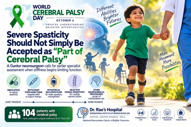
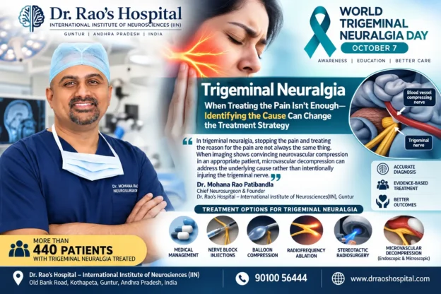

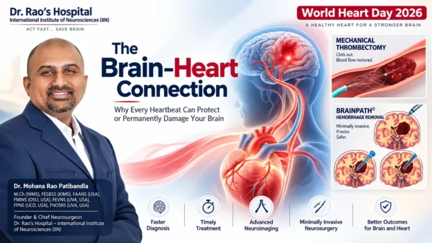


![[명의에게 묻다] '증상없는 뇌졸중'은 보험금 대상 아니다? | 연합뉴스](https://images.openai.com/static-rsc-4/aVoll6FeYTZjD0rYwDYBLsHAPNQAF74RTQlLJ3J6UJq_h8K9i-ZEVKORgMlIRj9fZyIwdsJ3s8OXwI7iQQOYrc_K6Bi_LS7uc4eGvkUp98dRPS2U7ihbIGXFX0ZIqjGuG19FHxJ1e1jEb9phN7KH4SPAJ8LUSWYjXN9i0YhWzDU?purpose=inline)






 Old Bank Street, Kothapeta, Guntur, Andhra Pradesh, India