Author Archives: Dr RAO

tarlovs-cyst-the-best-treatment-at-dr-raos-guntur

Tarlov’s cyst – the best treatment at Dr Raos, Guntur

Tarlov’s cyst – the best treatment at Dr Raos, Guntur

Introduction

A Tarlov’s cyst is a type of spinal cyst that can pressure the nerves in your spine. This can cause pain, tingling, or numbness in your legs or buttocks. You might also have weakness in your legs. Tarlov’s cysts are also called perineural cysts. They’re usually benign, which means they’re not cancerous. But they can still cause problems. Tarlov cysts are most common in women between 30 and 50. They’re more likely to happen if you have a family history of them. They can occur anywhere along your spine, but they’re most often found in the lower (lumbar) or sacral region. Looking for the best Tarlov’s treatment look no further than Dr Raos hospital, Guntur, Andhra Pradesh and Dr Rao is the best spine cord injury specialist. contact us @9010056444 or 9010057444.

causes

There are a number of possible causes of Tarlov cysts, though the exact cause is not yet known. It is thought that they may be the result of abnormal development of the spinal nerves or changes in the structure of the spine. They may also be caused by trauma to the spine or by infection.

Symptoms

Symptoms of Tarlov cysts can vary depending on the size and location of the cyst. Some people may not have any symptoms at all. Others may experience pain, numbness, or tingling in the affected area. The pain is often described as a dull ache that gets worse with sitting or standing for long periods. It may also get worse with coughing, sneezing, or straining. Sometimes, the pain can be severe enough to interfere with daily activities.

If the cyst is large or presses on the spinal cord or nerves, it can cause weakness, loss of sensation, or paralysis in the legs or arms. Urinary and bowel problems may also occur if the cyst presses on the nerves that control these functions.

diagnosis

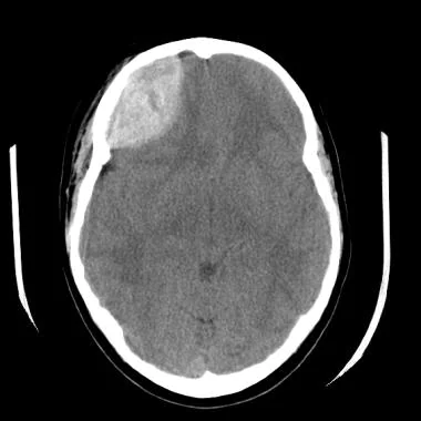
A Tarlov cyst is typically diagnosed using magnetic resonance imaging (MRI). This imaging technique can provide clear pictures of the spinal cord and nerves and any abnormal growths or fluid-filled areas. In some cases, a computed tomography (CT) scan may also be ordered to get a more detailed view of the cyst.

If a Tarlov cyst is suspected, your doctor will likely order an MRI of your spine. This imaging test can give your doctor a clear picture of your spinal cord and nerves and any abnormal growths or fluid-filled areas. In some cases, a CT scan may also be ordered to get a more detailed view of the cyst.

treatment – conservative and minimally invasive spine surgery

There are two main types of treatment for Tarlov cysts: conservative and minimally invasive spine surgery.

Conservative treatment includes pain medication, physical therapy, and epidural injections. This treatment is typically used to help relieve symptoms and improve quality of life. It is important to note that conservative treatment does not usually cure Tarlov cysts.

Minimally invasive spine surgery is a newer treatment that has shown promise in treating Tarlov cysts. This type of surgery is less invasive than traditional open spine surgery, which means there is less risk of complications and a shorter recovery time. Minimally invasive spine surgery typically involves placing a small tube called a shunt into the cyst to drain it of fluid. This can help to reduce pressure on the nerves and relieve symptoms.

outcome

The outcome of Tarlov cysts is generally good. Most people with Tarlov cysts do not experience any symptoms and do not require treatment. However, some people may experience pain, numbness, or weakness if the cyst presses on a nerve. In rare cases, the cyst may rupture and cause spinal cord damage. Treatment is typically only necessary if the cyst is causing symptoms.

Conclusion

The Tarlov cyst is a debilitating condition that can cause a great deal of pain and suffering for those affected by it. While there are treatments available, there is no cure for the condition and it can often recur. For this reason, it is important for patients to be as informed as possible about the condition and its potential treatments. Looking for the best Tarlov’s treatment look no further than Dr Raos hospital, Guntur, Andhra Pradesh and Dr Rao is the best spine cord injury specialist. contact us @9010056444 or 9010057444.

పరిచయం

టార్లోవ్ తిత్తి అనేది మీ వెన్నెముకలోని నరాలపై ఒత్తిడి తెచ్చే వెన్నెముక తిత్తి రకం. ఇది మీ కాళ్ళు లేదా పిరుదులలో నొప్పి, జలదరింపు లేదా తిమ్మిరిని కలిగిస్తుంది. మీ కాళ్ళలో కూడా బలహీనత ఉండవచ్చు. టార్లోవ్ సిస్ట్‌లను పెరిన్యురల్ సిస్ట్‌లు అని కూడా అంటారు. అవి సాధారణంగా నిరపాయమైనవి, అంటే అవి క్యాన్సర్ కావు. కానీ అవి ఇప్పటికీ సమస్యలను కలిగిస్తాయి.

30 మరియు 50 సంవత్సరాల మధ్య వయస్సు గల స్త్రీలలో టార్లోవ్ సిస్ట్‌లు సర్వసాధారణం. మీరు వారి కుటుంబ చరిత్రను కలిగి ఉన్నట్లయితే అవి సంభవించే అవకాశం ఉంది. అవి మీ వెన్నెముకలో ఎక్కడైనా సంభవించవచ్చు, కానీ అవి చాలా తరచుగా దిగువ (కటి) లేదా పవిత్ర ప్రాంతంలో కనిపిస్తాయి.

కారణాలు

టార్లోవ్ సిస్ట్‌లకు అనేక కారణాలు ఉన్నాయి, అయితే ఖచ్చితమైన కారణం ఇంకా తెలియలేదు. అవి వెన్నెముక నరాల యొక్క అసాధారణ అభివృద్ధి లేదా వెన్నెముక నిర్మాణంలో మార్పుల ఫలితంగా ఉండవచ్చని భావిస్తున్నారు. అవి వెన్నెముకకు గాయం లేదా ఇన్ఫెక్షన్ వల్ల కూడా సంభవించవచ్చు.

లక్షణాలు

తిత్తి పరిమాణం మరియు స్థానాన్ని బట్టి టార్లోవ్ సిస్ట్‌ల లక్షణాలు మారవచ్చు. కొందరిలో ఎలాంటి లక్షణాలు ఉండకపోవచ్చు. ఇతరులు ప్రభావిత ప్రాంతంలో నొప్పి, తిమ్మిరి లేదా జలదరింపును అనుభవించవచ్చు. నొప్పి తరచుగా నిస్తేజమైన నొప్పిగా వర్ణించబడింది, ఇది ఎక్కువసేపు కూర్చోవడం లేదా నిలబడటం వలన మరింత తీవ్రమవుతుంది. ఇది దగ్గు, తుమ్ములు లేదా ప్రయాసతో కూడా అధ్వాన్నంగా ఉండవచ్చు. కొన్ని సందర్భాల్లో, నొప్పి రోజువారీ కార్యకలాపాలకు అంతరాయం కలిగించేంత తీవ్రంగా ఉంటుంది.

తిత్తి పెద్దగా లేదా వెన్నుపాము లేదా నరాలపై నొక్కితే, అది బలహీనత, సంచలనాన్ని కోల్పోవడం లేదా కాళ్లు లేదా చేతుల్లో పక్షవాతం కలిగిస్తుంది. ఈ విధులను నియంత్రించే నరాలపై తిత్తి నొక్కితే మూత్ర మరియు ప్రేగు సమస్యలు కూడా సంభవించవచ్చు.

నిర్ధారణ

ఒక టార్లోవ్ తిత్తి సాధారణంగా మాగ్నెటిక్ రెసొనెన్స్ ఇమేజింగ్ (MRI)ని ఉపయోగించి నిర్ధారణ చేయబడుతుంది. ఈ ఇమేజింగ్ టెక్నిక్ వెన్నుపాము మరియు నరాల యొక్క స్పష్టమైన చిత్రాలను, అలాగే ఏదైనా అసాధారణ పెరుగుదలలు లేదా ద్రవంతో నిండిన ప్రాంతాలను అందిస్తుంది. కొన్ని సందర్భాల్లో, తిత్తి యొక్క మరింత వివరణాత్మక వీక్షణను పొందడానికి కంప్యూటెడ్ టోమోగ్రఫీ (CT) స్కాన్ కూడా ఆదేశించబడవచ్చు.

ఒక టార్లోవ్ తిత్తి అనుమానం ఉంటే, మీ వైద్యుడు మీ వెన్నెముక యొక్క MRIని ఆదేశించవచ్చు. ఈ ఇమేజింగ్ పరీక్ష మీ వైద్యుడికి మీ వెన్నుపాము మరియు నరాలు, అలాగే ఏదైనా అసాధారణ పెరుగుదలలు లేదా ద్రవం నిండిన ప్రాంతాల గురించి స్పష్టమైన చిత్రాన్ని అందిస్తుంది. కొన్ని సందర్భాల్లో, తిత్తి యొక్క మరింత వివరణాత్మక వీక్షణను పొందడానికి CT స్కాన్ కూడా ఆదేశించబడవచ్చు.

చికిత్స – సంప్రదాయవాద మరియు కనిష్ట ఇన్వాసివ్ వెన్నెముక శస్త్రచికిత్స

టార్లోవ్ తిత్తులకు రెండు ప్రధాన రకాల చికిత్సలు ఉన్నాయి: సంప్రదాయవాద మరియు కనిష్టంగా ఇన్వాసివ్ వెన్నెముక శస్త్రచికిత్స.

కన్జర్వేటివ్ చికిత్సలో నొప్పి మందులు, భౌతిక చికిత్స మరియు ఎపిడ్యూరల్ ఇంజెక్షన్లు ఉంటాయి. ఈ రకమైన చికిత్స సాధారణంగా లక్షణాల నుండి ఉపశమనం పొందేందుకు మరియు జీవన నాణ్యతను మెరుగుపరచడానికి ఉపయోగించబడుతుంది. సాంప్రదాయిక చికిత్స సాధారణంగా టార్లోవ్ తిత్తులను నయం చేయదని గమనించడం ముఖ్యం.

కనిష్టంగా ఇన్వాసివ్ వెన్నెముక శస్త్రచికిత్స అనేది టార్లోవ్ తిత్తుల చికిత్సలో వాగ్దానాన్ని చూపించే కొత్త రకమైన చికిత్స. ఈ రకమైన శస్త్రచికిత్స సాంప్రదాయ ఓపెన్ వెన్నెముక శస్త్రచికిత్స కంటే తక్కువ హానికరం, అంటే సమస్యల ప్రమాదం తక్కువగా ఉంటుంది మరియు తక్కువ రికవరీ సమయం ఉంటుంది. కనిష్టంగా ఇన్వాసివ్ వెన్నెముక శస్త్రచికిత్సలో సాధారణంగా ఒక షంట్ అని పిలువబడే ఒక చిన్న ట్యూబ్‌ను తిత్తిలోకి ద్రవం నుండి తీసివేయడం జరుగుతుంది. ఇది నరాల మీద ఒత్తిడిని తగ్గించడానికి మరియు లక్షణాల నుండి ఉపశమనం పొందేందుకు సహాయపడుతుంది.

ఫలితం

టార్లోవ్ సిస్ట్‌ల ఫలితం సాధారణంగా మంచిది. టార్లోవ్ తిత్తులు ఉన్న చాలా మంది వ్యక్తులు ఎటువంటి లక్షణాలను అనుభవించరు మరియు చికిత్స అవసరం లేదు. అయితే, కొంతమందికి నరాల మీద తిత్తి నొక్కినప్పుడు నొప్పి, తిమ్మిరి లేదా బలహీనతను అనుభవించవచ్చు. అరుదైన సందర్భాల్లో, తిత్తి చీలిపోయి వెన్నుపాము దెబ్బతింటుంది. తిత్తి లక్షణాలకు కారణమైతే మాత్రమే చికిత్స సాధారణంగా అవసరం.

Conclusion

టార్లోవ్ తిత్తి అనేది బలహీనపరిచే పరిస్థితి, దీని వలన ప్రభావితమైన వారికి చాలా నొప్పి మరియు బాధను కలిగిస్తుంది. చికిత్సలు అందుబాటులో ఉన్నప్పటికీ, పరిస్థితికి చికిత్స లేదు మరియు ఇది తరచుగా పునరావృతమవుతుంది. ఈ కారణంగా, రోగులకు పరిస్థితి మరియు దాని సంభావ్య చికిత్సల గురించి సాధ్యమైనంతవరకు తెలియజేయడం చాలా ముఖ్యం.

vision-problems-the-best-treatment-is-at-dr-raos-guntur

Vision Problems – the best treatment is at Dr Raos, Guntur

Vision Problems – the best treatment is at Dr Raos, Guntur

Introduction

The optic nerve is a bundle of nerve fibers that transmits visual information from the eye to the brain. The optic nerve is also known as cranial nerve II. The optic nerve is important for vision. If the optic nerve is damaged, it can cause decreased vision or even blindness. Looking for the best neurosurgery hospital or Best neurosurgeon or India’s best neurosurgeon or top ten best neurosurgeons, look no further than Dr Rao at Dr Rao’s hospital, the best spine surgery or best neurosurgery or best neurology hospital in Guntur, Andhra Pradesh, India. Call us at 9010056444 or 9010057444 for the best neurosurgeon or spine surgeon or neurologist in India. Dr Raos hospital is best for the visual blurring/ vertigo / numbness / weakness /tingling treatment in Guntur and India. In a recent deccanherald

causes

There are many potential causes of decreased vision. Some causes are more common than others, but all should be taken seriously. The most common cause of decreased vision is refractive error, which occurs when the eye does not focus light properly. This can be corrected with glasses or contact lenses. Other common causes include cataracts, glaucoma, and age-related macular degeneration. These conditions can often be treated with surgery or medication. Less common causes of decreased vision include diabetic retinopathy, stroke, and brain tumors. These conditions may require more aggressive treatment, such as surgery or radiation therapy.

Associated symptoms

There are several symptoms that may be associated with decreased vision. These include: -Blurry vision – Difficulty seeing at night – Difficulty reading – Eye fatigue – Headaches – Squinting

diagnsois

The first step in diagnosing decreased vision is to schedule an appointment with an eye doctor. During the appointment, the doctor will ask about your medical history and symptoms. They will also conduct a thorough eye exam. The eye exam will assess your vision and check for any underlying conditions that could be causing your decreased vision. The doctor may use a lighted instrument to examine your eyes and look for any damage to your optic nerve. They may also order tests, such as an MRI or CT scan, to get a better look at the structure of your eyes. Once the cause of your decreased vision has been determined, the doctor will develop a treatment plan. Treatment options vary depending on the underlying condition causing the decreased vision. In some cases, glasses or contact lenses can help improve vision. Other treatments may include surgery, medication, or lifestyle changes.

treatment

The most common treatment for decreased vision is to use corrective lenses. If the cause of the decreased vision is due to a refractive error, such as nearsightedness, farsightedness, or astigmatism, then glasses or contact lenses can be prescribed to help correct the problem. If the cause of the decreased vision is due to a more serious condition, such as cataracts, glaucoma, or macular degeneration, then surgery may be required. In some cases, medications may be used to treat the underlying condition causing the decreased vision.

prognosis

The prognosis for decreased vision caused by optic nerve damage is generally poor. However, if the damage is caught early and treated promptly, the chances for a better outcome are increased. In some cases, complete recovery is possible.

Conclusion

The optic nerve is an important part of the visual system, and any damage to it can lead to decreased vision. There are many possible causes of optic nerve damage, and associated symptoms can vary depending on the cause. Optic nerve damage can be diagnosed through a variety of methods, and treatment will vary depending on the underlying cause. The prognosis for patients with optic nerve damage depends on the severity of the damage and the underlying cause. Looking for the best neurosurgery hospital or Best neurosurgeon or India’s best neurosurgeon or top ten best neurosurgeons, look no further than Dr Rao at Dr Rao’s hospital, the best spine surgery or best neurosurgery or best neurology hospital in Guntur, Andhra Pradesh, India. Call us at 9010056444 or 9010057444 for the best neurosurgeon or spine surgeon or neurologist in India. Dr Raos hospital is best for the vertigo / numbness / weakness /tingling treatment in Guntur and India.

concussion-the-best-treatment-is-at-dr-raos-guntur

Concussion – the best treatment is at Dr Raos, Guntur

Concussion – the best treatment is at Dr Raos, Guntur

Introduction

A concussion is a type of traumatic brain injury (TBI) that is caused by a blow to the head or a jolt to the body. Concussions can occur in any type of accident, but they are most common in sports injuries and car accidents. A concussion is a serious medical condition that can cause long-term problems, so it is important to seek medical care as soon as possible after sustaining one. There are several different types of symptoms that can occur after a concussion, and they can range from mild to severe. The most common symptoms include headache, dizziness, nausea, vomiting, fatigue, difficulty concentrating, and difficulty remembering. Some people may also experience changes in mood, such as irritability or depression. In some cases, concussions can also cause seizures or coma. Most people who suffer from a concussion will recover within a few weeks with rest and proper medical care. However, some people may experience long-term effects from a concussion, such as problems with memory and concentration, headaches, dizziness, and sleep disorders. If you have any concerns about your health after sustaining a concussion, you should speak with your doctor. Looking for the best neurosurgery hospital or Best neurosurgeon or India’s best neurosurgeon for concussion treatment? Look no further than Dr Rao at Dr Raos hospital, Guntur.

When to seek medical care

If you think you or your child has a concussion, seek medical care right away. A concussion is a serious brain injury that can have lasting effects. Symptoms of a concussion can include:

• headache

• confusion

• memory problems

• nausea or vomiting

• dizziness or balance problems

• double or blurry vision

• sensitivity to light or noise

• feeling sluggish, hazy, foggy, or groggy

• concentration or memory problems

• changes in mood, such as feeling more irritable, sad, or anxious

Head Injury Prevention Tips

There are a number of things you can do to help prevent head injuries.

First, always wear a seatbelt when riding in a car. This will help protect your head and neck in the event of an accident.

Second, wear a helmet when participating in activities such as biking, skateboarding, or rollerblading. Helmets can help protect your head from serious injuries if you fall or are hit by another object.

Third, make sure the playing surface is safe before participating in any sport. If you see any potential hazards, such as loose gravel or potholes, avoid them.

Fourth, be aware of your surroundings at all times.

If you are walking near traffic, make sure to look both ways before crossing the street. By being aware of your surroundings, you can help avoid accidents that could lead to head injuries. Head injuries can happen anywhere, at any time.

By taking some simple precautions, you can help reduce your risk of sustaining a head injury.

Warning Signs of a Serious Brain Injury

If you or someone you know experiences any of the following symptoms after a head injury, it is important to seek medical attention immediately:

-Loss of consciousness

-Headache that gets worse or does not go away

-Repeated vomiting or nausea

-Slurred speech

-Drowsiness or inability to wake up

-Confusion

-Convulsions or seizures

-Weakness in one arm or leg

-Loss of coordination

Managing Concussions: The NFL Protocol

The National Football League (NFL) has a concussion protocol in place to protect players and ensure they receive the proper care and treatment if they suffer a head injury. The protocol is designed to standardize the evaluation and management of concussions, and it includes both sideline and locker room evaluations by team medical staff. If a player is suspected of having a concussion, they are removed from the game and cannot return until they are cleared by an independent medical consultant. The NFL’s concussion protocol has come under scrutiny in recent years, as some have criticized it for being too lenient and not doing enough to protect players. However, the league has made several changes to the protocol in recent years in an effort to improve player safety.

NCAA Concussion Update

The NCAA has updated its concussion protocol to require that any player who shows signs of a concussion must be removed from the game and cannot return until they are cleared by a medical professional. This is a major change from the previous protocol, which allowed players to stay in the game if they showed no symptoms of a concussion. The new protocol also requires that any player who is suspected of having a concussion must be evaluated by a medical professional at halftime or during the game. If the player is diagnosed with a concussion, they must be removed from the game and cannot return until they are cleared by a medical professional. The NCAA’s new concussion protocol is a major step forward in protecting athletes from serious brain injuries. This update will help to ensure that any player who suffers a concussion will receive the proper medical care and will not be allowed to return to play until it is safe for them to do so.

Conclusion

In conclusion, concussion is a serious medical condition that can have lasting effects. It is important to seek medical care if you think you or someone you know has suffered a concussion. There are also steps that can be taken to prevent head injuries. Be sure to watch for the warning signs of a serious brain injury and follow the appropriate protocol for managing concussions. Looking for the best neurosurgery hospital or Best neurosurgeon or India’s best neurosurgeon or top ten best neurosurgeons, look no further than Dr Rao at Dr Rao’s hospital, the best spine surgery or best neurosurgery or best neurology hospital in Guntur, Andhra Pradesh, India. Call us at 9010056444 or 9010057444 for the best neurosurgeon or spine surgeon or neurologist in India. Dr Raos hospital is best for the numbness / weakness /tingling treatment in Guntur and India.

కంకషన్

పరిచయం

ఒక కంకషన్ అనేది ఒక రకమైన బాధాకరమైన మెదడు గాయం (TBI), ఇది తలపై దెబ్బ లేదా శరీరానికి కుదుపు వల్ల వస్తుంది. ఏ రకమైన ప్రమాదంలోనైనా కంకషన్లు సంభవించవచ్చు, కానీ అవి స్పోర్ట్స్ గాయాలు మరియు కారు ప్రమాదాలలో సర్వసాధారణం. కంకషన్ అనేది తీవ్రమైన వైద్య పరిస్థితి, ఇది దీర్ఘకాలిక సమస్యలను కలిగిస్తుంది, కాబట్టి ఒకదానిని కొనసాగించిన తర్వాత వీలైనంత త్వరగా వైద్య సంరక్షణను పొందడం చాలా ముఖ్యం.

కంకషన్ తర్వాత సంభవించే అనేక రకాలైన లక్షణాలు ఉన్నాయి మరియు అవి తేలికపాటి నుండి తీవ్రమైన వరకు ఉంటాయి. అత్యంత సాధారణ లక్షణాలు తలనొప్పి, తల తిరగడం, వికారం, వాంతులు, అలసట, ఏకాగ్రత కష్టం మరియు గుర్తుంచుకోవడం కష్టం. కొంతమంది వ్యక్తులు చిరాకు లేదా నిరాశ వంటి మూడ్‌లో మార్పులను కూడా అనుభవించవచ్చు. కొన్ని సందర్భాల్లో, కంకషన్లు మూర్ఛలు లేదా కోమాకు కూడా కారణమవుతాయి.

కంకషన్‌తో బాధపడుతున్న చాలా మంది వ్యక్తులు విశ్రాంతి మరియు సరైన వైద్య సంరక్షణతో కొన్ని వారాలలో కోలుకుంటారు. అయినప్పటికీ, జ్ఞాపకశక్తి మరియు ఏకాగ్రత, తలనొప్పి, మైకము మరియు నిద్ర రుగ్మతలు వంటి సమస్యలు వంటి కంకషన్ నుండి కొంతమంది దీర్ఘకాలిక ప్రభావాలను అనుభవించవచ్చు. ఒక కంకషన్ తర్వాత మీ ఆరోగ్యం గురించి మీకు ఏవైనా ఆందోళనలు ఉంటే, మీరు మీ డాక్టర్తో మాట్లాడాలి.

ఎప్పుడు వైద్య సంరక్షణ పొందాలి

మీకు లేదా మీ బిడ్డకు కంకషన్ ఉందని మీరు భావిస్తే, వెంటనే వైద్య సంరక్షణ పొందండి. కంకషన్ అనేది తీవ్రమైన మెదడు గాయం, ఇది శాశ్వత ప్రభావాలను కలిగి ఉంటుంది.

కంకషన్ యొక్క లక్షణాలు వీటిని కలిగి ఉండవచ్చు:

• తలనొప్పి
• గందరగోళం
• జ్ఞాపకశక్తి సమస్యలు
• వికారం లేదా వాంతులు
• మైకము లేదా బ్యాలెన్స్ సమస్యలు
• డబుల్ లేదా అస్పష్టమైన దృష్టి
• కాంతి లేదా శబ్దానికి సున్నితత్వం
• నిదానంగా, మబ్బుగా, పొగమంచుగా లేదా గాఢంగా అనిపించడం
• ఏకాగ్రత లేదా జ్ఞాపకశక్తి సమస్యలు
• మరింత చిరాకుగా, విచారంగా లేదా ఆత్రుతగా అనిపించడం వంటి మానసిక స్థితి మార్పులు

తల గాయం నివారణ చిట్కాలు

తల గాయాలను నివారించడంలో మీరు సహాయం చేయగల అనేక అంశాలు ఉన్నాయి. మొదట, కారులో ప్రయాణించేటప్పుడు ఎల్లప్పుడూ సీటుబెల్ట్ ధరించండి. ప్రమాదం జరిగినప్పుడు ఇది మీ తల మరియు మెడను రక్షించడంలో సహాయపడుతుంది. రెండవది, బైకింగ్, స్కేట్‌బోర్డింగ్ లేదా రోలర్‌బ్లేడింగ్ వంటి కార్యకలాపాలలో పాల్గొనేటప్పుడు హెల్మెట్ ధరించండి. హెల్మెట్‌లు మీరు పడిపోతే లేదా మరొక వస్తువుతో కొట్టబడినప్పుడు మీ తలపై తీవ్రమైన గాయాల నుండి రక్షించడంలో సహాయపడతాయి. మూడవది, ఏదైనా క్రీడలో పాల్గొనే ముందు ఆడే ఉపరితలం సురక్షితంగా ఉందని నిర్ధారించుకోండి. మీరు వదులుగా ఉన్న కంకర లేదా గుంతలు వంటి ఏవైనా సంభావ్య ప్రమాదాలను చూసినట్లయితే, వాటిని నివారించండి. నాల్గవది, మీ పరిసరాల గురించి ఎల్లప్పుడూ తెలుసుకోండి. మీరు ట్రాఫిక్ దగ్గర నడుస్తుంటే, వీధిని దాటే ముందు రెండు వైపులా చూసుకోండి. మీ పరిసరాల గురించి తెలుసుకోవడం ద్వారా, తలకు గాయాలయ్యే ప్రమాదాలను నివారించడంలో మీరు సహాయపడవచ్చు.

తలకు గాయాలు ఎక్కడైనా, ఎప్పుడైనా జరగవచ్చు. కొన్ని సాధారణ జాగ్రత్తలు తీసుకోవడం ద్వారా, మీరు తలకు గాయం అయ్యే ప్రమాదాన్ని తగ్గించుకోవచ్చు.

తీవ్రమైన మెదడు గాయం యొక్క హెచ్చరిక సంకేతాలు

మీకు లేదా మీకు తెలిసిన ఎవరైనా తలకు గాయమైన తర్వాత క్రింది లక్షణాలలో దేనినైనా అనుభవిస్తే, వెంటనే వైద్య సంరక్షణను పొందడం చాలా ముఖ్యం:

-స్పృహ కోల్పోవడం
-అధ్వాన్నంగా లేదా తగ్గని తలనొప్పి

  • పదేపదే వాంతులు లేదా వికారం
  • అస్పష్టమైన ప్రసంగం
  • మగత లేదా మేల్కొలపడానికి అసమర్థత
  • గందరగోళం
  • మూర్ఛలు లేదా మూర్ఛలు
    -ఒక చేయి లేదా కాలులో బలహీనత
  • సమన్వయం కోల్పోవడం

కన్‌కషన్‌లను నిర్వహించడం: NFL ప్రోటోకాల్

నేషనల్ ఫుట్‌బాల్ లీగ్ (NFL) ఆటగాళ్లను రక్షించడానికి మరియు తలకు గాయం అయినట్లయితే వారికి సరైన సంరక్షణ మరియు చికిత్స అందుతుందని నిర్ధారించడానికి ఒక కంకషన్ ప్రోటోకాల్‌ను కలిగి ఉంది. ప్రోటోకాల్ కంకషన్ల మూల్యాంకనం మరియు నిర్వహణను ప్రామాణీకరించడానికి రూపొందించబడింది మరియు ఇది జట్టు వైద్య సిబ్బందిచే సైడ్‌లైన్ మరియు లాకర్ రూమ్ మూల్యాంకనాలను కలిగి ఉంటుంది. ఒక ఆటగాడికి కంకషన్ ఉన్నట్లు అనుమానించబడినట్లయితే, వారు ఆట నుండి తీసివేయబడతారు మరియు స్వతంత్ర వైద్య సలహాదారు ద్వారా క్లియర్ చేయబడే వరకు తిరిగి రాలేరు.

NFL యొక్క కంకషన్ ప్రోటోకాల్ ఇటీవలి సంవత్సరాలలో పరిశీలనలో ఉంది, కొంతమంది ఇది చాలా తేలికగా ఉందని మరియు ఆటగాళ్లను రక్షించడానికి తగినంతగా చేయలేదని విమర్శించారు. అయినప్పటికీ, ఆటగాళ్ల భద్రతను మెరుగుపరిచే ప్రయత్నంలో లీగ్ ఇటీవలి సంవత్సరాలలో ప్రోటోకాల్‌లో అనేక మార్పులు చేసింది.

NCAA కంకషన్ అప్‌డేట్

కన్‌కషన్ సంకేతాలను చూపించే ఏ ఆటగాడైనా తప్పనిసరిగా గేమ్ నుండి తీసివేయబడాలని మరియు వైద్య నిపుణుడిచే క్లియర్ చేయబడే వరకు తిరిగి రాకూడదని NCAA దాని కంకషన్ ప్రోటోకాల్‌ను నవీకరించింది. ఇది మునుపటి ప్రోటోకాల్ నుండి పెద్ద మార్పు, ఇది ఆటగాళ్ళు కంకషన్ లక్షణాలను చూపించకపోతే గేమ్‌లో ఉండటానికి అనుమతించింది.

కొత్త ప్రోటోకాల్ ప్రకారం ఏదైనా ఆటగాడు కంకషన్ ఉన్నట్లు అనుమానించబడినట్లయితే, హాఫ్‌టైమ్‌లో లేదా గేమ్ సమయంలో తప్పనిసరిగా వైద్య నిపుణులచే మూల్యాంకనం చేయబడాలి. ఆటగాడికి కంకషన్ ఉన్నట్లు నిర్ధారణ అయినట్లయితే, అతను తప్పనిసరిగా గేమ్ నుండి తీసివేయబడాలి మరియు వైద్య నిపుణులచే క్లియర్ చేయబడే వరకు తిరిగి రాకూడదు.

తీవ్రమైన మెదడు గాయాల నుండి అథ్లెట్లను రక్షించడంలో NCAA యొక్క కొత్త కంకషన్ ప్రోటోకాల్ ఒక ప్రధాన ముందడుగు. ఈ అప్‌డేట్ కంకషన్‌తో బాధపడే ఏ ఆటగాడికి సరైన వైద్య సంరక్షణ అందుతుందని నిర్ధారించడానికి సహాయం చేస్తుంది మరియు వారు అలా చేయడం సురక్షితంగా ఉండే వరకు తిరిగి ఆడేందుకు అనుమతించబడరు.

ముగింపు

ముగింపులో, కంకషన్ అనేది తీవ్రమైన వైద్య పరిస్థితి, ఇది శాశ్వత ప్రభావాలను కలిగి ఉంటుంది. మీరు లేదా మీకు తెలిసిన వారు కంకషన్‌కు గురయ్యారని మీరు భావిస్తే వైద్య సంరక్షణను పొందడం చాలా ముఖ్యం. తల గాయాలను నివారించడానికి కూడా తీసుకోగల చర్యలు ఉన్నాయి. తీవ్రమైన మెదడు గాయం యొక్క హెచ్చరిక సంకేతాల కోసం తప్పకుండా చూడండి మరియు కంకషన్లను నిర్వహించడానికి తగిన ప్రోటోకాల్‌ను అనుసరించండి.

spina bifida treatment

Spina bifida – the best treatment is at Dr Raos, Guntur

Spina bifida – the best treatment is at Dr Raos, Guntur

Introduction

Spina bifida is a birth defect that occurs when the spine and spinal cord do not form properly. It is the most common neural tube defect in the United States, affecting about 1,500 babies each year. There are three main types of spina bifida: meningocele, myelomeningocele, and lipomyelomeningocele. Meningocele is the least severe form, and only affects the meninges (the protective covering around the spinal cord). Myelomeningocele is more severe, and includes a sac-like protrusion of the spinal cord and meninges through an opening in the spine. Lipomyelomeningocele is also a severe form, and includes both a fatty tumor and a myelomeningocele. Most babies with spina bifida will have some type of disability, such as paralysis of the legs, urinary and bowel incontinence, hydrocephalus (build-up of fluid in the brain), and scoliosis (curvature of the spine). There is no cure for spina bifida, but treatment can improve quality of life and folic acid is important prevent measure. Surgery to close the opening in the spine is often recommended soon after birth or during the pregnancy, and may be followed by physical therapy, medications, and other treatments. Fortunately, with proper medical care, many children with spina bifida can lead active and productive lives. Twenty-year follow-up studies of children with spina bifida show that they enter college in the same proportion as the general population and many are actively employed. With recent advancements in medical care for these children, their outlook continues to improve. Looking for the best spina bifida treatment in Guntur, look no further than Dr. Raos hospital or Dr. Rao, the best neurosurgeon for spina bifida. @9010056444.

Spina bifida awareness month is October – pre conceptional Folic acid prevents 70% of the spina bifida cases

risk factors

There are many different factors that can contribute to an individual developing spina bifida, and it is often difficult to determine exactly what causes the condition. However, there are certain risk factors that have been identified which may increase an individual’s chances of developing spina bifida.

These include:

– A family history of spina bifida or other neural tube defects

– Exposure to certain environmental toxins or chemicals, such as those used in pesticides

– Maternal diabetes

– Obesity

– Use of certain medications during pregnancy, such as valproic acid

types

Spina bifida is of four kinds, occulta, meningocele, myelomeningocele, Lipomyelomeningocele

There are four main types of spina bifida: 1. Spina bifida occulta: This is the mildest form of the condition, and is often not diagnosed until adulthood. The spinal cord and nerves are usually normal, although there may be some malformation of the bones of the spine. 2. Meningocele: This form of spina bifida results in a sac-like protrusion of the meninges (the membranes that surround the spinal cord) through a defect in the vertebrae. The spinal cord and nerves are usually not affected. 3. Myelomeningocele: This is the most severe form of spina bifida, and is characterized by a sac-like protrusion of the meninges and spinal cord through a defect in the vertebrae. This can result in paralysis and loss of sensation below the level of the lesion. 4. Lipomyelomeningocele: This rare form of spina bifida is characterized by a fatty mass attached to the spinal cord, which may or may not be covered by a thin layer of skin.

symptoms

The most common symptom of spina bifida is a small lump or bulge on the back, usually at the level of the lower spine. This bulge may be covered by skin or it may be open and covered by a thin membrane. Other symptoms may include:

-Weakness or paralysis of the legs

-Loss of sensation in the legs

-Urinary incontinence or difficulty urinating

-Constipation

-Abnormalities in the structure of the spine

diagnosis

  • Maternal serum alpha-fetoprotein test (Physican may order follow-up tests for confirmation including amniocentesis)
  • 20-week ultrasound
  • Infant ultrasound
  • Fetal MRI

A diagnosis of spina bifida or meningocele can be made prenatally by fetal ultrasound, which may show evidence of a mass in the lumbosacral region, hydrocephalus, or both. After birth, the diagnosis is typically made by physical examination, which may reveal a sac-like protrusion from the spinal column, an absence of skin over the lesion, and/or neurological deficits. In some cases, magnetic resonance imaging (MRI) may be used to further evaluate the extent of the lesion and associated abnormalities. A biopsy of the lesion is generally not necessary for diagnosis but may be performed to rule out other conditions.

treatment

Prenatal or Fetal surgery

Infant surgery

There are a number of different treatment options available for those with spina bifida, depending on the severity of the condition. In some cases, surgery may be necessary to close the opening in the spine and prevent further damage to the spinal cord. In other cases, treatment may focus on managing symptoms and improving quality of life. For example, people with spina bifida may need to take medication to control seizures or pain. They may also need physical therapy to help them improve their mobility and strength. In some cases, people with spina bifida may need to use a wheelchair or other assistive devices. The goal of treatment is to improve quality of life and help people with spina bifida live as independently as possible.

Conclusion

In conclusion, spina bifida is a birth defect that can cause many different problems. There are different types of spina bifida, and the severity of the condition can vary from person to person. There are many risk factors for spina bifida, and it is important to be aware of them. If you think you or your child may have spina bifida, it is important to see a doctor so that you can get the proper diagnosis and treatment. Fortunately, with proper medical care, many children with spina bifida can lead active and productive lives. Twenty-year follow-up studies of children with spina bifida show that they enter college in the same proportion as the general population and many are actively employed. With recent advancements in medical care for these children, their outlook continues to improve. Looking for the best spina bifida treatment in Guntur, look no further than Dr. Raos hospital or Dr. Rao, the best neurosurgeon for spina bifida. @9010056444.

Dr. Rao receives award in EENADU: "ACT FAST SAVE BRAIN AND SPINE" - promoting urgent action for neurological health.

Vertigo – the best treatment is at Dr Raos, Guntur

Vertigo – the best treatment is at Dr Raos, Guntur


Introduction

Vertigo is a type of dizziness that can cause a person to feel as if they are spinning or moving even when they are not. It can be a very unsettling feeling and can cause nausea, vomiting, and balance problems. Vertigo can be caused by many things, including inner ear infections, head injuries, and certain medications. It is important to see a doctor if you are experiencing vertigo so that the cause can be found and treated. Looking for the best neurosurgery hospital or Best neurosurgeon or India’s best neurosurgeon or top ten best neurosurgeons, look no further than Dr Rao at Dr Rao’s hospital, the best spine surgery or best neurosurgery or best neurology hospital in Guntur, Andhra Pradesh, India. Call us at 9010056444 or 9010057444 for the best neurosurgeon or spine surgeon or neurologist in India. Dr Raos hospital is best for the vertigo / numbness / weakness /tingling treatment in Guntur and India.

classification

There are two types of vertigo: peripheral and central.

Peripheral vertigo is caused by a problem in the inner ear or vestibular nerve, while central vertigo is caused by a problem in the brain. Peripheral vertigo is further divided into two types: vestibular neuritis and labyrinthitis. Vestibular neuritis is caused by inflammation of the vestibular nerve, while labyrinthitis is caused by inflammation of the inner ear.

Central vertigo is also further divided into two types: migrainous vertigo and vertebrobasilar insufficiency. Migrainous vertigo is caused by migraines, while vertebrobasilar insufficiency is caused by a decrease in blood flow to the brain.

Symptoms

There are many different symptoms that can be associated with vertigo. These can include a feeling of dizziness, lightheadedness, nausea, vomiting, and a loss of balance. Vertigo can also cause problems with vision, hearing, and coordination. People who suffer from vertigo may feel as if they are spinning or that their surroundings are spinning around them.

diagnosis

There are a few different ways to diagnose vertigo. The most common is through a physical examination and a review of your symptoms. Your doctor may also use special tests, such as the Dix-Hallpike maneuver, to help diagnose vertigo. If you have vertigo, your doctor will likely ask you about your symptoms and perform a physical examination. He or she may also use special tests to help diagnose vertigo, such as the Dix-Hallpike maneuver. The Dix-Hallpike maneuver is used to help diagnose positional vertigo. To do this test, your doctor will have you sit in a chair with your head turned to one side. Then, he or she will quickly move you from the sitting position to lying down on your back with your head still turned to the side. This maneuver can provoke dizziness and nystagmus (abnormal eye movements).

treatment

There is no one-size-fits-all approach to treating vertigo, as the condition can be caused by a variety of underlying factors. However, there are some general treatment strategies that can be effective in managing the symptoms of vertigo. One common approach is vestibular rehabilitation therapy (VRT), which is a type of physical therapy designed to help retrain the brain to better process information from the vestibular system. VRT exercises can help improve balance and reduce dizziness and nausea. Medications can also be used to treat vertigo, although they will not cure the underlying condition. For example, antihistamines can be used to treat dizziness caused by allergies, while anticholinergics can be used to treat dizziness caused by an inner ear disorder. In some cases, surgery may be necessary to correct the underlying cause of vertigo. For example, if vertigo is caused by a problem with the inner ear, a surgical procedure called labyrinthectomy may be needed to remove the affected portion of the inner ear.

Living with it

Living with vertigo can be extremely difficult. The constant feeling of dizziness and nausea can make it hard to focus on anything else. There are a few things that can help make life easier though. First, try to avoid any sudden movements. This can trigger an episode of vertigo and make the symptoms worse. Second, try to keep your head still as much as possible. This can be difficult, but it will help lessen the symptoms. Finally, stay hydrated. Drinking plenty of fluids will help keep your body healthy and may help reduce the frequency of vertigo episodes.

Conclusion

Vertigo can be a debilitating condition that significantly impacts one’s quality of life. However, with the right treatment, many people are able to manage their symptoms and live relatively normal lives. If you are suffering from vertigo, it is important to seek medical help so that you can get the treatment you need. Looking for the best neurosurgery hospital or Best neurosurgeon or India’s best neurosurgeon or top ten best neurosurgeons, look no further than Dr Rao at Dr Rao’s hospital, the best spine surgery or best neurosurgery or best neurology hospital in Guntur, Andhra Pradesh, India. Call us at 9010056444 or 9010057444 for the best neurosurgeon or spine surgeon or neurologist in India. Dr Raos hospital is best for the Vertigo/numbness / weakness /tingling treatment in Guntur and India.

తలతిరగడం

పరిచయం

వెర్టిగో అనేది ఒక రకమైన తలతిరగడం. ఇది చాలా కలవరపెట్టే అనుభూతి మరియు వికారం, వాంతులు మరియు సమతుల్య సమస్యలను కలిగిస్తుంది. వెర్టిగో అనేది లోపలి చెవి ఇన్ఫెక్షన్లు, తల గాయాలు మరియు కొన్ని మందులతో సహా అనేక విషయాల వల్ల సంభవించవచ్చు. మీరు వెర్టిగోను ఎదుర్కొంటుంటే వైద్యుడిని సంప్రదించడం చాలా ముఖ్యం, తద్వారా కారణాన్ని కనుగొని చికిత్స చేయవచ్చు.

వర్గీకరణ

వెర్టిగోలో రెండు రకాలు ఉన్నాయి: పెరిఫెరల్ మరియు సెంట్రల్. పెరిఫెరల్ వెర్టిగో అనేది లోపలి చెవి లేదా వెస్టిబ్యులర్ నరాల సమస్య వల్ల వస్తుంది, అయితే సెంట్రల్ వెర్టిగో మెదడులోని సమస్య వల్ల వస్తుంది.

పెరిఫెరల్ వెర్టిగో రెండు రకాలుగా విభజించబడింది: వెస్టిబ్యులర్ న్యూరిటిస్ మరియు లాబిరింథిటిస్. వెస్టిబ్యులర్ న్యూరిటిస్ అనేది వెస్టిబ్యులర్ నరాల వాపు వల్ల వస్తుంది, అయితే లాబ్రింథిటిస్ లోపలి చెవి యొక్క వాపు వల్ల వస్తుంది.

సెంట్రల్ వెర్టిగో కూడా రెండు రకాలుగా విభజించబడింది: మైగ్రేనస్ వెర్టిగో మరియు వెర్టెబ్రోబాసిలర్ ఇన్సఫిసియెన్సీ. మైగ్రేనస్ వెర్టిగో మైగ్రేన్‌ల వల్ల వస్తుంది, అయితే వెర్టెబ్రోబాసిలర్ లోపం మెదడుకు రక్త ప్రసరణ తగ్గడం వల్ల వస్తుంది.

లక్షణాలు

వెర్టిగోతో అనుబంధించబడే అనేక విభిన్న లక్షణాలు ఉన్నాయి. వీటిలో తలతిరగడం, తలతిరగడం, వికారం, వాంతులు మరియు సమతుల్యత కోల్పోవడం వంటివి ఉంటాయి. వెర్టిగో దృష్టి, వినికిడి మరియు సమన్వయంతో కూడా సమస్యలను కలిగిస్తుంది. వెర్టిగోతో బాధపడుతున్న వ్యక్తులు తాము తిరుగుతున్నట్లు లేదా వారి చుట్టూ తమ చుట్టూ తిరుగుతున్నట్లు భావించవచ్చు.

నిర్ధారణ

వెర్టిగోని నిర్ధారించడానికి కొన్ని విభిన్న మార్గాలు ఉన్నాయి. శారీరక పరీక్ష మరియు మీ లక్షణాల సమీక్ష ద్వారా సర్వసాధారణం. మీ వైద్యుడు వెర్టిగోను నిర్ధారించడంలో సహాయపడటానికి డిక్స్-హాల్‌పైక్ యుక్తి వంటి ప్రత్యేక పరీక్షలను కూడా ఉపయోగించవచ్చు.

మీకు వెర్టిగో ఉంటే, మీ వైద్యుడు మీ లక్షణాల గురించి మిమ్మల్ని అడగవచ్చు మరియు శారీరక పరీక్ష చేస్తారు. అతను లేదా ఆమె డిక్స్-హాల్‌పైక్ యుక్తి వంటి వెర్టిగోను నిర్ధారించడంలో సహాయపడటానికి ప్రత్యేక పరీక్షలను కూడా ఉపయోగించవచ్చు.

డిక్స్-హాల్‌పైక్ యుక్తి స్థాన వెర్టిగోను నిర్ధారించడంలో సహాయపడటానికి ఉపయోగించబడుతుంది. ఈ పరీక్ష చేయడానికి, మీ డాక్టర్ మిమ్మల్ని కుర్చీలో కూర్చోబెట్టి, మీ తల ఒక వైపుకు తిప్పుతారు. అప్పుడు, అతను లేదా ఆమె మిమ్మల్ని కూర్చున్న స్థానం నుండి మీ తల వైపుకు తిప్పి మీ వెనుకభాగంలో పడుకునేలా చేస్తుంది. ఈ యుక్తి మైకము మరియు నిస్టాగ్మస్ (అసాధారణ కంటి కదలికలు) రేకెత్తిస్తుంది.

చికిత్స

వెర్టిగో చికిత్సలో అందరికీ సరిపోయే విధానం లేదు, ఎందుకంటే ఈ పరిస్థితి వివిధ కారణాల వల్ల సంభవించవచ్చు. అయినప్పటికీ, వెర్టిగో యొక్క లక్షణాలను నిర్వహించడంలో ప్రభావవంతంగా ఉండే కొన్ని సాధారణ చికిత్సా వ్యూహాలు ఉన్నాయి.

వెస్టిబ్యులర్ రిహాబిలిటేషన్ థెరపీ (VRT) అనేది ఒక సాధారణ విధానం, ఇది వెస్టిబ్యులర్ సిస్టమ్ నుండి సమాచారాన్ని మెరుగ్గా ప్రాసెస్ చేయడానికి మెదడుకు తిరిగి శిక్షణ ఇవ్వడంలో సహాయపడే ఒక రకమైన భౌతిక చికిత్స. VRT వ్యాయామాలు సమతుల్యతను మెరుగుపరచడంలో సహాయపడతాయి మరియు మైకము మరియు వికారం తగ్గుతాయి.

వెర్టిగో చికిత్సకు మందులు కూడా ఉపయోగించవచ్చు, అయినప్పటికీ అవి అంతర్లీన పరిస్థితిని నయం చేయవు. ఉదాహరణకు, యాంటిహిస్టామైన్‌లను అలెర్జీల వల్ల వచ్చే మైకము చికిత్సకు ఉపయోగించవచ్చు, అయితే యాంటీకోలినెర్జిక్స్ లోపలి చెవి రుగ్మత వల్ల వచ్చే మైకానికి చికిత్స చేయడానికి ఉపయోగించవచ్చు.

కొన్ని సందర్భాల్లో, వెర్టిగో యొక్క మూల కారణాన్ని సరిచేయడానికి శస్త్రచికిత్స అవసరం కావచ్చు. ఉదాహరణకు, లోపలి చెవి సమస్య వల్ల వెర్టిగో ఏర్పడినట్లయితే, లోపలి చెవి యొక్క ప్రభావిత భాగాన్ని తొలగించడానికి లాబిరింథెక్టమీ అనే శస్త్రచికిత్సా ప్రక్రియ అవసరమవుతుంది.

దానితో జీవించడం

వెర్టిగోతో జీవించడం చాలా కష్టం. మైకము మరియు వికారం యొక్క స్థిరమైన భావన మరేదైనా దృష్టి పెట్టడం కష్టతరం చేస్తుంది. అయితే జీవితాన్ని సులభతరం చేయడంలో సహాయపడే కొన్ని అంశాలు ఉన్నాయి.

మొదట, ఏదైనా ఆకస్మిక కదలికలను నివారించడానికి ప్రయత్నించండి. ఇది వెర్టిగో యొక్క ఎపిసోడ్‌ను ప్రేరేపిస్తుంది మరియు లక్షణాలను మరింత తీవ్రతరం చేస్తుంది. రెండవది, మీ తలను వీలైనంత వరకు ఉంచడానికి ప్రయత్నించండి. ఇది కష్టంగా ఉంటుంది, కానీ ఇది లక్షణాలను తగ్గించడంలో సహాయపడుతుంది. చివరగా, హైడ్రేటెడ్ గా ఉండండి. పుష్కలంగా ద్రవాలు తాగడం మీ శరీరాన్ని ఆరోగ్యంగా ఉంచడంలో సహాయపడుతుంది మరియు వెర్టిగో ఎపిసోడ్‌ల ఫ్రీక్వెన్సీని తగ్గించడంలో సహాయపడుతుంది.

ముగింపు

వెర్టిగో అనేది ఒకరి జీవన నాణ్యతను గణనీయంగా ప్రభావితం చేసే బలహీనపరిచే పరిస్థితి. అయినప్పటికీ, సరైన చికిత్సతో, చాలా మంది వ్యక్తులు తమ లక్షణాలను నిర్వహించగలుగుతారు మరియు సాపేక్షంగా సాధారణ జీవితాలను గడపగలుగుతారు. మీరు వెర్టిగోతో బాధపడుతున్నట్లయితే, వైద్య సహాయం తీసుకోవడం చాలా ముఖ్యం, తద్వారా మీకు అవసరమైన చికిత్సను పొందవచ్చు.

the-best-spinal-infection-treatment-is-at-dr-raos-guntur

Spinal infection – the best treatment is at Dr Raos, Guntur

Spinal infection – the best treatment is at Dr Raos, Guntur

Introduction

Spinal infection treatment is critical because the spine is a vital part of the human body, providing structure and support for the entire upper torso. Because of its importance, any infection or inflammation of the spine can have serious consequences. Spinal infections can occur anywhere along the length of the spine, from the neck to the lower back. There are several different types of spinal infections, each with its own set of symptoms and treatment options. The most common type of spinal infection is vertebral osteomyelitis, which is an infection of the bone tissue. Other types of spinal infections include intervertebral disc space infections and spinal canal infections. Symptoms of a spinal infection may include pain, stiffness, and fever. In some cases, neurological problems such as weakness or paralysis can also occur. Diagnosis of a spinal infection typically involves a combination of physical examination, imaging tests, and laboratory tests. Treatment for a spinal infection typically involves antibiotics to clear the infection, as well as pain relief and rest. In some cases, surgery may also be necessary to remove infected tissue or to stabilize the spine. If you’re looking for the best spine surgery center for spinal infection, look no further than Dr. Rao’s hospital, the best spine surgery center in India. Our experienced neurosurgeon and spine specialist will work with you to find the best treatment option for your condition. Call us today to schedule a consultation @9010056444.

risk factors

There are many risk factors for developing a spinal infection, including:

• Having a weakened immune system

• Having diabetes

• Having cancer

• Having a history of drug abuse

• Having had recent surgery on the spine or another area of the body

types

There are four main types of vertebral osteomyelitis:

1. Infection of the vertebral body: This is the most common type of vertebral osteomyelitis, accounting for approximately 60% of all cases. It usually occurs as a result of bacteria spreading from another infection site in the body, such as the lungs or urinary tract.

2. Infection of the intervertebral disc space: This type of vertebral osteomyelitis accounts for approximately 20% of all cases. It usually occurs as a result of bacteria spreading from another infection site in the body, such as the lungs or urinary tract.

3. Infection of the spinal canal: This type of vertebral osteomyelitis accounts for approximately 10% of all cases. It usually occurs as a result of bacteria spreading from another infection site in the body, such as the lungs or urinary tract.

4. Infection of the paravertebral tissues: This type of vertebral osteomyelitis accounts for approximately 10% of all cases. It usually occurs as a result of bacteria spreading from another infection site in the body, such as the lungs or urinary tract.

symptoms

The symptoms of a spinal infection can vary depending on the location and severity of the infection. However, some common symptoms include back pain, stiffness, and fever. If the infection is in the spinal cord, additional symptoms may include weakness, numbness, and difficulty walking. If the infection is left untreated, it can spread to other parts of the body and cause life-threatening complications.

diagnosis

There are a few different ways that doctors can diagnose a spinal infection. The first way is through a physical examination. Your doctor will feel for any areas of tenderness or pain in your spine. They will also look for any signs of redness or swelling. The second way to diagnose a spinal infection is through imaging tests. These tests can include an X-ray, MRI, or CT scan. These tests will help your doctor to see if there is any damage to the bones or tissues in your spine. The third way to diagnose a spinal infection is through a blood test. This test can show if there are any bacteria or viruses present in your blood. Once your doctor has diagnosed a spinal infection, they will then be able to start you on the proper treatment plan.

treatment

The mainstay of treatment for vertebral osteomyelitis is antibiotics. Antibiotics are typically given intravenously (through a vein) for four to six weeks. Surgery is sometimes needed to remove infected bone or tissue. In some cases, a longer course of antibiotics may be needed.

Conclusion

In conclusion, spinal infections are serious and potentially life-threatening conditions that require prompt diagnosis and treatment. Early diagnosis and treatment is essential to prevent permanent damage to the spine and nervous system. If you suspect that you or someone you know may have a spinal infection, it is important to seek medical attention immediately. If you’re looking for the best spine surgery center for spinal infection, look no further than Dr. Rao’s hospital, the best spine surgery center in India. Our experienced neurosurgeon and spine specialist Dr. Rao will work with you to find the best treatment option for your condition. Call us today to schedule a consultation @9010056444.

the-best-intraventricular-hemorrhage-treatment-is-at-dr-raos-guntur

Intraventricular Hemorrhage – the best treatment is at Dr Raos, Guntur

Intraventricular Hemorrhage – the best treatment is at Dr Raos, Guntur

Introduction

Intraventricular hemorrhage (IVH) is a type of bleeding that occurs inside the ventricles of the brain. The ventricles are the four cavities in the brain that contain cerebrospinal fluid (CSF). IVH can occur in both infants and adults, but it is most common in premature babies and newborns. IVH is a serious condition that can lead to neurological problems and even death. Early diagnosis and treatment are essential for the best possible outcome. If you are looking for complete care of your cerebral or intracranial or intraventricular or brain hemorrhage, look no further than Dr. Raos hospital, the best neurosurgery, spine surgery, and neurology hospital in Guntur. Dr Rao is the best fellowship trained dual cerebrovascular neurosurgeon practicing in Guntur, and Call us @ 9010056444 or 9010057444 for your loved ones’ care.

intraventricular hemorrhage Risk factors

There are several risk factors for developing intraventricular hemorrhage (IVH).

These include:

-Prematurity: Babies born before 37 weeks gestation are at increased risk for IVH.

-Low birth weight: Babies who weigh less than 5.5 pounds (2.5 kg) at birth are at increased risk.

-Maternal age: Women over the age of 35 are more likely to have a baby with IVH.

-Preeclampsia: This is a condition that can occur during pregnancy and is characterized by high blood pressure and protein in the urine. Preeclampsia is a leading cause of IVH.

-Abnormalities of the placenta: If the placenta does not develop properly, it can lead to IVH.

-Infection: Infections during pregnancy can increase the risk of IVH.

Intraventricular hemorrhage causes

There are many possible causes of intraventricular hemorrhage (IVH), but the most common cause is head trauma.

Other possible causes include:

-Brain tumors

-Cerebral aneurysms

-Arteriovenous malformations (AVMs)

-Hypertensive encephalopathy

-Coagulopathies

-Infections of the central nervous system (CNS)

-Vascular abnormalities

Intraventricular hemorrhage Symptoms

The most common symptom of intraventricular hemorrhage (IVH) is a sudden onset of headache.

Other symptoms may include:

-Nausea and vomiting

-Sensitivity to light

-Blurred vision

-Seizures

-Coma

Intraventricular hemorrhage diagnosis

There are a few different ways that doctors can diagnose an intraventricular hemorrhage.

One way is through a computed tomography (CT) scan of the head, which will show any areas of bleeding in the brain.

Another way is through an magnetic resonance imaging (MRI) scan, which can provide more detailed information about the location and extent of the bleed.

In some cases, a lumbar puncture (spinal tap) may be done in order to collect cerebrospinal fluid (CSF) for analysis. This can help to rule out other causes of bleeding, such as an infection or a tumor.

If an intraventricular hemorrhage is suspected, it is important to seek medical attention immediately so that proper diagnosis and treatment can be started as soon as possible.

Intraventricular hemorrhage treatment

There are two main types of treatment for intraventricular hemorrhage: medical and surgical.

Medical treatment involves the use of medications to control the bleeding. This may include stopping the use of blood thinners, such as heparin, or anti-platelet medications, such as aspirin.

Surgery may be necessary to remove the blood clot and relieve pressure on the brain. This can be done through a procedure called a craniotomy, which involves making an incision in the skull to access the brain or through external ventricular drain placement.

The type of treatment that is best for a person with intraventricular hemorrhage will depend on the severity of their condition. In some cases, medical treatment may be all that is needed to control the bleeding. However, in more severe cases, surgery may be necessary to remove the blood clot and relieve pressure on the brain.

Conclusion

Intraventricular hemorrhage (IVH) is a serious complication that can occur after a head injury. Although the exact cause of IVH is not known, it is thought to be related to the rupture of small blood vessels in the brain. IVH can lead to significant neurological impairment and even death. Early diagnosis and treatment of IVH is essential for the best possible outcome. If you are looking for complete care of your cerebral or intracranial or intraventricular or brain hemorrhage, look no further than Dr. Raos hospital, the best neurosurgery, spine surgery, and neurology hospital in Guntur. Dr Rao is the best fellowship trained dual cerebrovascular neurosurgeon practicing in Guntur, and Call us @ 9010056444 or 9010057444 for your loved ones’ care.

the-best-extradural-hematoma-treatment-is-at-dr-raos-guntur

Extradural hematoma – the best treatment is at Dr Raos, Guntur

Extradural hematoma – the best treatment is at Dr Raos, Guntur

Introduction

Extradural Hematoma – Epidural hemorrhage (EDH) is a serious condition that can occur when blood vessels in the brain rupture. This can happen after a head injury, or it may be the result of a medical condition such as an aneurysm. EDH can lead to brain damage and even death if it is not treated quickly. Symptoms of EDH include headache, nausea, vomiting, dizziness, and confusion. If you experience any of these symptoms after a head injury, it is important to seek medical help immediately. EDH is diagnosed with a CT scan or MRI of the brain. Treatment for EDH typically involves surgery to repair the damaged blood vessels. If you are looking for complete care of your intracerebral or intracranial or brain hemorrhage, look no further than Dr. Raos hospital, the best neurosurgery, spine surgery, and Best neurology hospital in Guntur. Call us @ 9010056444 or 9010057444 for your loved ones’ care.

Epidural hemorrhage Risk factors

There are several risk factors associated with epidural hemorrhage. One of the most significant is head trauma. This can occur during a car accident, a fall, or any other type of impact to the head. Other risk factors include:

– Use of blood thinners or anticoagulants

– History of bleeding disorders

– Pregnancy

– Advanced age

– Alcohol use

Epidural hemorrhage causes: Extradural Hematoma

There are many potential causes of epidural hemorrhage. One common cause is head trauma, which can damage blood vessels in the brain and lead to bleeding. Other causes include brain tumors, aneurysms, and strokes. In some cases, the exact cause of the hemorrhage is unknown.

Epidural hemorrhage Symptoms

Symptoms of an epidural hemorrhage may include:

-Sudden, severe headache

-Neck stiffness

-Nausea and vomiting

-Sensitivity to light

-Loss of consciousness

Epidural hemorrhage diagnosis

The diagnosis of EDH is made by CT scan in most cases. The classic finding on CT scan is a hyperdense hematoma in the epidural space. The hematoma may be homogeneous or heterogeneous in density. A heterogeneous density is often seen when there is active bleeding. The hematoma may compress the brain and cause mass effect.

Epidural hemorrhage treatment

There are two main types of treatment for epidural hemorrhage: medical and surgical. Medical treatment involves the use of medications to control the bleeding. This may include stopping the use of blood thinners, such as heparin, or anti-platelet drugs, such as aspirin. Surgery is usually only necessary if the bleeding is severe and does not respond to medical treatment. Surgical treatment involves the removal of the blood clot and the repair of any damaged blood vessels. This can be done through a variety of methods, including open surgery, endoscopic surgery, or angiography. The choice of treatment will depend on the severity of the hemorrhage and the health of the patient. In most cases, medical treatment is successful in controlling the bleeding and preventing further damage.

Conclusion

There are many factors that contribute to the development of epidural hemorrhage, and it is important to be aware of the risks. Early diagnosis and treatment is essential to preventing serious complications. If you are looking for complete care of your cerebral or intracranial or brain hemorrhage, look no further than Dr. Raos hospital, the best neurosurgery, spine surgery, and neurology hospital in Guntur. Call us @ 9010056444 or 9010057444 for your loved ones’ care.

Brain Path - minimally invasive clot removal for intracerebral-hemorrhage-the-best-treatment-at-dr-raos-guntur by Dr Rao

Intracerebral hemorrhage – the best treatment at Dr Raos, Guntur

Intracerebral hemorrhage – the best treatment at Dr Raos, Guntur

Introduction

Intracerebral hemorrhage (ICH) is a type of stroke that occurs when a blood vessel in the brain ruptures and bleeds into the surrounding tissue. ICH can be caused by a number of different conditions, including head trauma, hypertension, and aneurysms. Symptoms of ICH include headache, nausea, vomiting, seizures, and paralysis. ICH is a medical emergency and can often be fatal. Treatment for ICH typically involves surgery to remove the blood clot and relieve pressure on the brain. In some cases, medication may also be used to control symptoms. If you are looking for complete care of your cerebral or intracranial or brain hemorrhage, look no further than Dr. Raos hospital, the best neurosurgery, spine surgery, and neurology hospital in Guntur. Call us @ 9010056444 or 9010057444 for your loved ones’ care.

Risk factors

There are many risk factors for intracerebral hemorrhage (ICH), and they can be divided into two main categories: modifiable and non-modifiable risk factors.

Modifiable risk factors are those that can be changed or controlled, such as high blood pressure, diabetes, and smoking.

Non-modifiable risk factors are those that cannot be changed or controlled, such as age, family history, and ethnicity.

Some of the most common modifiable risk factors for ICH include:

– High blood pressure: This is the most important modifiable risk factor for ICH, and it is thought to be responsible for up to 50% of all cases.

– Diabetes: This condition increases the risk of ICH by 2-4 times.

– Smoking: Smoking doubles the risk of ICH.

– Alcohol use: Excessive alcohol use is a major risk factor for ICH.

– illicit drug use: Cocaine and other illicit drugs are major risk factors for ICH.

causes

There are many possible causes of intracerebral hemorrhage (ICH), but the most common is high blood pressure. Other causes include head injury, aneurysms, and blood vessel abnormalities. High blood pressure is the most common cause of ICH. When blood pressure is too high, it can damage the walls of your blood vessels. This can cause them to leak or rupture, which can lead to bleeding in your brain. Head injury can also cause ICH. If you hit your head hard enough, it can cause bleeding inside your skull. This is more likely to happen if you have a pre-existing condition that makes your blood vessels weaker, such as high blood pressure. Aneurysms are another possible cause of ICH. These are abnormal bulges in your blood vessels that can rupture and bleed into your brain. Aneurysms are often caused by atherosclerosis, which is the buildup of plaque in your arteries. Blood vessel abnormalities can also lead to ICH. These abnormalities can include things like an abnormal connection between arteries and veins (arteriovenous malformation), or a blockage in a vein (thrombosis).

Symptoms

The symptoms of intracerebral hemorrhage (ICH) can vary depending on the location and size of the bleed. ICH can cause a sudden, severe headache that is often described as the “worst headache of my life.” Other symptoms may include nausea and vomiting, confusion, dizziness, and weakness or paralysis on one side of the body. If you experience any of these symptoms, it is important to seek medical attention immediately.

diagnosis

The diagnosis of intracerebral hemorrhage (ICH) is made by a combination of medical history, physical examination, and neuroimaging. The most important neuroimaging modality for diagnosing ICH is noncontrast computed tomography (CT) of the head, which can detect hematomas as small as 2-3 mL.

treatment

The most common treatment for intracerebral hemorrhage is surgery. This involves removing the blood clot and any damaged tissue. In some cases, a shunt may be placed to drain excess fluid from the brain. Medications may also be prescribed to help reduce swelling and pressure in the brain.

Conclusion

In conclusion, intracerebral hemorrhage is a serious condition that can be caused by a number of different factors. It is important to be aware of the symptoms of ICH so that you can seek treatment as soon as possible. If you think you may be at risk for ICH, it is important to speak with your doctor so that you can make an informed decision about your treatment options. If you are looking for complete care of your intracerebral or intracranial or brain hemorrhage, look no further than Dr. Raos hospital, the best neurosurgery, spine surgery, and neurology hospital in Guntur. Dr Rao is the best fellowship dual trained neurosurgeon in India available at Guntur. Call us @ 9010056444 or 9010057444 for your loved ones’ care.

the-best-subdural-hemorrhage-treatment-at-dr-raos

Subdural hemorrhage – the best treatment is at Dr RAOS, Guntur

Subdural hemorrhage – the best treatment is at Dr RAOS, Guntur

Introduction

A subdural hemorrhage is a type of brain bleed. It happens when blood vessels rupture and leak blood into the space between the brain and the thin tissue that covers it. A subdural hemorrhage can be life-threatening. There are two types of subdural hemorrhages:

Acute: This is a medical emergency. It happens suddenly and gets worse quickly.

Chronic: This type develops over time. It happens slowly and can get worse over days, weeks, or months.

Looking for the best neuro trauma or subdural hemorrhage treatment in Guntur or Andhra Pradesh, look no further than Dr. Raos hospital Guntur, the best neurosurgery hospital with UpToDate technology, Dr Rao is the best neurosurgeon in Guntur and India. Call us @9010056444 or 9010057444 for appointment.

Risk factors

There are several risk factors associated with subdural hemorrhage. One of the most significant is head trauma. This can occur from a fall, car accident, or other type of impact.

Other risk factors include:

-Bleeding disorders

-Brain tumors

-Cerebral aneurysms

-Drug abuse (particularly intravenous drug use)

-Hypertension

-Use of blood thinners

causes

There are many possible causes of subdural hemorrhage. One common cause is head trauma, which can cause bleeding from torn blood vessels. Other causes include brain tumors, cerebral aneurysms, and strokes. In some cases, the exact cause of the hemorrhage is unknown.

Symptoms

Symptoms of a subdural hemorrhage can range from mild to severe, and may include:

– headache

– dizziness

– nausea

– vomiting

– confusion

– drowsiness

– seizures

– paralysis

diagnosis

A diagnosis of subdural hemorrhage can be made based on a person’s symptoms and a physical examination. A CT scan or MRI of the head can also be helpful in diagnosing this condition.

treatment

There are two main types of treatment for subdural hemorrhage: medical and surgical.

Medical treatment involves the use of medications to control the bleeding. This may include stopping the use of blood thinners, such as heparin, or anti-platelet drugs, such as aspirin.

Surgery may be necessary to remove the blood clot and relieve pressure on the brain. Surgical treatment usually involves a craniotomy, which is a procedure in which a surgeon removes a portion of the skull to access the brain. The surgeon will then remove the blood clot and repair any damage to the brain tissue. According to mid-day

Conclusion

Subdural hemorrhage is a serious condition that can have life-threatening consequences. Early diagnosis and treatment is essential for the best possible outcome.

Looking for the best trauma or subdural hemorrhage treatment in Guntur or Andhra Pradesh, look no further than Dr. Raos hospital Guntur, the best neurosurgery hospital with UpToDate technology, Dr Rao is the best neurosurgeon in Guntur and India. Call us @9010056444 or 9010057444 for appointment.