Seizures – the best treatment is at Dr Raos, Guntur
Introduction
Epilepsy is a chronic neurological disorder that affects the nervous system. It is characterized by recurrent seizures that can range from brief and nearly undetectable to long and debilitating. Seizures are caused by abnormal electrical activity in the brain. Epilepsy can be caused by a variety of factors, including genetic predisposition, head trauma, stroke, and brain tumors. In many cases, the cause is unknown. Epilepsy affects people of all ages, but is most commonly diagnosed in children and young adults. It is estimated that 1 in 26 people will develop epilepsy at some point in their lifetime. There are many different types of seizures, and they can vary in severity. Some people with epilepsy only experience occasional seizures that do not interfere with their daily lives, while others may have frequent or severe seizures that can be disabling. There is no cure for epilepsy, but it can be managed with medication and other treatments. In some cases, surgery may be an option. With proper treatment, most people with epilepsy are able to live normal, healthy lives. Looking for the best seizure treatment in Guntur, look no further than Dr Raos hospital, Dr Rao is the best neurosurgeon and epileptologist in Guntur and India.
causes
There are many possible causes of seizures. Sometimes, the cause is unknown. Possible causes include: • Genetic conditions. Some people are born with a higher risk for seizures because of a family history of epilepsy or a genetic disorder. • Brain injuries. A head injury from a car accident or other trauma can cause damage to the brain and lead to seizures. • Infections. Infections such as meningitis or encephalitis can cause inflammation in the brain and lead to seizures. • Stroke. A stroke occurs when blood flow to the brain is interrupted. This can cause damage to the brain and lead to seizures. • Brain tumors. Tumors in the brain can put pressure on surrounding tissue and lead to seizures.
symptoms
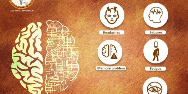
There are many different types of seizures, and the symptoms can vary depending on the type. Some people may experience a change in their vision, while others may have muscle spasms or convulsions. Some people may even lose consciousness during a seizure.
Diagnosis
A diagnosis of seizures generally begins with a medical history and physical examination. If your doctor suspects you have seizures, he or she may refer you to a neurologist, a doctor who specializes in disorders of the nervous system. The neurologist will likely ask about your family history, as well as your personal medical history. He or she will also perform a neurological exam, which assesses your mental status, reflexes, muscle strength, sensation and coordination. If the neurologist suspects you have seizures, he or she may order one or more of the following tests: • Blood tests. These tests can help rule out other conditions that may cause seizure-like symptoms, such as low blood sugar (hypoglycemia) or an infection. • Imaging tests. An MRI (magnetic resonance imaging) scan or a CT (computed tomography) scan can provide detailed images of your brain to look for abnormalities that may be causing your seizures. • Electroencephalography (EEG). This test records electrical activity in your brain using sensors (electrodes) attached to your scalp. An EEG can help diagnose epilepsy and determine what type of seizure disorder you have. • Neuropsychological testing. This testing assesses thinking, memory and behavior problems that can be caused by a seizure disorder. • Sleep studies. A sleep study may be recommended if your doctor suspects you have nighttime seizures or if you have daytime sleepiness that might be related to seizures.
treatment
There are many different types of seizures, and therefore, there is not just one type of treatment. The most common type of seizure is the grand mal seizure, which is characterized by loss of consciousness and muscle spasms. There are many different medications that can be used to treat this type of seizure, and the most common one is called phenytoin. This medication works by reducing the amount of electrical activity in the brain. There are also many other types of seizures that do not involve loss of consciousness. These types of seizures are called partial seizures, and they can be treated with a variety of different medications. The most common type of partial seizure is the temporal lobe seizure, which is characterized by changes in behavior or sensation. There are many different medications that can be used to treat this type of seizure, and the most common one is called carbamazepine. This medication works by reducing the amount of electrical activity in the brain. In some cases, surgery may be necessary to treat seizures. The most common type of surgery for seizures is called a corpus callosotomy, which involves cutting the connection between the two hemispheres of the brain. This surgery is usually only done when other treatments have failed.
Epilepsy surgery
Epilepsy surgery is a treatment option for people with epilepsy who have not been able to control their seizures with medication. Epilepsy surgery is usually only considered when other treatments have failed and the person’s seizures are significantly impacting their quality of life. The goal of epilepsy surgery is to remove the part of the brain that is causing the seizures while preserving as much normal brain tissue as possible. Epilepsy surgery is a very serious decision and should only be made after careful consideration and consultation with a team of experts. There are several different types of epilepsy surgery, and the type that is right for each person depends on many factors, including the type of seizures they have, where the seizures originate in the brain, and the person’s overall health. After epilepsy surgery, most people experience a significant reduction in their seizure frequency. In some cases, seizures may stop completely. It is important to note that epilepsy surgery does not cure epilepsy, but it can greatly improve quality of life for those who are unable to control their seizures with medication.
Conclusion
In conclusion, seizures are a serious medical condition that can have a profound impact on an individual’s life. It is important to be aware of the potential causes and symptoms of seizures in order to seek prompt medical attention. There are a variety of treatment options available, and epilepsy surgery can be an effective option for some people with intractable seizures. be an option. With proper treatment, most people with epilepsy are able to live normal, healthy lives. Looking for the best seizure treatment in Guntur, look no further than Dr Raos hospital, Dr Rao is the best neurosurgeon and epileptologist in Guntur and India.