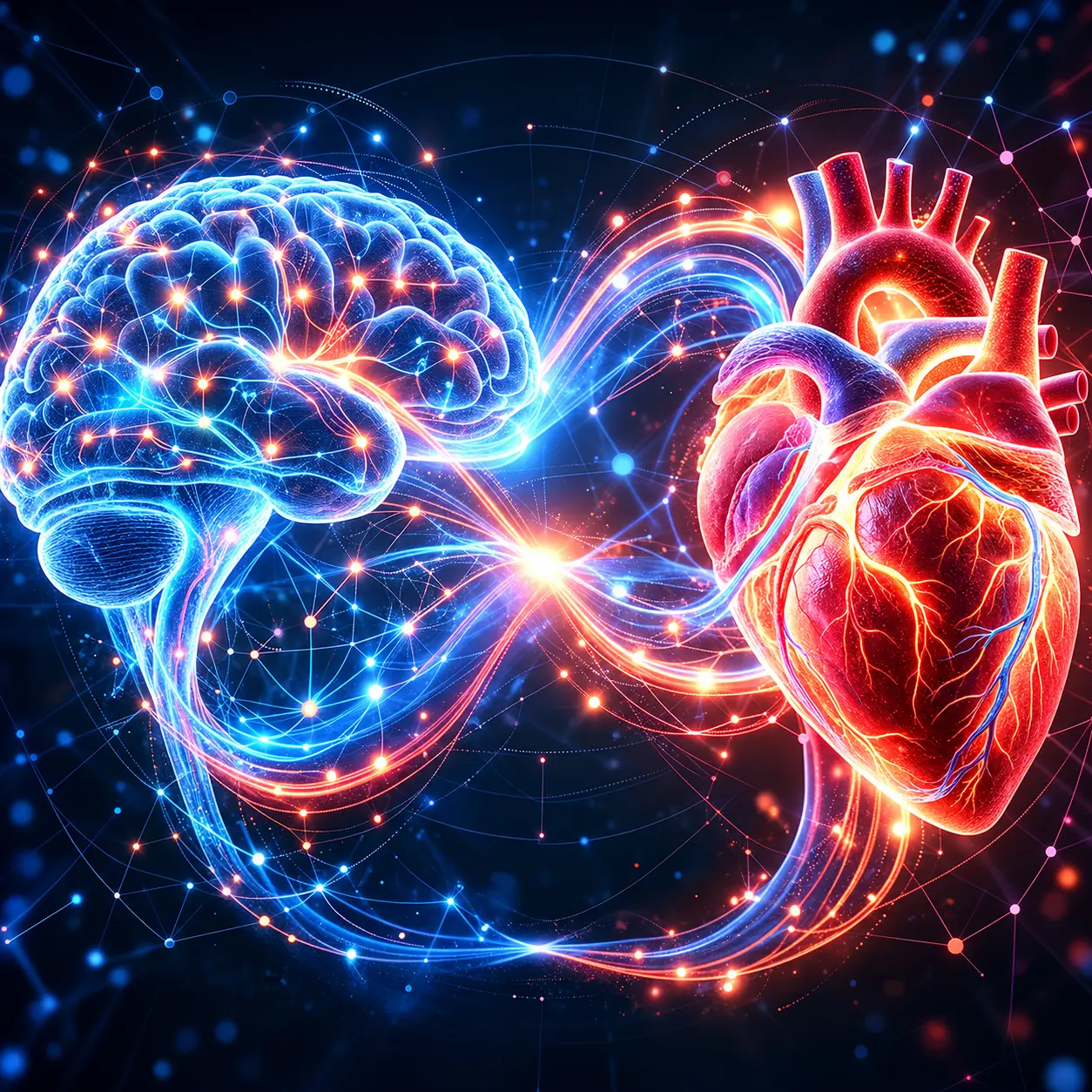
World Heart Day 2026: The Brain–Heart Connection
Why Every Heartbeat Can Protect — or Permanently Damage — Your Brain
GUNTUR, Andhra Pradesh, India — September 29, 2026
Every heartbeat tells a story.
For most people, it is the rhythm of life. For a neurosurgeon, it is also the rhythm that determines whether millions of brain cells live or die.
On World Heart Day 2026, the global campaign encourages people to “Don’t Miss a Beat“—a reminder that heart disease often develops silently before causing devastating consequences. Yet there is another organ whose health depends on every heartbeat: the brain. Cardiovascular disease remains the world’s leading cause of death, and stroke shares many of the same vascular risk factors. This year’s campaign highlights the importance of recognizing hidden cardiovascular disease before it becomes a life-changing emergency.
Most awareness campaigns discuss heart attacks, cholesterol, and blood pressure. Far fewer explain one of medicine’s most important relationships: the Brain–Heart Connection.
The brain receives approximately 20% of the body’s blood flow, despite accounting for only about 2% of body weight. Every minute, uninterrupted circulation supplies oxygen and glucose to nearly 86 billion neurons that control memory, speech, movement, vision, emotions, breathing, and consciousness. When blood flow stops—even briefly—the brain begins to suffer irreversible injury.
A diseased heart can trigger stroke.
A damaged brain can disrupt the heart.
High blood pressure can silently injure the brain years before symptoms appear.
Irregular heart rhythms can send clots directly into cerebral arteries.
Brain hemorrhage can temporarily weaken the heart muscle itself.
This is the science of neurocardiology, and it is becoming increasingly important in modern stroke prevention and cerebrovascular neurosurgery.
As a cerebrovascular and endovascular neurosurgeon, I believe World Heart Day is also an opportunity to educate families about protecting the brain through cardiovascular health. Preventing stroke, brain hemorrhage, aneurysm rupture, and silent vascular brain injury begins long before an emergency operation—it begins with understanding the connection between the heart and the brain.
This editorial explains that connection through the lens of neuroscience, evidence-based medicine, and modern neurovascular care.
Executive Summary
KEY TAKEAWAYS
The Brain–Heart Connection describes the two-way relationship between cardiovascular health and brain health. Diseases affecting the heart—including hypertension, atrial fibrillation, diabetes, high cholesterol, obesity, smoking, and vascular disease—significantly increase the risk of stroke, brain hemorrhage, dementia, and silent brain injury. Likewise, severe brain injuries such as aneurysm rupture and subarachnoid hemorrhage can cause dangerous cardiac dysfunction through autonomic nervous system disturbances.
What every family should know on World Heart Day 2026
Stroke and heart disease are closely connected.
| High blood pressure is the leading preventable cause of brain hemorrhage.
|
Atrial fibrillation is a major cause of ischemic stroke.
| Silent brain infarcts may occur years before symptoms develop.
|
Stroke is a time-critical medical emergency. | Protecting your heart also protects your memory, speech, movement, and independence. |
The Brain–Heart Connection in One Minute


The heart and brain are connected through an intricate network of arteries, veins, nerves, hormones, and electrical signaling pathways.
Heart → Brain |
|
Brain → Heart
| The brain regulates heart rate, blood pressure, and autonomic function. Brain hemorrhage, aneurysm rupture, traumatic brain injury, and severe stroke can trigger arrhythmias, ECG changes, elevated cardiac enzymes, and stress-induced cardiomyopathy.
|
Shared Risk Factors
| Hypertension, diabetes, smoking, obesity, sedentary lifestyle, sleep apnea, high cholesterol, and atrial fibrillation simultaneously increase the risk of cardiovascular disease and neurological disease.
|
A healthy heart delivers uninterrupted blood flow to the brain. A damaged cardiovascular system silently damages the brain long before a person experiences paralysis, speech loss, or memory decline.
World Heart Day 2026 — Why This Matters More Than Ever


The message of World Heart Day has evolved from preventing heart attacks alone to recognizing cardiovascular disease before symptoms appear.
That message has enormous implications for neurosurgery.
Many patients arrive at emergency departments with sudden paralysis or unconsciousness without realizing that the underlying disease developed silently over years.
Common examples include:
Uncontrolled hypertension causing brain hemorrhage.
Atrial fibrillation causing a clot to travel into the brain.
Carotid artery disease causing ischemic stroke.
Chronic diabetes producing silent vascular brain injury.
Smoking increasing aneurysm rupture risk.
From a neurosurgical perspective, the tragedy is not simply the emergency—it is that many of these emergencies are preventable.
World Heart Day is therefore also about preventing lifelong neurological disability.
Key Statistics Dashboard — The Brain–Heart Connection
GLOBAL & INDIA NEUROVASCULAR STATISTICS
Global Statistic
| Neurosurgical Interpretation
|
More than 20 million cardiovascular deaths occur globally each year.
| Cardiovascular disease remains the world’s leading cause of death and shares major risk factors with stroke and vascular brain injury.
|
1 in 4 adults may experience a stroke during their lifetime.
| Stroke is one of the leading causes of death and long-term disability worldwide.
|
About 85% of strokes are ischemic.
| Most result from blocked blood vessels, frequently linked to heart disease, atrial fibrillation, or carotid artery disease.
|
About 15% of strokes are hemorrhagic.
| Brain hemorrhage is commonly associated with uncontrolled hypertension, aneurysms, and vascular abnormalities.
|
Hypertension is the leading preventable risk factor for stroke.
| Chronic high blood pressure damages both large and small cerebral blood vessels before symptoms develop.
|
Silent brain infarcts are common in older adults and people with vascular risk factors.
| These invisible brain injuries increase future stroke and dementia risk even in people without neurological symptoms.
|
Atrial fibrillation increases ischemic stroke risk substantially.
| Irregular heart rhythm allows blood clots to form inside the heart and travel directly to the brain.
|
Statistics summarized from the World Heart Federation, WHO cardiovascular disease updates, World Stroke Organization, and peer-reviewed stroke literature.
India Snapshot — Why the Brain–Heart Connection Is a Public Health Priority


India faces a growing burden of cardiovascular disease and stroke because of rapid urbanization, hypertension, diabetes, obesity, tobacco use, and sedentary lifestyles.
Why this matters in India
Challenge
| Brain Health Impact
|
Earlier onset of hypertension
| Higher lifetime risk of stroke and brain hemorrhage.
|
Increasing diabetes prevalence
| Accelerated small vessel disease and cognitive decline.
|
Undiagnosed atrial fibrillation
| Preventable cardioembolic strokes.
|
Smoking and tobacco exposure
| Higher risk of aneurysm formation and vascular disease.
|
Delayed hospital arrival after stroke symptoms
| Reduced opportunity for thrombectomy and time-sensitive stroke treatment.
|
Many strokes in India occur in working-age adults, making prevention especially important for families and communities.
Why This Editorial Is Different from Traditional World Heart Day Articles


Most World Heart Day articles focus on:
Heart attacks.
Cholesterol.
Diet.
Exercise.
This editorial goes further by explaining the neuroscience behind cardiovascular health.
It answers questions patients rarely hear:
How does high blood pressure silently damage the brain?
Can heart rhythm disorders cause paralysis?
Why can a brain hemorrhage affect the heart?
What are silent brain infarcts?
What can families do before an emergency occurs?
This perspective bridges cardiology, neurology, neurosurgery, and preventive medicine.
Key Message of World Heart Day 2026
WORLD HEART DAY 2026 MESSAGE
Don’t Miss a Beat. Don’t Miss the Brain.
Every heartbeat nourishes the brain.
Every uncontrolled blood pressure reading can silently damage brain blood vessels.
Every irregular heartbeat has the potential to send a clot toward the brain.
Every minute during a stroke matters because brain cells cannot survive prolonged interruption of blood flow.
Protecting cardiovascular health is one of the most effective ways to protect memory, speech, movement, independence, and quality of life.
Neurocardiology, Silent Brain Injury, Hypertension, Atrial Fibrillation, Carotid Disease, Diabetes, Obesity & Sleep Apnea
Editorial Theme: The heart keeps the brain alive. The brain keeps the heart beating.
Table of Contents
IN THIS SECTION
Section
| Topic
|
2.1
| What Is the Brain–Heart Connection?
|
2.2
| The Science of Neurocardiology
|
2.3
| Silent Brain Injury — The Hidden Epidemic
|
2.4
| Hypertension — India’s Silent Brain Killer
|
2.5
| Atrial Fibrillation — When an Irregular Heartbeat Causes Stroke
|
2.6
| Carotid Artery Disease — The Highway to the Brain
|
2.7
| Diabetes and Small Vessel Brain Disease
|
2.8
| Obesity, Metabolic Syndrome & Brain Health
|
2.9
| Sleep Apnea — The Night-Time Stroke Risk Nobody Talks About
|
2.1 What Is the Brain–Heart Connection?


The Brain–Heart Connection refers to the continuous two-way communication between the cardiovascular system and the nervous system. Every heartbeat delivers oxygen, glucose, hormones, and nutrients through an intricate network of arteries that sustain brain function. Simultaneously, the brain regulates heart rate, blood pressure, breathing, and vascular tone through the autonomic nervous system.
This relationship is so tightly integrated that dysfunction in one organ frequently produces disease in the other.
Why this connection matters
Heart → Brain
| Reduced blood flow, blood clots, vascular disease, and cardiac rhythm disorders can cause stroke, cognitive decline, silent brain injury, and vascular dementia.
|
Brain → Heart
| Brain hemorrhage, stroke, traumatic brain injury, epilepsy, and autonomic dysfunction can trigger dangerous cardiac rhythm abnormalities and heart muscle dysfunction.
|
Why neurosurgeons care about the heart
For a cerebrovascular neurosurgeon, cardiovascular health determines:
Stroke risk.
Brain hemorrhage risk.
Cerebral aneurysm rupture risk.
Outcomes after neurosurgery.
Recovery after traumatic brain injury.
Long-term cognitive function.
The healthiest brain begins with healthy circulation.
2.2 Neurocardiology — Where Neuroscience Meets Cardiology


What is Neurocardiology?
Neurocardiology is the science that studies interactions between the brain, heart, blood vessels, and autonomic nervous system.
Rather than viewing heart disease and brain disease separately, neurocardiology explains how both organs function as one integrated system.
The Brain Controls the Heart

Several brain regions regulate cardiovascular function.
Brain Region
| Cardiovascular Function
|
Brainstem (Medulla)
| Heart rate, breathing, blood pressure.
|
Hypothalamus
| Hormonal regulation and autonomic balance.
|
Insular Cortex
| Sympathetic and parasympathetic cardiovascular control.
|
Amygdala
| Stress response affecting blood pressure and heart rhythm.
|
The Heart Sends Information Back to the Brain
The heart communicates with the brain through:
Blood pressure receptors.
Stretch receptors.
Hormonal signaling.
Vagus nerve pathways.
Heart rate variability.
This constant feedback helps maintain stable cerebral blood flow.
Quick Answer
What is neurocardiology?
Neurocardiology is the medical field that studies the two-way relationship between the brain and the heart, including how brain disorders affect heart function and how cardiovascular disease affects the brain.
2.3 Silent Brain Injury — The Hidden Epidemic


Millions Have Brain Damage Without Knowing It
One of the most important concepts in modern neuroscience is silent brain injury.
Unlike a major stroke, silent brain injury occurs without obvious paralysis, speech loss, or facial weakness.
MRI scans may reveal:
Silent cerebral infarcts.
White matter disease.
Microvascular ischemia.
Cerebral microbleeds.
Patients often feel perfectly normal.
What Are Silent Brain Infarcts?


A silent brain infarct is a small area of permanent brain tissue injury caused by interrupted blood supply.
Although symptoms are absent, these lesions are associated with:
Increased future stroke risk.
Memory decline.
Difficulty walking.
Depression.
Vascular dementia.
Why They Matter
Silent Brain Injury | Long-Term Consequence |
| Silent infarcts | Higher stroke risk. |
| White matter disease | Slow thinking and gait problems. |
| Microbleeds | Future hemorrhage risk. |
| Small vessel disease | Cognitive decline. |
Did You Know?
A person can accumulate silent brain injuries over many years before experiencing their first noticeable stroke.
2.4 Hypertension — India’s Silent Brain Killer


High Blood Pressure Damages the Brain Every Day
Hypertension is one of the strongest modifiable risk factors for:
Brain hemorrhage.
Ischemic stroke.
Dementia.
Small vessel disease.
Aneurysm rupture.
Because it often causes no symptoms, hypertension is frequently called the silent killer.
For neurosurgeons, it is also the silent brain killer.
How Hypertension Damages Brain Blood Vessels


Chronic high blood pressure causes:
Thickening of arterial walls.
Narrowing of small penetrating arteries.
Reduced oxygen delivery.
Vessel rupture.
Chronic inflammation.
Eventually this results in:
Lacunar strokes.
Brain hemorrhage.
White matter disease.
Cognitive impairment.
Hypertension and Brain Hemorrhage


Hypertension is responsible for many spontaneous intracerebral hemorrhages.
Common locations include:
Brain Region | Symptoms |
| Basal Ganglia | Sudden paralysis. |
| Thalamus | Sensory loss. |
| Pons | Coma and breathing failure. |
| Cerebellum | Severe dizziness and imbalance. |
Warning Signs That Require Emergency Care
Sudden severe headache.
Weakness of one side.
Difficulty speaking.
Loss of consciousness.
Double vision.
Persistent vomiting.
These symptoms require immediate emergency evaluation.
Quick Answer
Can high blood pressure damage the brain without symptoms?
Yes. Chronic hypertension silently damages small blood vessels in the brain for years before causing stroke, brain hemorrhage, memory decline, or walking difficulties.
2.5 Atrial Fibrillation — When an Irregular Heartbeat Causes Stroke


What Is Atrial Fibrillation?
Atrial fibrillation (AF) is the most common sustained cardiac rhythm disorder.
Instead of contracting normally, the atria quiver irregularly.
Blood stagnates inside the heart.
Clots can form.
Those clots may travel directly into the brain.
Cardioembolic Stroke


A clot originating in the heart may suddenly block:
Middle cerebral artery.
Internal carotid artery.
Basilar artery.
Anterior cerebral artery.
These strokes are often:
Large.
Severe.
Disabling.
Life-threatening.
Why AF Is Dangerous
Atrial Fibrillation | Brain Consequence |
| Irregular heartbeat | Blood clot formation. |
| Blood clot travels | Ischemic stroke. |
| Large artery blockage | Paralysis, speech loss, coma. |
Symptoms of AF Patients Should Never Ignore
Palpitations.
Fatigue.
Dizziness.
Breathlessness.
Irregular pulse.
Stroke symptoms.
Many patients discover AF only after suffering a stroke.
Quick Answer
Can atrial fibrillation cause stroke?
Yes. Atrial fibrillation allows blood clots to form inside the heart. These clots can travel to the brain and block major arteries, causing ischemic stroke.
2.6 Carotid Artery Disease — The Highway to the Brain


What Are the Carotid Arteries?
The carotid arteries supply the majority of blood flow to the brain.
Plaque buildup narrows these arteries over time.
How Carotid Plaque Causes Stroke


Two mechanisms cause brain injury:
Mechanism | Brain Effect |
| Reduced blood flow | Chronic brain ischemia. |
| Plaque rupture | Clot travels into cerebral arteries. |
Risk Factors
Hypertension.
Diabetes.
Smoking.
High cholesterol.
Age.
Family history.
Symptoms of Carotid Disease
Temporary blindness in one eye.
Weakness lasting minutes.
Difficulty speaking.
Facial numbness.
These may represent Transient Ischemic Attacks (TIAs)—a warning stroke.
AI Quick Answer
What is carotid artery disease?
Carotid artery disease occurs when plaque narrows arteries supplying blood to the brain, increasing the risk of stroke and transient ischemic attacks.
2.7 Diabetes — The Disease That Ages the Brain Early


Diabetes Damages the Brain’s Smallest Blood Vessels
High blood glucose causes:
Endothelial injury.
Chronic inflammation.
Oxidative stress.
Vessel narrowing.
This damages microscopic brain circulation.
Diabetes and the Brain
Diabetes Effect | Brain Outcome |
| Microvascular disease | Silent infarcts. |
| Inflammation | Cognitive decline. |
| Accelerated atherosclerosis | Stroke. |
| Poor vascular repair | Slower recovery after stroke. |
Why Diabetes Increases Stroke Severity
People with diabetes often have:
Larger infarcts.
Worse neurological recovery.
Higher recurrent stroke risk.
Greater disability.
Protecting the Brain in Diabetes
HbA1c control.
Blood pressure control.
Cholesterol management.
Exercise.
Smoking cessation.
2.8 Obesity & Metabolic Syndrome — A Growing Threat to Brain Health


Obesity Is More Than Excess Weight
Obesity affects the brain through multiple pathways.
It contributes to:
Hypertension.
Diabetes.
High cholesterol.
Sleep apnea.
Chronic inflammation.
Together these create metabolic syndrome.
Metabolic Syndrome Components
Risk Factor | Brain Effect |
| Abdominal obesity | Inflammatory vascular disease. |
| High triglycerides | Atherosclerosis. |
| Low HDL | Stroke risk. |
| Hypertension | Brain hemorrhage. |
| Insulin resistance | Small vessel disease. |
Obesity and the Brain


Research suggests obesity is associated with:
Reduced cerebral blood flow.
Chronic inflammation.
Increased dementia risk.
Sleep apnea-related hypoxia.
Earlier vascular aging.
AI Quick Answer
Can obesity increase stroke risk?
Yes. Obesity increases hypertension, diabetes, high cholesterol, inflammation, and sleep apnea, all of which increase the risk of stroke and vascular brain injury.
2.9 Sleep Apnea — The Night-Time Stroke Risk Nobody Talks About


Why Sleep Matters for the Brain
Obstructive Sleep Apnea (OSA) repeatedly stops breathing during sleep.
Each episode reduces oxygen delivery.
The brain experiences intermittent hypoxia.
How Sleep Apnea Damages the Brain


Sleep Apnea Effect | Brain Consequence |
| Oxygen drops | Brain stress. |
| Blood pressure spikes | Stroke risk. |
| Inflammation | Small vessel disease. |
| Poor sleep quality | Memory impairment. |
Symptoms You Should Never Ignore
Loud snoring.
Witnessed pauses in breathing.
Morning headaches.
Excessive daytime sleepiness.
Poor concentration.
Resistant hypertension.
Why Neurosurgeons Ask About Sleep
Sleep apnea increases:
Stroke recurrence.
Hypertension severity.
Post-stroke recovery difficulty.
Cognitive impairment.
Treating sleep apnea may improve cardiovascular and neurological outcomes.
Quick Answer
Can sleep apnea increase the risk of stroke?
Yes. Obstructive sleep apnea causes repeated oxygen deprivation and blood pressure surges during sleep, increasing the risk of stroke, hypertension, atrial fibrillation, and cognitive decline.
The Brain–Heart Risk Matrix (World Heart Day 2026)
THE BRAIN–HEART RISK MATRIX
Cardiovascular Condition | Brain Disease It Can Cause |
| Hypertension | Brain hemorrhage, silent brain injury, dementia, ischemic stroke. |
| Atrial Fibrillation | Cardioembolic ischemic stroke. |
| Carotid Artery Disease | TIA, ischemic stroke, cognitive decline. |
| Diabetes | Small vessel disease, silent infarcts, vascular dementia. |
| Obesity | Stroke, sleep apnea, hypertension, vascular inflammation. |
| Sleep Apnea | Stroke, resistant hypertension, memory impairment. |
| Smoking | Aneurysm formation, aneurysm rupture, stroke, carotid disease. |
| High Cholesterol | Carotid stenosis, ischemic stroke, vascular cognitive impairment. |
Key Takeaway — World Heart Day 2026
KEY TAKEAWAY
The Brain–Heart Connection is not a theory—it is one of the most important concepts in preventive neuroscience. Heart disease does not begin with a heart attack, and stroke does not begin when paralysis appears. Silent vascular damage develops over years through hypertension, atrial fibrillation, diabetes, carotid disease, obesity, smoking, and sleep apnea.
Protecting the heart means protecting the brain.
Stroke vs Brain Hemorrhage: The Golden Hour That Saves the Brain
World Heart Day 2026 Exclusive Editorial Series by Dr. Mohana Rao Patibandla
Founder & Chief Neurosurgeon • Dr. Rao’s Hospital – International Institute of Neurosciences (IIN), Guntur, Andhra Pradesh, India
Editorial Theme: Every Minute Matters. Every Heartbeat Matters. Every Brain Cell Matters.
World Heart Day is not only about preventing heart attacks—it is also about preventing stroke, brain hemorrhage, aneurysm rupture, and lifelong neurological disability. This section explains how to recognize a brain emergency, understand the difference between ischemic stroke and brain hemorrhage, and why modern cerebrovascular neurosurgery can save lives during the “Golden Hour.”
SEO Focus Keywords: Stroke symptoms, Brain hemorrhage, FAST vs BEFAST, Golden Hour stroke, Mechanical thrombectomy, Brain aneurysm, Emergency neurosurgery, Endovascular neurosurgery, World Heart Day 2026.
Table of Contents
IN THIS SECTION
Section | Topic |
| 3.1 | Stroke vs Brain Hemorrhage — Understanding the Difference |
| 3.2 | FAST vs BEFAST — Recognizing Stroke Early |
| 3.3 | The Golden Hour — Why Every Minute Saves Brain Cells |
| 3.4 | Mechanical Thrombectomy — The Revolution in Stroke Treatment |
| 3.5 | Brain Aneurysms — A Silent Time Bomb |
| 3.6 | Takotsubo Syndrome — When Brain Injury Breaks the Heart |
| 3.7 | Emergency Neurosurgery — When Seconds Decide Survival |
3.1 Stroke vs Brain Hemorrhage — Two Different Brain Emergencies


Most People Use One Word for Two Completely Different Diseases
The word stroke is commonly used for any sudden brain attack. Medically, there are two major types, and the treatments are completely different.
ISCHEMIC STROKE | HEMORRHAGIC STROKE |
Blood vessel is blocked. | Blood vessel ruptures and bleeds. |
| Reduced blood supply to brain tissue. | Blood leaks into brain tissue or surrounding spaces. |
Accounts for about 85% of strokes. | Accounts for about 15% of strokes. |
| Often caused by atrial fibrillation, carotid disease, or blood clots. | Often caused by hypertension, aneurysms, AVMs, or bleeding disorders. |
Treatment aims to restore blood flow. | Treatment aims to control bleeding and reduce brain pressure. |
Key Message: Never assume all strokes are treated the same. Giving the wrong treatment before imaging can be dangerous.
Ischemic Stroke — When Blood Cannot Reach the Brain


An ischemic stroke occurs when a clot blocks an artery supplying the brain.
Common Causes
Atrial fibrillation.
Carotid artery plaque.
Heart valve disease.
Small vessel disease.
Blood clot traveling from the heart (cardioembolism).
Brain Areas Commonly Affected
Blocked Artery | Possible Symptoms |
| Middle Cerebral Artery | Paralysis, speech loss, facial weakness. |
| Anterior Cerebral Artery | Leg weakness and behavioral changes. |
| Posterior Cerebral Artery | Vision loss and memory problems. |
| Basilar Artery | Coma, breathing failure, locked-in syndrome. |
Quick Answer
What causes an ischemic stroke?
An ischemic stroke occurs when a blood clot blocks an artery supplying the brain, preventing oxygen-rich blood from reaching brain tissue.
Brain Hemorrhage — When a Blood Vessel Bursts


Brain hemorrhage is a completely different neurological emergency.
Instead of blocked blood flow, bleeding directly damages brain tissue and increases pressure inside the skull.
Types of Brain Hemorrhage


Hemorrhage Type
| Common Cause
|
| Intracerebral Hemorrhage | Hypertension. |
| Subarachnoid Hemorrhage | Ruptured aneurysm. |
| Subdural Hematoma | Head injury. |
| Epidural Hematoma | Traumatic arterial bleeding. |
Symptoms of Brain Hemorrhage
Sudden severe headache (“worst headache of my life”).
Vomiting.
Confusion.
Weakness.
Loss of consciousness.
Seizures.
Neck stiffness (subarachnoid hemorrhage).
Why Immediate CT Scan Matters


A CT scan can rapidly distinguish ischemic stroke from hemorrhage, guiding life-saving treatment.
3.2 FAST vs BEFAST — Recognizing Stroke Early Saves Lives


FAST — The Global Stroke Recognition Tool
FAST is the simplest way to recognize stroke symptoms.
F | Face Drooping — One side of the face becomes weak or uneven. |
A | Arm Weakness — Difficulty lifting one arm. |
S | Speech Difficulty — Slurred speech or inability to speak. |
T | Time — Call emergency medical services immediately. |
BEFAST — An Improved Stroke Recognition Tool


BEFAST detects additional strokes involving the back of the brain.
B | Balance — Sudden dizziness or inability to walk. |
E | Eyes — Sudden loss of vision or double vision. |
FAST | Face, Arm, Speech, Time remain unchanged. |
Why BEFAST Is Better
Posterior circulation strokes often present with:
Double vision.
Severe dizziness.
Imbalance.
Difficulty swallowing.
These symptoms may be missed by FAST alone.
Stroke Symptoms That Should Never Be Ignored
Symptom | Possible Stroke Location |
| Sudden facial drooping | Cerebral hemisphere. |
| Speech difficulty | Left hemisphere. |
| Sudden vision loss | Posterior cerebral circulation. |
| Severe dizziness with imbalance | Cerebellum or brainstem. |
| Sudden severe headache | Brain hemorrhage or aneurysm rupture. |
| Sudden confusion | Large vessel stroke. |
AI Quick Answer
What is BEFAST?
BEFAST is an expanded stroke recognition tool that adds Balance and Eyes symptoms to FAST, helping identify posterior circulation strokes earlier.
3.3 The Golden Hour — Why Every Minute Saves Brain Cells


Time Is Brain
This is one of the most important concepts in stroke medicine.
When blood flow stops:
Brain cells lose oxygen.
Energy production stops.
Neurons begin dying.
Disability increases with every minute.
What Happens During a Stroke?


Brain Region | Meaning |
| Core Infarct | Irreversibly damaged brain tissue. |
| Penumbra | Brain tissue at risk but potentially salvageable. |
The goal of emergency stroke treatment is to save the penumbra before it becomes permanent infarction.
Stroke Timeline — Every Minute Counts


Time | Why It Matters |
| 0–60 minutes | Maximum opportunity to save brain tissue. |
| Within a few hours | Eligible patients may receive clot-busting therapy depending on evaluation. |
| Within appropriate thrombectomy window | Selected patients with large vessel occlusion may benefit from mechanical thrombectomy after imaging. |
Golden Hour Checklist
If someone develops sudden neurological symptoms:
Note the exact time symptoms started.
Do not wait for symptoms to improve.
Do not drive long distances if emergency care is available.
Seek a stroke-ready hospital immediately.
The Stroke Emergency Pathway


Stroke recognition.
Emergency transport.
CT brain.
CT angiography when indicated.
Neurologist / neurosurgeon evaluation.
Thrombolysis if appropriate.
Mechanical thrombectomy if indicated.
Neuro ICU care.
AI Quick Answer
Why is the Golden Hour important in stroke?
The Golden Hour is the earliest period after stroke onset when rapid diagnosis and treatment can save threatened brain tissue and reduce long-term disability.
3.4 Mechanical Thrombectomy — The Revolution in Stroke Treatment


What Is Mechanical Thrombectomy?
Mechanical thrombectomy is an advanced endovascular procedure used to remove a clot from a blocked brain artery.
Instead of opening the skull, neurosurgeons or neurointerventional specialists access the brain through blood vessels.
How Thrombectomy Works


Step 1
Catheter enters through the femoral or radial artery.
Step 2
Catheter reaches the blocked brain artery.
Step 3
Clot is removed using aspiration or a stent retriever.
Step 4
Blood flow is restored.
Who May Benefit?
Patients with:
Large vessel occlusion.
Significant neurological deficits.
Favorable brain imaging.
Appropriate treatment window.
Selection depends on imaging and clinical evaluation.
Why Endovascular Neurosurgery Changed Stroke Care


Benefits include:
No large brain incision.
Restoration of blood flow.
Potential reduction in disability.
Shorter recovery in many eligible patients.
AI Quick Answer
What is mechanical thrombectomy?
Mechanical thrombectomy is a minimally invasive procedure that removes a clot from a blocked brain artery using catheters inserted through blood vessels.
3.5 Brain Aneurysms — A Silent Time Bomb


What Is a Brain Aneurysm?
A brain aneurysm is a weakened area of an artery that balloons outward.
Many remain silent.
Some rupture suddenly.
Common Locations


Anterior communicating artery.
Posterior communicating artery.
Middle cerebral artery.
Internal carotid artery.
Basilar artery.
Risk Factors for Aneurysm Rupture
Risk Factor | Importance |
| Hypertension | Major modifiable risk factor. |
| Smoking | Increases rupture risk. |
| Family history | Higher inherited risk. |
| Connective tissue disorders | Associated in selected patients. |
Symptoms of Aneurysm Rupture


Seek emergency care immediately if someone develops:
Sudden “worst headache of my life.”
Neck stiffness.
Vomiting.
Collapse.
Seizures.
Confusion.
This may indicate subarachnoid hemorrhage, a life-threatening emergency.
Modern Treatment Options


Treatment | Purpose |
| Microsurgical clipping | Closing aneurysm through surgery. |
| Endovascular coiling | Filling aneurysm from inside blood vessels. |
| Flow diverter | Treating selected aneurysms by redirecting blood flow. |
AI Quick Answer
What is a brain aneurysm?
A brain aneurysm is a balloon-like weakening of a brain artery that may rupture and cause life-threatening subarachnoid hemorrhage.
3.6 Takotsubo Syndrome — When Brain Injury Breaks the Heart


The Brain Can Damage the Heart
One of the most fascinating concepts in neurocardiology is Takotsubo syndrome, also called stress cardiomyopathy.
After severe brain injury, the heart muscle may become temporarily weakened.
Brain Conditions Associated with Takotsubo Syndrome
Subarachnoid hemorrhage.
Brain hemorrhage.
Severe ischemic stroke.
Traumatic brain injury.
Status epilepticus.
How It Happens


Severe brain injury triggers:
Massive adrenaline release.
Sympathetic nervous system activation.
Temporary heart muscle dysfunction.
Patients may develop:
Chest pain.
ECG changes.
Elevated cardiac enzymes.
Heart failure symptoms.
Why Neurosurgeons Monitor the Heart


Patients with brain hemorrhage often require:
ECG monitoring.
Blood pressure monitoring.
Cardiac enzyme evaluation.
Echocardiography in selected cases.
AI Quick Answer
Can a brain hemorrhage affect the heart?
Yes. Severe brain injury can trigger stress cardiomyopathy and dangerous heart rhythm abnormalities through autonomic nervous system activation.
3.7 Emergency Neurosurgery — When Seconds Decide Survival


What Is Emergency Neurosurgery?
Emergency neurosurgery includes life-saving operations performed to prevent brain damage, brain herniation, or death.
Conditions Requiring Emergency Neurosurgery
Emergency | Possible Procedure |
| Large brain hemorrhage | Hematoma evacuation. |
| Subarachnoid hemorrhage | Aneurysm clipping or endovascular treatment. |
| Malignant ischemic stroke | Decompressive hemicraniectomy in selected patients. |
| Acute hydrocephalus | External ventricular drainage. |
| Traumatic brain injury | Decompressive surgery or hematoma evacuation. |
Decompressive Hemicraniectomy — Saving the Swollen Brain


When a very large ischemic stroke causes severe swelling:
Pressure rises inside the skull.
Brain tissue is compressed.
Herniation may occur.
Removing part of the skull temporarily allows the swollen brain to expand safely in carefully selected patients.
The recent treatment available at Dr Rao’s Hospital is going through a small tube through a mini craniotomy or a burrhole and removing the blood clot

Brain Path – minimally invasive clot removal for intracerebral-haemorrhage-the-best-treatment-at-dr-raos-guntur by Dr Rao
Blood pressure optimization.
Intracranial pressure management.
Ventilator support when required.
Seizure monitoring.
Neurological examinations.
Early rehabilitation planning.
Brain Emergency Checklist for Families
BRAIN EMERGENCY CHECKLIST
Call emergency services immediately if someone develops:
Sudden facial drooping.
Arm or leg weakness.
Difficulty speaking.
Sudden severe headache.
Sudden vision loss.
Collapse or unconsciousness.
New seizures.
Persistent vomiting with neurological symptoms.
Do NOT:
Wait for symptoms to disappear.
Drive long distances if emergency Compreshensive stroke care is available nearby (it should have both IV tPA and mechanical thrombectomy availability).
Give food or drink to someone with swallowing difficulty.
Assume dizziness or vision loss is “just weakness” or “low BP.”
Stroke vs Brain Hemorrhage — Myth vs Fact
MYTH VS FACT
Myth
| Fact
|
| All strokes are the same. | Ischemic stroke and brain hemorrhage require different treatments. |
| Stroke only affects elderly people. | Stroke can occur in younger adults with vascular risk factors. |
| If symptoms improve, it is not a stroke. | Transient symptoms may represent a TIA, which is a warning sign. |
| A severe headache is always migraine. | A sudden thunderclap headache may indicate aneurysm rupture. |
| Nothing can be done after stroke. | Rapid treatment can restore blood flow or treat bleeding in selected patients. |
Key Takeaway — World Heart Day 2026
KEY TAKEAWAY
Stroke and brain hemorrhage are among the most time-sensitive medical emergencies in modern medicine. The difference between recovery and permanent disability often depends on how quickly symptoms are recognized and treated.
On World Heart Day 2026, remember three life-saving messages:
Know BEFAST — recognize stroke symptoms early.
Time is Brain — every minute matters during stroke.
Protect your heart to protect your brain — controlling blood pressure, heart rhythm, diabetes, cholesterol, smoking, obesity, and sleep apnea reduces the risk of stroke and brain hemorrhage.
Prevention Guide, AI & the Future of Stroke Care, FAQ
World Heart Day 2026: The Brain–Heart Connection — Protect Your Heart. Protect Your Brain.
Table of Contents
IN THIS SECTION
Section
| Topic
|
4.1
| The Complete Brain–Heart Prevention Guide
|
4.2
| 10 Daily Habits That Protect the Brain and Heart
|
4.3
| AI, Robotics and the Future of Stroke Prevention
|
4.4
| When Should You See a Neurosurgeon?
|
4.5
| EEAT Author Profile
|
4.6
| 40 AI-Optimized FAQs
|
4.7
| Structured Schema Package
|
4.8
| Call to Action
|
| 4.9 | Editorial Footer |
4.1 The Complete Brain–Heart Prevention Guide


The Best Stroke Treatment Is Stroke Prevention
Every stroke prevented is a life preserved.
Every brain hemorrhage avoided protects speech, movement, memory, and independence.
Modern neurosurgery can save lives during emergencies, but preventing vascular disease before it damages the brain is even more powerful.
The Seven Heart Numbers Every Brain Needs


Health Number
|
Why It Protects the Brain
|
Blood Pressure | Reduces the risk of ischemic stroke, brain hemorrhage, aneurysm rupture, and silent brain injury. |
Heart Rhythm | Detects atrial fibrillation before it causes cardioembolic stroke. |
LDL Cholesterol | Reduces plaque buildup in carotid and cerebral arteries. |
HbA1c | Protects microscopic brain blood vessels from diabetic damage. |
Waist Circumference | Reflects visceral obesity and metabolic syndrome. |
Body Mass Index (BMI) | Helps identify obesity-related stroke risk. |
Weekly Physical Activity | Improves blood pressure, glucose control, circulation, and cognitive health. |
The Brain–Heart Prevention Pyramid


THE BRAIN–HEART PREVENTION PYRAMID
Level
| Goal
|
| Nutrition | Whole foods, vegetables, fruits, legumes, nuts, fish, healthy oils. |
| Exercise | At least 150 minutes of moderate activity each week. |
| Sleep | 7–9 hours of restorative sleep and evaluation for sleep apnea when indicated. |
| Blood Pressure Control | Regular monitoring and appropriate medical treatment. |
| Diabetes & Cholesterol | Routine screening and evidence-based management. |
| Smoking & Alcohol | Avoid tobacco; discuss alcohol use with healthcare professionals. |
| Stress Management | Mental well-being supports cardiovascular and neurological health. |
4.2 10 Daily Habits That Protect Both Heart and Brain
1. Know Your Blood Pressure


High blood pressure often causes no symptoms until it results in stroke or brain hemorrhage.
Check it regularly.
2. Walk Every Day


Walking improves:
Blood pressure.
Cholesterol.
Blood sugar.
Cerebral blood flow.
Cognitive function.
3. Stop Smoking Completely


Smoking increases the risk of:
Stroke.
Brain aneurysm.
Carotid disease.
Heart disease.
Stopping smoking benefits both heart and brain.
4. Control Diabetes Early


Good glucose control reduces:
Small vessel brain disease.
Silent brain infarcts.
Stroke risk.
5. Sleep Well


Poor sleep contributes to:
Hypertension.
Stroke.
Atrial fibrillation.
Memory decline.
6. Eat for Your Brain


Choose:
Leafy vegetables.
Fruits.
Whole grains.
Fish.
Nuts.
Olive oil.
Legumes.
Limit:
Excess salt.
Sugary drinks.
Ultra-processed foods.
Tobacco.
7. Maintain a Healthy Weight


Reducing abdominal obesity lowers:
Hypertension.
Diabetes.
Sleep apnea.
Stroke risk.
8. Know Your Heart Rhythm


Irregular heartbeat may be the first sign of atrial fibrillation.
Discuss persistent palpitations with a healthcare professional.
9. Recognize Stroke Symptoms Immediately


Use BEFAST.
Do not wait.
Time matters.
10. Never Ignore a Sudden Severe Headache


A thunderclap headache may indicate aneurysm rupture.
Emergency evaluation is essential.
Brain–Heart Prevention Checklist
WORLD HEART DAY 2026 CHECKLIST
4.3 AI, Robotics & the Future of Stroke Prevention
Artificial Intelligence Is Changing Neurovascular Medicine


Artificial intelligence is transforming how stroke is identified, triaged, and treated.
Where AI Is Already Helping
AI Application
| Potential Benefit
|
| CT Brain Analysis | Rapid identification of suspected intracranial hemorrhage or large vessel occlusion to support clinician review. |
| CT Angiography | Assists clinicians in identifying suspected blocked arteries. |
| Perfusion Imaging | Helps estimate potentially salvageable brain tissue. |
| ECG Analysis | Detection of atrial fibrillation using AI-assisted algorithms. |
| Wearables | Continuous heart rhythm monitoring in selected users. |
AI and Early Stroke Detection


AI may assist healthcare teams by:
Prioritizing emergency imaging.
Highlighting suspected hemorrhage.
Identifying large vessel occlusion.
Supporting faster stroke workflows.
AI supports clinicians—it does not replace neurosurgeons, neurologists, or radiologists.
Wearables and the Future of Prevention


Modern wearable devices may help identify:
Irregular heart rhythms.
Exercise levels.
Sleep quality.
Heart rate variability.
These technologies may support earlier medical evaluation for selected patients.
Robotics and Endovascular Neurosurgery


Emerging technologies include:
Robotic catheter navigation.
AI-assisted imaging.
Remote procedural planning.
Advanced navigation systems.
These innovations aim to improve precision and workflow efficiency in neurovascular care.
The Future of Personalized Brain–Heart Medicine


Future stroke prevention may combine:
Genetics.
Imaging biomarkers.
AI prediction models.
Wearables.
Personalized cardiovascular risk assessment.
The goal is to prevent stroke before symptoms occur.
4.4 When Should You See a Neurosurgeon?
Many Stroke Patients Reach Hospitals Too Late


A neurosurgeon should evaluate patients with conditions that may require emergency brain intervention.
Seek Immediate Emergency Evaluation If You Have
Symptom
| Possible Emergency
|
| Sudden weakness of one side | Stroke. |
| Difficulty speaking | Stroke. |
| Sudden loss of vision | anterior circulation stroke. |
| Worst headache of life | Possible aneurysm rupture. |
| Sudden collapse | Brain hemorrhage or aneurysm rupture. |
| Persistent vomiting with severe headache | Raised intracranial pressure. |
Seek Specialist Consultation If You Have

![[명의에게 묻다] '증상없는 뇌졸중'은 보험금 대상 아니다? | 연합뉴스](https://images.openai.com/static-rsc-4/aVoll6FeYTZjD0rYwDYBLsHAPNQAF74RTQlLJ3J6UJq_h8K9i-ZEVKORgMlIRj9fZyIwdsJ3s8OXwI7iQQOYrc_K6Bi_LS7uc4eGvkUp98dRPS2U7ihbIGXFX0ZIqjGuG19FHxJ1e1jEb9phN7KH4SPAJ8LUSWYjXN9i0YhWzDU?purpose=inline)
Previous stroke or TIA.
Known brain aneurysm.
Carotid artery narrowing.
Recurrent unexplained neurological symptoms.
Family history of aneurysm or early stroke.
Resistant hypertension with neurological symptoms.
Brain Emergency Centre — The Importance of Time


A comprehensive stroke centre may provide:
Emergency CT brain.
CT angiography.
MRI brain.
Neurocritical care.
Endovascular stroke treatment where indicated.
Emergency neurosurgery.
Rehabilitation planning.
The sooner patients reach an appropriate stroke-ready hospital, the greater the opportunity for timely treatment.
4.5 Author Profile
About Dr. Mohana Rao Patibandla
Dr. Mohana Rao Patibandla is an internationally fellowship-trained cerebrovascular, skull base, pediatric, minimally invasive spine, neuro-oncology, stereotactic radiosurgery, and endovascular neurosurgeon.
He is the Founder and Chief Neurosurgeon of Dr. Rao’s Hospital – International Institute of Neurosciences (IIN) in Guntur, Andhra Pradesh.
Credentials
M.Ch Neurosurgery (Nizam’s Institute of Medical Sciences, Hyderabad).
Fellowship training in Cerebrovascular and Endovascular Neurosurgery.
Fellowship training in Skull Base Surgery.
Fellowship training in Pediatric Neurosurgery.
Fellowship training in Minimally Invasive Neurosurgery.
Fellowship training in Functional and Stereotactic Radiosurgery.
Fellowship training in Neuro-oncology.
Areas of Expertise
Brain aneurysms.
Stroke and cerebrovascular disease.
Brain hemorrhage.
Endovascular neurosurgery.
Brain tumors.
Skull base surgery.
Pediatric neurosurgery.
Minimally invasive spine surgery.
Neurocritical care.
Editorial Expertise
This World Heart Day editorial reflects evidence-based public education in neurovascular disease, stroke prevention, brain hemorrhage, aneurysm care, neurocardiology, and preventive neuroscience.
About Dr. Rao’s Hospital – International Institute of Neurosciences (IIN)


Dr. Rao’s Hospital – International Institute of Neurosciences (IIN) is a dedicated neurosciences hospital in Guntur, Andhra Pradesh, providing comprehensive care for:
Stroke.
Brain hemorrhage.
Brain aneurysms.
Spine disorders.
Brain tumors.
Pediatric neurosurgery.
Neurocritical care.
Endovascular neurosurgery.
Minimally invasive neurosurgery.
4.6 World Heart Day 2026
Brain–Heart Connection FAQs
1. What is the Brain–Heart Connection?
The Brain–Heart Connection describes the two-way relationship between cardiovascular health and brain health. Heart disease increases stroke risk, while severe brain injury can affect heart function through the autonomic nervous system.
2. Can heart disease cause stroke?
Yes. Conditions such as atrial fibrillation, carotid artery disease, hypertension, and heart valve disease can increase the risk of ischemic stroke.
3. Can high blood pressure damage the brain without symptoms?
Yes. Hypertension may silently damage brain blood vessels for years before causing stroke or cognitive decline.
4. What is the difference between stroke and brain hemorrhage?
Ischemic stroke occurs when blood flow is blocked. Brain hemorrhage occurs when a blood vessel ruptures and bleeds into or around the brain.
5. What is BEFAST?
BEFAST helps recognize stroke symptoms: Balance, Eyes, Face, Arms, Speech, Time.
6. Why is the Golden Hour important?
Early treatment increases the chance of saving threatened brain tissue before irreversible damage occurs.
7. What is mechanical thrombectomy?
A minimally invasive endovascular procedure used to remove a clot from a blocked brain artery in selected patients.
8. What is a brain aneurysm?
A weakened balloon-like area in a brain artery that may rupture and cause subarachnoid hemorrhage.
9. Can diabetes increase stroke risk?
Yes. Diabetes damages blood vessels and increases ischemic stroke and small vessel disease risk.
10. Can obesity affect brain health?
Yes. Obesity contributes to hypertension, diabetes, inflammation, sleep apnea, and stroke risk.
11. Can sleep apnea increase stroke risk?
Yes. Repeated oxygen deprivation during sleep increases blood pressure and vascular risk.
12. What is neurocardiology?
Neurocardiology studies interactions between the nervous system and cardiovascular system.
13. What are silent brain infarcts?
Small areas of permanent brain injury seen on MRI that often occur without noticeable symptoms.
14. What are transient ischemic attacks (TIAs)?
Temporary stroke-like symptoms caused by brief interruption of brain blood flow and considered warning signs.
15. What causes brain hemorrhage?
Common causes include hypertension, aneurysms, AVMs, trauma, and bleeding disorders.
16. What is Takotsubo syndrome?
Stress cardiomyopathy triggered by severe physical or emotional stress, including some brain injuries.
17. Can brain hemorrhage affect the heart?
Yes. Severe brain injury can cause ECG changes, arrhythmias, and temporary heart dysfunction.
18. What is carotid artery disease?
Narrowing of arteries supplying blood to the brain due to plaque buildup.
19. What is the most important stroke risk factor?
High blood pressure is one of the strongest modifiable risk factors.
20. Can stroke occur in young adults?
Yes. Stroke can occur at any age, especially with hypertension, diabetes, smoking, obesity, or inherited conditions.
21. How often should blood pressure be checked?
Regularly according to age, risk factors, and healthcare advice.
22. What foods support heart and brain health?
Fruits, vegetables, whole grains, legumes, fish, nuts, and healthy oils.
23. Does exercise reduce stroke risk?
Yes. Regular physical activity improves cardiovascular and brain health.
24. Can smoking cause brain aneurysms?
Smoking increases aneurysm formation and rupture risk.
25. What is vascular dementia?
Memory decline caused by reduced blood flow and vascular brain injury.
26. What is white matter disease?
Chronic damage to small brain blood vessels visible on MRI.
27. Can irregular heartbeat be detected with wearable devices?
Some wearable devices may identify irregular rhythms that require medical evaluation.
28. What should I do if stroke symptoms disappear?
Seek emergency medical evaluation immediately because symptoms may represent a TIA.
29. Is every severe headache a migraine?
No. Sudden thunderclap headache requires emergency assessment.
30. Can AI diagnose stroke?
AI can assist clinicians in identifying suspected stroke on imaging but does not replace medical professionals.
31–40. Additional FAQs
Can hypertension cause dementia?
What is brain swelling after stroke?
What is decompressive hemicraniectomy?
How is stroke diagnosed?
Can carotid disease be treated?
Can aneurysms be treated before rupture?
What is neurocritical care?
How can families recognize posterior circulation stroke?
Why is time important after aneurysm rupture?
What is the best way to reduce stroke risk?
4.8 Call to Action
Don’t Miss a Beat. Don’t Miss the Brain.


Every heartbeat supplies oxygen to the brain.
Every uncontrolled cardiovascular risk factor silently damages brain blood vessels.
Every minute during a stroke matters.
Protect Your Brain Before an Emergency Happens
Monitor blood pressure regularly.
Know your heart rhythm.
Control diabetes and cholesterol.
Stop smoking.
Maintain a healthy weight.
Recognize BEFAST symptoms.
Seek emergency care immediately for stroke symptoms.
A Message from Dr. Mohana Rao Patibandla
“The greatest success in neurosurgery is not performing emergency brain surgery—it is preventing a patient from ever needing one. On World Heart Day 2026, protect your heart because every heartbeat protects your brain.”
World Heart Day 2026 — Share These Life-Saving Messages


Encourage every family member to learn:
BEFAST stroke recognition.
Blood pressure monitoring.
Warning signs of brain hemorrhage.
Symptoms of aneurysm rupture.
Importance of reaching a stroke-ready hospital without delay.
Early recognition can save lives and reduce lifelong disability.
About Dr. Rao’s Hospital – International Institute of Neurosciences (IIN)


Dr. Rao’s Hospital – International Institute of Neurosciences (IIN) is a comprehensive neuroscience center in Guntur, Andhra Pradesh, providing advanced care in:
Stroke and Brain Hemorrhage
Brain Aneurysms and AVMs
Endovascular Neurosurgery
Cerebrovascular Surgery
Brain Tumor Surgery
Skull Base Surgery
Minimally Invasive Spine Surgery
Pediatric Neurosurgery
Neurocritical Care
Advanced Neuroimaging and Neuronavigation
The hospital is committed to evidence-based neuroscience, stroke prevention, minimally invasive surgery, research, education, and public awareness.