-
What Is Awake Craniotomy?
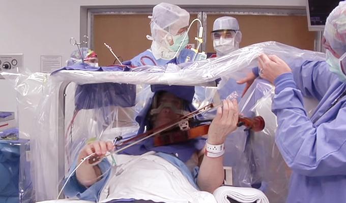
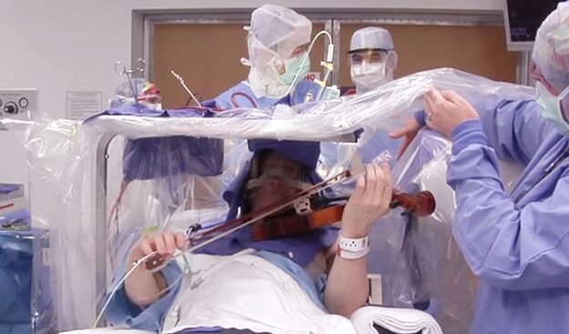
- An awake craniotomy is a surgical procedure performed while the patient is awake. It allows neurosurgeons to interact with the patient during brain surgery, mainly when the area being operated on is critical for speech and language functions.
- This technique is commonly used for surgeries involving eloquent areas of the brain, which are essential for sensorimotor and linguistic processing.
- Dr. Rao’s hospital does an awake craniotomy for language mapping.
-
Why Awake Craniotomy?
- Awake craniotomies are best suited for cases where the tumor or lesion is located in or near eloquent areas. These areas include regions responsible for:
- Motor control
- Speech production
- Language comprehension
- Vision
- The goal is to maximize safe resection while preserving critical functions1.
- Awake craniotomies are best suited for cases where the tumor or lesion is located in or near eloquent areas. These areas include regions responsible for:
-
The Role of Language Testing:
- Preoperative Assessment:
- Prior to surgery, patients undergo language testing to establish baselines. This assessment includes evaluating motor aspects of elocution, linguistic processing, and vision.
- Standardized tests are used to assess speech and language functions.
- Intraoperative Assessment:
- During surgery, the patient is awake, and direct electrical stimulation is applied to different brain areas.
- Language tasks are performed, such as counting, naming objects, assessing speech arrest, and identifying specific language disturbances.
- The surgeon uses this information to guide the surgical approach and avoid critical functional areas.
- Post-Operative Expectations:
- Patients may experience temporary speech changes after surgery, but recovery is expected.
- Awake mapping helps tailor the surgical corridor, ensuring maximal safe resection while respecting normal brain tissue1.
- Preoperative Assessment:
-
Team and Equipment:
- The surgical team includes:
- Patient
- Surgeon and residents
- Speech-language pathologist (SLP)
- Neurophysiologist
- Neuromonitoring technician
- Anesthesiologist
- The equipment includes:
- Microscope
- Electrical stimulation tools
- Neuronavigation system
- And lots more!
- The patient is positioned comfortably, and the surgical steps involve incision, craniotomy, dural opening, and awake assessment1.
- The surgical team includes:
-
Language Testing Tasks:
- During awake mapping, the patient performs various language tasks:
- Common Language Tasks During Mapping:
- Counting: Assess numerical fluency.
- Naming: Identify objects.
- Anarthria / Speech Arrest: Observe speech disruptions.
- Dysarthria: Evaluate speech clarity.
- Anomia: difficulty recalling words.
- Paraphasia: substituting incorrect words.
- Articulatory Disturbance: Problems with Speech Sounds.
- Common Language Tasks During Mapping:
- The surgeon uses this information to create a tailored surgical approach that respects functional brain areas while maximizing tumor resection1.
- During awake mapping, the patient performs various language tasks:
-
Frequently Asked Questions:
- Does it hurt? No, the patient doesn’t feel pain during direct stimulation.
- Will speech recover if it worsens after surgery? Yes, most patients experience improvement.
- What if a seizure occurs during surgery? Historical evidence shows that trepanation (burr hole) was used for seizures thousands of years ago1.
In summary, awake craniotomy with language testing is a remarkable approach that allows surgeons to navigate the intricate landscape of the brain while preserving critical functions. 🧠💡


