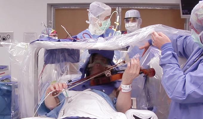
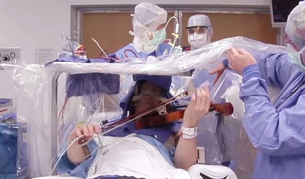
Awake Craniotomy at Dr. Rao’s Hospital | Brain Surgery and Speech/Language Testing at Dr Rao’s Hospital
What Is an Awake Craniotomy?
An awake craniotomy is a specialized neurosurgical procedure performed while the patient is awake. It allows surgeons to map and preserve critical functional areas of the brain during tumor resection, epileptic focus removal, or vascular lesion treatment. The goal is to maximize safe tumor removal while minimizing damage to essential brain regions. As per India Today
Ideal Candidates for Awake Craniotomy
Patients who are ideal candidates for this procedure typically have tumors in or near “eloquent areas”—regions essential for sensorimotor and linguistic processing. These areas include:
Motor cortex: Responsible for movement control.
Language centers:
Broca’s area: Associated with speech production.
Wernicke’s area: Involved in language comprehension.
The Evaluation Process
Preoperative Assessment:
Baseline assessments are conducted a few days before surgery.
Patients undergo local scalp anesthesia or pin block.
The patient’s brain is mapped while asleep to establish reference points.
Intraoperative Mapping:
During surgery, the patient is positioned laterally (on their side) with an unobstructed line of sight.
The surgical team performs cortical stimulation while the patient is awake.
Tasks include counting, naming objects, assessing speech, and identifying specific language deficits.
Direct electrical stimulation helps identify critical areas and guides surgical decisions.
Post-Operative Considerations:
Questions addressed:
Does it hurt during mapping?
Will speech recover if affected?
What if a seizure occurs during surgery?
Tailored surgical corridors are designed based on anatomical knowledge and tumor morphology.
The extent of resection is optimized while preserving functional tissue.
Collaborators in Awake Craniotomy:
Patient
Surgeon/Resident(s)
RN Circulator
Scrub Technician(s)
Anesthesiologist/Resident(s)/CRNA(s)
Speech-Language Pathologist (SLP) Team
Neurophysiologist
Neuromonitoring Technician
Adjunct Device/Drug Representatives
Neuronavigation Representative
Microscope (bigger than a full-size kangaroo)
Key Takeaways:
Awake craniotomy allows precise mapping of functional brain areas.
It ensures safer tumor resection while preserving critical functions.
Patients are assessed during surgery using various language tasks.
The eloquent functional areas are interconnected beneath the brain surface.
Remember, this procedure combines cutting-edge science with meticulous care, all while keeping the patient’s well-being at the forefront. ????????
Dr. Rao’s Contact Information:
- Phone:9010056444, 9010057444
- Email: info@drraoshospitals.com
- Address: Old Bank St, GV Thota, Beside AK Biryani Point, Guntur, Andhra Pradesh 522001
- Website: Dr. Rao’s Hospital


