Author Archives: Dr RAO

brain-tumor-causes-and-treatmentdr-raos-hospital

Brain Tumor – Causes and the best Treatment | Dr Raos Hospital

 Brain Tumor: Causes and Treatment.

Have you or someone in your family been diagnosed with a brain tumor? You have a lot of doubts that need to be answered and assured that there are treatments for brain tumors. Speak to your neurosurgeon in Andhra Pradesh. Your doctor is your best guide in these times.

Brain tumors might compromise brain function if it grows large enough to push on nearby nerves, blood vessels, or tissue. Your recovery from the tumor largely depends on:

  • The tumor’s type, grade, and location.
  • The success of tumor removal surgery.
  • Your age and overall health.

Dr. Mohana Rao Patibandla, the founder of Dr. Rao’s Hospital, is amongst the best neurosurgeons in Andhra Pradesh.

Dr. Rao’s Hospital is a state-of-the-art neurology hospital in Guntur, with an aim to give comprehensive treatment facilities to patients suffering from brain, spine, and nerve damage or abnormality. 

If you or anyone in your family is looking for the best brain tumor treatment in Guntur, we have some of the best neurologists and neurosurgeons working with us.

To know more about brain tumor treatment, please continue reading this article.

Brain Tumors and Brain Cancer | Johns Hopkins Medicine

First, let’s know,

What is a brain tumor?

Cells that have grown abnormally in or around the brain is called a brain tumor. 

Brain tumors can be either benign (non-cancerous) or malignant (cancerous). Some tumors develop swiftly, while others grow slowly.

Only approximately a third of all brain tumors are malignant. Even if they are benign, brain tumors can affect brain function by impinging on surrounding nerves, blood vessels, and tissue. 

Primary tumors are those that form in the brain.

The Most Common Brain Tumor: 5 Things You Should Know | Johns Hopkins  Medicine

Secondary tumors or metastatic tumors have moved to the brain growing from another section of the body.

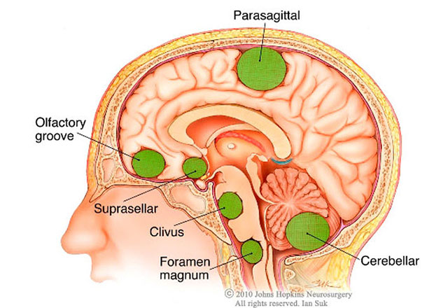
Here, we will get some information on primary tumors. Primary brain and spinal cord tumors come in a variety of shapes and sizes.

Types of Primary Brain Tumors:

Tumors of the brain and central nervous system are classified by their location and the type of cells they affect.

Benign brain tumors:

Vestibular schwannomas, also named acoustic neuromas, is a tumor that develops on the vestibular nerve (the nerve that leads from the inner ear to the brain).

Pineocytoma: Pineocytoma is a slowly progressing tumor of the pineal gland, situated in the deep brain, and secretes melatonin hormone.

Gangliocytoma: These tumors develop in neurons (nerve cells) and affect the central nervous system.

Meningioma: A tumor in the meningeal layer that protects the brain and spinal cord. They are mostly non-cancerous.

Pituitary adenoma: These tumors develop in the pituitary gland near the base of the brain. 

Chordoma: Chordomas are slow-growing tumors that usually start at the base of the skull or the bottom of the spine. They’re mostly harmless (not cancerous).

Malignant brain tumors:

Glioma: Glioma is a type of tumor that develops in the glial cells surrounding and aid nerve cells. Gliomas are classified as follows:

Astrocytomas are tumors that develop in the glial cells known as astrocytes.

Glioblastoma: Glioblastomas are astrocytomas that grow aggressively.

Oligodendroglioma: These rare malignancies start in myelin-producing cells (a layer of insulation around nerves in the brain).

Medulloblastomas are rapidly growing tumors that originate at the base of the skull. They mostly prevail in children.


What causes brain tumors? 

A variety of factors causes a brain tumor.

  • Unusual cell propagation is caused by a genetic mutation.
  • Radiation from X-rays. 
  • Chemicals in the environment. 
  • Family history.

What signs and symptoms do you have if you have a brain tumor?

A brain or central nervous system tumor can cause no symptoms in some persons. Doctors may detect cancer while treating another problem in some situations.

A brain tumor may create symptoms when it grows and presses on nearby nerves or blood vessels. The location, nature, and size of a brain tumor and the affected portion of the brain influence the signs and symptoms. They may include the following:

  • Headaches that are persistent or severe, or that begin first in the morning or disappear after vomiting.
  • Changes in behavior or personality.
  • Confusion.
  • Problems with balance and coordination.
  • Trouble concentrating.
  • Nausea and vomiting.
  • Tingling, numbness,  or weakness in one side or section of the body or face.
  • Vision, hearing, or speech difficulties.
  • Seizures.
  • In ordinary sleepiness
  • Thinking, memory, speaking, and understanding language problems

Diagnosis of Brain Tumour: 

Doctors use several tests to confirm the presence of a brain tumor. These tests include the following:

Physical examination and medical history: Your doctor will do a general physical examination to search for signs of sickness. Your doctor will also inquire about your past and present surgeries and medical treatments and family history of diseases.

Blood test: To see if there are any tumor markers.

Biopsy: A tissue sample is taken and sent for testing.

PET scans, CT, MRI, and SPECT are imaging techniques that assist doctors in finding tumors and deciding whether they are malignant or benign. 

Spinal tap for the presence of a malignant tumor in the central nervous system.

A tumor is graded 1-4 based on how fast it is growing and likely to spread.

Treatment option for brain tumor:

The location, size, and kind of tumor determine the course of treatment. To treat a tumor, most neurosurgeons in Hyderabad frequently utilize a mix of medicines, surgeries, and radiation.

The surgeon decides the approach of the surgery to limit harm to functional parts of the brain.

They work very cautiously, sometimes performing surgery while the patient is conscious. 

Sometimes, the surgeons have to perform a craniotomy, where they have to surgically cut out a fragment of the skull to reach the tumor.

Types of Craniotomy:

  • Extended Bifrontal Craniotomy
  • Retro-Sigmoid “Keyhole” Craniotomy
  • Translabyrinthine Craniotomy
  • Minimally Invasive Supra-Orbital “Eyebrow” Craniotomy
  • Orbitozygomatic Craniotomy

Other Treatment Methods

Radiation:

High dosages of X-rays are used to kill brain tumor cells or reduce the tumor. Before surgery, some people receive radiation to reduce a brain tumor so that the surgeon has to remove less tissue.

Chemotherapy:

Anti-cancer medications destroy cancer cells in the brain and throughout the body during chemotherapy. Chemotherapy can be injected into a vein or taken orally. Sometimes, doctors will use chemotherapy before surgery to shrink the tumor. Following surgery, your doctor may recommend chemotherapy to eliminate any cancer cells that remain or to prevent tumor cells from developing.

Immunotherapy:

Immunotherapy, often known as biological therapy, is a cancer treatment that uses your body’s own immune system to fight the disease. The primary goal of the treatment is to activate the immune system.

Targeted therapy:

Drugs that target specific traits in cancer cells without hurting healthy cells are known as targeted treatments. If you are having problems with the adverse effects of chemotherapy, such as exhaustion and nausea, your doctor may offer targeted therapy.

Laser thermal ablation

Laser thermal ablation is a treatment that kills tumor cells with lasers.

Active follow-ups. A doctor uses regular tests to keep an eye on the tumor for symptoms of growth.

How may a brain tumor be prevented?

A brain tumor is unavoidable. Avoiding smoking and excessive radiation exposure can help you prevent having a brain tumor.

What is the prognosis for those who have a brain tumor?

Different people and brain tumors have different outcomes. The type, grade, and location of the tumor; effective removal of the entire tumor; and your age and overall health are all factors that can affect your prognosis.

In many patients, the surgeon can successfully treat a brain tumor.

Brain tumors can come back in some persons following therapy. To keep the tumor from developing or spreading, these people may need to continue treatments such as chemotherapy or radiation. Following brain tumor therapy, you should see your doctor regularly.

the-effects-of-osteoporosis-on-the-spine

The best Osteoporosis Spine treatment in Guntur

Osteoporosis is a bone disease that affects both men and women. Millions of people around the world have it or are at greater risk of getting it.

The best Osteoporosis Spine treatment in Guntur

Osteoporosis affects roughly 40 million Americans | Information provided by  Spine Nevada in Reno, Sparks and Carson City

Image Source: Spine Nevada

A healthy bone has microscopic gaps on the inside like a honeycomb. Osteoporosis enlarges these gaps, causing the bone to deteriorate in strength and density. Furthermore, the exterior of the bone becomes thinner and weaker. 

This condition can strike anyone at any age, but it is more common in the elderly, particularly women. Osteoporosis makes people more prone to fractures, especially in the spine, known as vertebral compression fractures.

Early diagnosis and treatment can be of great help as they prevent further compression and irreversible change in the spine. You can consult Dr. Mohana Rao Patibandla, one of the best neurosurgeons in Guntur, at Dr. Rao’s Hospital for advanced surgical intervention. 

What are the symptoms of spine fractures?

A compression fracture can result in back or neck pain as well as loss of height. It is a  fracture in one of your neck or back vertebrae that is so weak that it breaks under normal spinal pressure.

The fracture can cause symptoms such as:

  • Severe sudden back pain
  • Have problems in twisting or bending your body
  • Pain that increases when you walk or stand
  • Curved shape spine
  • There is far more significant pain and discomfort in moving if the fracture is in the lower spine

What are the causes of osteoporosis?

Osteoporosis and Spinal Fractures - OrthoInfo - AAOS

Image Source: OrthoInfo – AAOS

Osteoporosis is a natural aging process in which our bones weaken as we get older. The vertebrae in the spine might narrow and flatten due to it. This can result in a hump or rounded back in older adults giving them a bent forward look. 

The weaker vertebrae are particularly vulnerable to fracture. When too much pressure is applied to a weak vertebra, its front cracks and loses height, resulting in a vertebral compression fracture. They are most commonly caused by falls. However, persons with osteoporosis can sustain a fracture just by coughing, twisting, or sneezing.

How is osteoporosis diagnosed?

Your doctor will evaluate your medical history and do a physical exam to determine if you have osteoporosis. They may also conduct blood and urine tests to look for disorders that could cause bone loss. Tests to check the levels of calcium and vitamin D are also included. In a recent mayoclinic.

To determine whether you have a spine fracture, your doctor will recommend an X-ray, MRI, or CT scan to examine your bones more closely.

Further, he will ask you to undergo a bone density test called bone densitometry, or dual-energy X-ray absorptiometry (DEXA).

Treatment for vertebral compression fracture

1. Nonsurgical treatment

Most of the people with a spinal compression fracture recover within three months without needing to have the fracture fixed. Simple measures, such as a brief period of rest and the use of pain medications, when necessary, are all that is required. The doctor may ask the patients to wear a brace to limit their movement and allow the spinal compression fracture to heal.

The doctor may also recommend lifestyle changes and prescribe calcium, Vitamin D, bisphosphonates, and anabolic bone drugs.

2. Surgical Treatment

Surgery may be recommended if you have significant pain that does not respond to nonsurgical treatment. Patients with recent spinal compression fractures are the best candidates for surgery. Your doctor will discuss which operation is best for you based on the type of spinal compression fracture you have. 

Dr. Rao’s Hospital is one of the best neurology hospitals in Guntur, where you are offered advanced minimally invasive vertebral augmentation procedures.

3. Kyphoplasty

During this procedure, the surgeon inserts a balloon tamp into the fractured vertebra. The balloon is then inflated, which helps in the restoration of the vertebra’s height and shape. The small cavity is left behind by the device that the doctor fills with special bone cement.

4. Vertebroplasty

Here, the surgeon directly injects the bone cement into the narrowed vertebra to keep it stable. It helps to alleviate discomfort and help avoid additional vertebral fractures and a bent spine.

5. Cemented augmented fenestrated screws

In patients with low bone quality due to osteoporosis, fenestrated screws for cement augmentation provided effective and long-lasting outcomes.

They are used for spine stabilization in the elderly, allowing for rapid functional recovery in fractures and deformities. 

It also overcomes the problem of screws loosening in individuals with inadequate bone stock due to osteoporosis and causing morbidity. 

Contact Dr. Mohana Rao Patibandla, one of the best neurosurgeons in Guntur, Andhra Pradesh, for superior treatment options for spinal compression fractures due to osteoporosis.

best-functional-neurosurgery-in-andhra-pradesh

The Best Functional Neurosurgery In Andhra Pradesh

The Best Functional Neurosurgery In Andhra Pradesh

The goal of functional neurosurgery is to restore neurological health and function. We treat chronic neurological problems with various procedures at Dr. Rao’s Hospital in Guntur, Andhra Pradesh, including deep brain stimulation and neuromodulation. This is one of the fastest-growing subspecialties in neurosurgery. It offers considerable promise to patients with previously untreatable neurological diseases.

At our Guntur hospital, every patient is evaluated correctly. Patients are frequently evaluated by a big interdisciplinary team that specializes in a specific condition and uses cutting-edge diagnostic techniques. A comprehensive assessment is obtained using a full spectrum of diagnostic means, and therapeutic procedures are implemented with state-of-the-art technology under the direction of Dr. Mohana Pratibandla, the best neurosurgeon in Guntur and the only neurosurgeon to have studied all the subspecialties of Neurosciences and received extensive training in the USA.

Diagnosis and Comprehensive Assessment

Clinical: Our multidisciplinary team includes neurosurgeons, neurologists, psychologists, and physiologists. As a result, we can accurately detect neurological illnesses and assess how they influence the patient’s health, quality of living, and cognition.  

Imaging: The neural axis can be viewed using a variety of cutting-edge imaging techniques. MRI and CT and functional imaging such as PET, SPECT, and fMRI are examples of imaging used.

Electrophysiology: Other than nervous system testing with EMG and EEG, precise electrophysiological assessments are required when applying focused therapy. Single cellular microelectrode recordings of the basal ganglia and mapping of neurons in eloquent cortical areas of the brain are among the tests performed.

Disorders of the Nervous System

Many previously thought-to-be-untreatable chronic neurological illnesses are now treated in our department successfully. Regimens are begun as usual, but now successful surgical therapies may also be administered – and even done early in the course of the disease – before adverse effects from frequent medication use occur.

At Dr. Rao’s Hospital in Guntur, functional neurosurgeons are presently treating:

• Parkinson’s disease, dystonia, tremor, hemifacial spasm, Tourette’s syndrome, among others

• Epilepsy

• Spasticity originating in the brain (Spinal cord injury and MS)

• Neuropsychiatric disorders, such as depression and OCD

• Chronic pain caused by neuropathic injuries or disorders, and cancer

• Nerve damage, such as trigeminal neuralgia and postherpetic neuralgia.

Are you suffering from any of the above conditions?

You are not alone! Many people in Andhra Pradesh are suffering from some or other disorder stated above.

But do not worry! Now is the time to seek the solution to your neurological problems by visiting our renowned hospital in Guntur, Andhra Pradesh.

At our hospital in Guntur, we have a team of experts that provide the best functional neurosurgery in Andhra Pradesh.

You might be interested to know the neurosurgical treatments offered by us!

Below we have mentioned the modern neurosurgical treatments performed at our hospital by the best functional neurosurgeon in Andhra Pradesh.

What are the modern neurosurgical treatments offered at Dr. Rao’s Hospital in Guntur?

The following are some of the modern neurosurgical treatments performed by our experienced neurosurgeon in Guntur-

Neuromodulation

Neuromodulation allows a neurosurgeon to modify or modulate nerve activity using electricity or pharmaceutical drugs in a problematic area.

When treating parts of the spinal cord, peripheral nervous system, and brain that are crucial to the patient’s normal functioning, neuromodulation allows for precise targeting and is relatively safe.

Deep Brain Stimulation (DBS) 

Source-www.recoveryanswers.org

Deep Brain Stimulation is used to treat essential tremors, Parkinson’s disease, epilepsy, OCD, and dystonia. Electrodes are placed in specific brain locations, and electrical impulses are generated to control the brain’s abnormal processes.

Chemical imbalances that produce abnormalities can be corrected with the help of these electrodes. At the upper chest, the programmable generator is hidden beneath the skin.

 Stereotactic neurosurgical treatment

The use of stereotactic neurosurgery can help locate regions in the brain’s deepest recesses. To detect areas deep within the brain for treatment, such as deep-seated brain cancers, a neurosurgeon uses a specialized 3-dimensional coordinate system with MRIs or CT scans.

During stereotactic procedures, the patient is awake and responsive, which reduces the risk of adverse effects on the brain. This procedure can treat epilepsy, deep-seated brain tumors and deliver radiation and drugs to the innermost sections of the brain.

What are the advantages of functional neurosurgery?

Some of the advantages of functional neurosurgery are as follows:

• Functional neurosurgery is gaining traction as a safe and effective therapy choice for neurological patients.

• Because of advances in neuroimaging and surgical processes, functional neurosurgery accuracy has substantially improved.

• The neurosurgeon can work more accurately, focusing on the regions that need treatment while avoiding crucial regions or those that don’t need treatment.

• Functional neurosurgery offers a rapid recovery time for patients.

• Successful functional neurosurgery provides significant pain relief and minimizes patients’ need for painkillers.

Wow! Isn’t functional neurosurgery a beneficial treatment option?

Then what’s stopping you from availing it?

Visit Dr. Rao’s Hospital in Guntur to get the best functional neurosurgery treatment in Andhra Pradesh! 

You are just a visit away from your solution! 

Dr. Rao and his treating staff at Dr. Rao’s Hospital understand the importance of precise and timely diagnosis, consistent monitoring, complication management, and patient compliance. We place a stronger emphasis on these areas. And thus, we are known to provide the best functional neurosurgery in Andhra Pradesh. 

So, what are you waiting for? 

Visit now!

epilepsy-precautions-and-treatment-in-andhra-pradesh

EPILEPSY PRECAUTIONS AND THE BEST TREATMENT IN ANDHRA PRADESH

EPILEPSY PRECAUTIONS AND THE BEST TREATMENT IN ANDHRA PRADESH

Do you often feel anxious?

Do you experience contraction and jerking of muscles? 

Do you often lose consciousness and feel weak? 

If yes, it is likely you have epilepsy. Epilepsy is a neurological disorder in which brain activity becomes abnormal, causing seizures or periods of unusual behavior, sensations, and sometimes loss of awareness. It is quite common in Andhra Pradesh. 

But do not worry! Every problem comes with a solution. The issue of epilepsy can be treated by getting the best epilepsy treatment in Andhra Pradesh. Dr. Rao’s Hospital provides the best epilepsy treatment in Guntur, Andhra Pradesh. Dr. Mohana Rao Patibandla, our Medical Director, is a highly regarded neurosurgeon in Andhra Pradesh. He is one of the few neurosurgeons who has undergone training in the United States and is a neuroscience specialist and provides the best Epilepsy Treatment in Andhra Pradesh. Keep reading this article to know the treatment options offered by our top neurosurgeon in Andhra Pradesh to treat epilepsy. This article also states the precautions one should take to control seizures.

What are the treatment options available in Guntur to treat epilepsy?

Source-medcraveonline.com

Medications are usually used to treat epilepsy first. If drugs don’t work, doctors may recommend surgery or another treatment option.

Medication

The majority of people with epilepsy can get rid of seizures by using just 1 anti-seizure drug, often known as an anti-epileptic drug. Others may find that taking a combination of drugs reduces the intensity and frequency of their seizures.

Children with epilepsy who do not have symptoms of epilepsy can eventually stop taking their drugs and live a life free of seizures. After 2 or more years of being seizure-free, many adults can stop taking their medications. Your neurosurgeon in Guntur will let you know when is the right time to discontinue your drugs.

It can be challenging to find the right drug and dosage. When deciding which medicine to prescribe, our neurosurgeon in Guntur will evaluate your condition, your age, the frequency of your seizures, and other considerations. Our experienced neurosurgeon will also go over any other medications you’re taking to make sure they don’t interact with the anti-epileptic drugs.

Your doctor would most likely start you on a single medicine at a low dose and progressively raise the dosage until the seizures are under control.

There are about 20 different anti-seizure drugs on the market. The type of epilepsy medicine prescribed by your doctor is determined by the kind of seizures you have and other criteria like your age and other medical conditions.

Follow these measures to get the most excellent seizure control possible with medication:

·Follow the directions on your prescription carefully.

• Before switching to a generic version of your medication or using other medicines, over-the-counter drugs, or herbal therapies, always consult your doctor.

• Do not discontinue your medication without first consulting your physician.

• If you detect new or increasing thoughts of suicide and depression or experience changes in your behavior or mood, contact your doctor right away.

• If you suffer from migraines, tell your doctor. Your doctor may prescribe Anti-epileptic drugs to treat epilepsy and prevent migraines. 

With initial medication, at least 50% of persons newly diagnosed with epilepsy will be seizure-free. If anti-epileptic meds aren’t working, our neurosurgeon may recommend surgery or some other treatments. Regular follow-up appointments will be scheduled to monitor your medications and condition. 

Surgery

When medicine fails to keep seizures under control, surgery can be a helpful alternative. Our neurosurgeon in Guntur removes the part of your brain that causes seizures during epilepsy surgery.

Surgery is frequently performed when examinations reveal that:

• The source of your seizures is a limited, well-defined area of your brain.

• The operation site in your brain does not interfere with essential functions, including language, speech, vision, hearing, or motor function. 

When an open operation is too hazardous for some kinds of epilepsy, minimally invasive techniques like MRI-guided stereotactic laser ablation may be a useful therapeutic option. In these procedures, our neurosurgeons use a thermal laser probe to damage the tissue in the brain that causes seizures to manage the seizures better.

Therapies

Stimulation of the vagus nerve: 

Doctors implant a device called a vagus nerve stimulator beneath the skin of your chest, similar to a heart pacemaker, in vagus nerve stimulation. The vagus nerve in your neck is attached to wires from the stimulator.

The battery-operated device sends electrical energy bursts to your brain via the vagus nerve. It’s unclear how this gadget prevents seizures; however, it can usually decrease seizures by twenty to forty percent.

Ketogenic diet: 

The ketogenic diet is a high-fat, low-carbohydrate diet. The body burns down fats instead of carbohydrates for energy with a ketogenic diet. Some children with epilepsy have reduced their seizures by adhering to a rigorous low-carbohydrate, high-fat diet.

Deep brain stimulation:

 Deep brain stimulation involves the implantation of electrodes into a specific area of the brain, usually the thalamus. The electrodes are attached to a generator that is surgically placed in your chest. The generator provides timed electrical pulses to your brain regularly, which may help to lessen your seizures. This therapy is frequently used to treat persons who have seizures that do not respond to medicines.

Responsive neurostimulation: 

These pacemaker-like devices, which can be implanted, can help significantly reduce the number of seizures that occur. These devices scan brain activity patterns to identify seizures early on and provide an electrical charge or a medication to end the seizure before it leads to any harm. According to research, this therapy appears to have fewer side effects and can lead to long-term relief from seizures.

Now, let us discuss the precautions to control seizures.

What are some of the precautions to control seizures?

Some of the precautions to control seizures are as follows- 

  • Good compliance with medications 
  • Following a proper sleep schedule
  • Avoid alcohol

The best neurosurgeon in Andhra Pradesh advises epilepsy patients with severe and uncontrolled seizures to take the following precautions-

  • Avoid driving
  • Avoid unsupervised swimming
  • Avoid high impact sports activities

So, now is the time to tell the problem of epilepsy goodbye by getting the best solution for epilepsy at our renowned hospital in Guntur. 

We are the key to the door of neurological solutions. We help you access the best solutions to all your neurological problems. At our hospital in Guntur, we have effective and the best solutions for treating all your neurological issues. 

So, what are you waiting for? 

Visit Dr. Rao’s Hospital in Guntur, Andhra Pradesh now! You are just a visit away from the solution to your epilepsy problem or other neurological problems!

get-the-proper-treatment-for-neurotrauma-in-andhra-pradesh

Get The best Treatment For Neurotrauma In Andhra Pradesh

Get The best Treatment For Neurotrauma In Andhra Pradesh

A brain, spine, or nerve injury is known as neurological trauma. Accidents involving autos, sports, bikes, or falls can result in this sort of injury. It can impact the organs, bones, muscles, and blood vessels, among other body parts.

But do not worry! At Dr. Rao’s Hospital in Guntur, we have a team of professionals who provide the best neurotrauma treatment in Guntur. Our Medical Head, Dr. Mohana Rao Patibandla, is the best neurotrauma surgeon in Guntur. He is among the few neurosurgeons who are excellent in neurosciences and trained in the USA. 

Before we come to the treatment options provided by the best neurology hospital in Guntur to treat neurotrauma, let us discuss more in detail about neurotrauma, its causes, and symptoms.

Neurotrauma is a term that encompasses two distinct conditions:

1. ABI (acquired brain injury) and

2. SCI (spinal cord injury)

Acquired brain injury can cause issues with concentration, planning, and social interaction, whereas an injury to the spinal cord damage can leave you paralyzed for the rest of your life.

Individuals with ABI or SCI may require assistance with daily tasks.

ABI and SCI have also been associated with severe mental health disorders such as depression and behavioral problems.

What are the causes of traumatic brain injury?

Source-link.springer.com

A blow to the head or other severe injury to the body can result in traumatic brain injury. The severity of the injury can be determined by several factors, including the nature of the injury and the power of impact.

The following are examples of common incidents that result in traumatic brain injury:

  1. Falls:

     The most prevalent cause of traumatic brain injury in older adults and children are falling from bed or a ladder, downstairs, in the bath, and other falls.

  1. Vehicle-related crashes:

TBI is commonly caused by collisions involving vehicles, bicycles, motorbikes, and pedestrians engaged in such events.

  1. Violence: 

Common causes include gunshot wounds, child abuse, domestic violence, and other assaults. A shaken baby syndrome is traumatic brain damage in neonates induced by forceful shaking.

  1. Sports injury: 

Sports injuries are prevalent among children and teenagers. Injuries from various sports, such as boxing, football, baseball, lacrosse, skateboarding, hockey, and other high-impact or intense activities, can result in traumatic brain injuries.

  1. Explosive blasts and other combat injuries: 

In active-duty military troops, explosive blasts are a common cause of traumatic brain injury.

What are the signs and symptoms of neurotrauma?

The following are the immediate and delayed signs of neurotrauma:

• Confusion

• Concentration difficulties

• Vision that is hazy

• Infants exhibit irritation and continuous crying.

What are the neurotrauma therapy alternatives accessible in Guntur, Andhra Pradesh?

At Dr. Rao’s Hospital, a top neurology hospital in Guntur, the following treatment methods are used by our neurotrauma surgeon to treat neurotrauma-

Rest, medicine, and surgery are all part of neurotrauma treatment in Andhra Pradesh. Neuropsychological therapy, physical therapy, speech therapy, occupational therapy, and rehabilitation drugs are also available, depending on your needs. The following are the various types of surgery used to treat neurotrauma at our neurology hospital in Guntur:

External ventricular drain placement

A small tube called an external ventricular drain (EVD) is used to drain excess cerebrospinal fluid (CSF) from the brain. Illness or injury may be the cause of the additional liquid. The pressure in and around the brain rises when there is too much fluid—an EVD aids in lowering brain pressure to a safe level.

Lumbar drain placement

A lumbar drain is a tiny tube inserted into your lower back and spinal column to drain or collect cerebral spinal fluid (CSF).

Spinal decompression

A surgical treatment called spinal decompression relieves pressure on the spinal cord or a compressed nerve root. The bone channels through which the spinal cord and nerves flow are opened by spinal therapy. This allows them to move about freely.

Spinal fusion

Spinal fusion is a procedure that permanently joins two or more vertebrae in your spine, preventing them from moving. Spinal fusion permanently joins two or more vertebrae in your spine to improve stability, relieve pain, or rectify deformities.

Techniques for spinal fusion are aimed to mirror the natural healing process of damaged bones. Our experienced neurosurgeon fills the area between 2 spinal vertebrae with bone or a bonelike substance during spinal fusion. To hold the vertebrae together until they recover into one solid unit, rods, screws, and metal plates may be utilized.

Decompressive craniectomy

A decompressive craniectomy is a type of brain surgery in which a part of the skull is removed. When the brain expands due to an accident, the pressure inside the skull can build up, causing more damage. Removing a part of the skull can lessen the chance of severe brain injury, and in some cases, it can even save a person’s life.

Cranioplasty

The surgical repair of a bone defect in the skull caused by a previous operation or accident is known as cranioplasty. Different types of cranioplasties exist, but the majority entail raising the scalp and reconstructing the contour of the skull with the original skull piece or a custom-molded graft manufactured from titanium, synthetic bone substitute, or other materials.

Minimally invasive spine instrumentation, halo stabilization, complex craniofacial repair, kyphoplasty/vertebroplasty, sacropelvic spine instrumentation, and fusion are among the numerous other surgical techniques performed at our hospital in Guntur to treat neurotrauma.

So, what are you waiting for?

Visit Dr. Rao’s hospital in Guntur, Andhra Pradesh, to get the best neurotrauma treatment in Andhra Pradesh.

The solution for neurotrauma is just a visit away!

Biplane Cath Lab - cerebral-angiography-the-best-at-dr-raos-guntur

Causes, Prevention and The Best Treatment of Cerebrovascular Disorders

Causes, Prevention and The Best Treatment of Cerebrovascular Disorders

A stroke, also known as a brain attack, occurs when the brain’s blood supply is cut off, depriving the brain of oxygen and causing brain cells to die. When cells die, the damaged part of the brain loses control of whatever part of the body it controls. It affects people of all ages, genders, and races. It is the leading cause of disability.

Although stroke is a brain disease, it can impact the entire body. The consequences of a stroke are determined by which section of the brain is affected and how serious the injury is. 

Some of the common disabling symptoms of a stroke are:

  • Paralysis or issues controlling movement. 
  • Sensory abnormalities, including pain. 
  • Problems using or comprehending language. 
  • Problems with thinking and memory. 
  • Emotional disturbances. 

Stroke can have fatal consequences. Thanks to innovative and sophisticated procedures and therapies, life-long impairment or death is no longer addressed.

At Dr. Rao’s Hospital, one of the best neurology hospital in Guntur, Andhra Pradesh, highly qualified and experienced neurosurgeons collaborate to ensure that patients receive the care they require, whether it’s urgent care, brain surgery, cutting-edge diagnostics and treatment, cutting-edge clinical trials, or rehabilitation.

Because time is of the essence when treating stroke, our emergency department teams are prepared to deliver immediate, high-quality, and best stroke and cerebrovascular treatment in Andhra Pradesh. We also have a rapid response team that assesses patients and initiates particular procedures to determine the etiology of the stroke.

Now, let’s know,

What Causes a Stroke?

High blood pressure (hypertension): 

Itis a primary cause of stroke.

Vascular diseases: 

Vascular illnesses, such as clogged arteries, heart valve malfunction, irregular heartbeat, heart infection, and heart abnormalities, can lead to strokes, especially in the elderly.

Smoking or chewing tobacco: 

Smoking can cause several health problems, including fatty deposits in the neck artery, high blood pressure, and blood thickening. Blood clots in the blood vessels can result due to this. Even exposure to second hand smoke can put you at risk for a stroke.

Obesity: 

Being obese or overweight increases the risk of having a stroke.

Diabetes: 

Obesity, high blood pressure, and diabetes are all variables that increase the chances of stroke.

Family history: 

Family genes can also increase the chance of stroke, heart attack, or other major risk factors such as diabetes or high blood pressure.

Now, let’s know more about,

Acute Treatment Strategies for Stroke

If someone has a stroke, they should seek emergency medical attention from a neurologist in Guntur. You can be treated for a stroke with treatments like mechanical thrombectomy.

Neurosurgeons in Guntur use the following equipment to perform mechanical thrombectomy to remove blood clots.

1. Stentriever Mechanical Thrombectomy

A stentriever is the most recent mechanical device. A self-expanding mesh tube is linked to a wire-guided through a catheter (thin tube). The surgeon places the catheter in a groin artery and guides it through numerous blood channels until it reaches the blood clot in the brain.

2. Aspiration Thrombectomy 

Manual cluster yearning or the use of catheter equipment to suck the coagulation out of the vessel are both used in aspiration thrombectomy.

Now, let’s see,

Procedure of Mechanical Thrombectomy

Mechanical thrombectomy is a procedure used to remove blood clots. Neurosurgeons remove blood clots using a vacuum or mechanical gear in mechanical thrombectomy. Therefore, the blood flow is restored, and the stroke is cured.

To achieve the finest results, it’s best to seek mechanical thrombectomy treatment from the best neurology hospital in Guntur.

What are the Advantages of Mechanical Thrombectomy?

  • Mechanical thrombectomy treatment allows patients to recover from a stroke within a 24-hour window following the commencement of the stroke. This will assist more individuals in recovering from stroke and avoiding the adverse effects of stroke. 
  • For patients who have had a stroke, mechanical thrombectomy offers a gleam of hope. Mechanical thrombectomy provides them hope and bravery in their battle against stroke. It resurrects their long-dead hopes and gives them the strength to battle stroke. It is a potent tool that aids patients in overcoming dangerous strokes.
  • Mechanical thrombectomy also speeds up the recovery of patients with stroke. Though mechanical thrombectomy is a valuable technique for treating stroke, it is only available at a few neurology hospitals in Guntur.

Let’s discuss,

Stroke Prevention Strategies

Regardless of family history or age, there are several steps one can take to reduce the risk of having a stroke:

1. Keeping an eye on the blood pressure

One of the leading causes of stroke is high blood pressure. To improve vascular health, an individual should regularly monitor blood pressure and treat it if it rises.

2. Keeping weight in check

Obesity is connected to comorbidities such as diabetes and hypertension, which increase the risk of a stroke. Reduce your calorie consumption and increase your physical activity. Consult your doctor to develop a personal weight-loss plan that works for you.

3. Regular exercise is essential

Daily exercise will help you stay in shape and better control your weight and blood pressure, minimizing your risk of stroke.

4. Avoiding smoking and consuming alcohol

Smoking increases plaque build-up and blood thickening in the neck arteries, which can lead to clotting. Drinking increases the risk of stroke by causing high blood pressure, diabetes, obesity, liver damage, and various other disorders. Reduce your risk of stroke by eating a healthy diet and quitting smoking. In addition, if you consume alcohol, do it in moderation.

5. Keeping an eye on the blood sugar level

Exercise, drugs, and a balanced diet can all help you keep track of your blood sugar levels. Diabetes can cause blood arteries to rupture and clot inside them over time.

Consult our expert neurosurgeons at Dr. Rao’s Hospital if you need cerebrovascular disease treatment or a stroke specialist in Andhra Pradesh.

Spinal-Cord-Tumors

Get The best Treatment For Spinal Cord Tumors

Get The Best Treatment For Spinal Cord Tumors at Dr Rao’s Hospital

Spinal Cord Tumors, on occasion, probably go unnoticed because they may no longer be unusual, and their signs resemble those of more significant, not uncommon, conditions. Therefore, your physician must recognize your clinical record and perform each fashionable bodily and neurological exam.

Dr. Mohan Rao Patibandla, one of Guntur’s most renowned and best neurosurgeons, has years of expertise in treating spinal tumors. You can consult with him and get all the suitable guidance regarding the process of treating a spinal tumor. In a recent  mid-day. 

If your physician suspects a spinal tumor, those checks can assist in verifying the prognosis and pinpoint the tumor’s location:

Spinal magnetic resonance imaging (MRI).

 MRI uses an effective magnetic area and radio waves to correctly photograph your spine, spinal cord, and nerves. MRI is usually the preferred way to diagnose spinal cord tumors and surrounding tissue tumors. An evaluation agent that ensures that the tissues and systems can be injected right into a vein on your hand or forearm throughout the take-a-look.

Computerized tomography (CT)

This test uses a thin beam of radiation to provide specific photographs of your spine. Sometimes, it is mixed with an injected contrast dye to make unusual modifications inside the spinal canal or spinal cord less challenging to see. A CT test is hardly ever used to assist in diagnosing spinal tumors.

Biopsy

The simplest way to determine the precise form of a spinal tumor is to study a small tissue sample (biopsy) under a microscope. The biopsy results will assist in deciding on treatment options.

Treatment

Ideally, a spinal tumor remedy aims to cure the tumor. However, this aim can be complex, using the threat of everlasting harm to the spinal cord tumors and surrounding nerves. Doctors additionally have to forget your age and normal health.

The sort of tumor and whether or not it arises from the systems of the backbone or spinal canal or has unfolded on your backbone from someplace else to your frame additionally have to be taken into consideration in figuring out a remedy plan.

Treatment alternatives for maximum spinal tumors include:

1. Monitoring

Some spinal tumors can be found earlier than they cause symptoms—frequently while you are being evaluated for every other condition. If small tumors are not developing or urgent on surrounding tissues, looking at them cautiously can be all that is needed.

2. Surgery

This is frequently the remedy of preference for tumors that may be eliminated with an appropriate chance of spinal cord tumors or nerve damage. Newer strategies and devices permit neurosurgeons to attain tumors that have been inaccessible as soon as taken into consideration. The high-powered microscopes utilized in microsurgery make differentiating tumors from wholesome tissue simpler.

Doctors can also screen the features of the spinal wire and different crucial nerves for the duration of the surgical operation, minimizing the threat of wounding them. In a few instances, very high-frequency sound waves are probably used during the surgical operation to interrupt tumors and do away with the fragments.

3. Radiation therapy.

This can be used to dispose of the remnants of tumors that continue to be after surgical procedure, to deal with inoperable tumors or to deal with the ones tumors in which surgical procedure is simply too risky.

 Medications may also assist in easing a number of the aspect results of radiation, which include nausea and vomiting.

4. Chemotherapy.

A fashionable remedy for plenty of varieties of most cancers, chemotherapy makes use of medicinal drugs to damage most cancer cells or forestall them from growing. Your health practitioner can decide whether or not chemotherapy is probably helpful for you, both on your own or in a mixture with radiation therapy.

 Side outcomes may also include fatigue, nausea, vomiting, an accelerated threat of infection, and hair loss.

5. Other drugs.

Because surgical procedures and radiation remedies, in addition to tumors themselves, can irritate the spinal cord, doctors, once in a while, prescribe corticosteroids to lessen the swelling, both after a surgical procedure or at some point in radiation treatments.

Although corticosteroids lessen irritation, they may be commonly used for quick intervals to avoid extreme aspects, such as muscle weakness, osteoporosis, excessive blood pressure, diabetes, and a multiplied susceptibility to infection.

Dr. Rao’s Contact Information:

the best Head-Injury treatment in andhra Pradesh by Dr Rao at Dr Rao's hospital

Head Injury Precautions and Treatments

Head Injury Precautions and Treatments

A knock on the head and a skull fracture are all examples of head injuries. Some severe head traumas might result in brain damage or even death.

Knowing the signs and symptoms of a concussion and some basic first aid can help individuals respond promptly to a head injury, decreasing the risk of consequences. In a recent India today article.

Dr. Rao’s hospital in Guntur has successfully performed many head injury surgeries. It is one of the most renowned and best neurology hospitals in Guntur, with expertise over the years.

Types

There are different types of brain injuries, each of which can be more or less devastating.

 Traumatic brain injury (TBI) occurs when the brain is damaged, usually as a result of an accident. TBI can occur when an object hits a person’s head or when an object enters the brain through the skull.

 A high-velocity ball that hits the head, a massive fall to the head, or a gunshot wound are examples.

Shaking the head vigorously can also cause SHT because the brain can jump or spin in the skull. Whiplash is a common cause of these injuries.

Traumatic brain injury (TBI) is damage to the brain that frequently results in blood clots or bruises. An intracranial hematoma (ICH) is a blood clot in the brain. Depending on its size and location in the brain, the ego can be moderate or heavy.

A concussion is a type of traumatic brain injury (TBI) in which the brain temporarily ceases to function normally. Concussion symptoms are usually not severe or long-lasting, but they can be bothersome.

The brain is protected by the skull, which is formed of thick, sturdy bone. A powerful strike, on the other hand, has the potential to crack or break the skull.

An open head wound, or a penetrating head wound, occurs when the skull is fractured and a bone or other item pierces the brain.

When should you see a doctor?

Both moderate and severe head injuries require medical attention right away. If the symptoms of a mild head injury persist for more than two weeks, get medical help.

The signs and symptoms of a concussion do not usually show right away. A person’s symptoms may appear days or weeks after an incident.

Always proceed with caution while dealing with a head injury. Even after a minor mishap, if someone is worried about their symptoms, they should see a doctor.

First aid and treatment

Mild head injuries are frequently treated at home. Swelling can be reduced by applying a cold pack to the affected region.

Tylenol can also be used, but non-steroidal anti-inflammatory medications (NSAIDs), such as ibuprofen and aspirin, should only be used if a doctor has prescribed them. Someone should check on a person periodically over the first 24 hours following a moderate head injury.

It is critical to seek medical attention if a person loses consciousness or exhibits indications of disorientation or memory loss.

After a brain injury, a person should avoid using drugs or alcohol, driving, or participating in contact sports. They may have to miss work or school.

Severe head injuries necessitate medical care right away. Anyone who encounters someone with severe head injury symptoms should dial 9010056444 or their local emergency number.

It is advisable not to move a person who has suffered a severe head injury to prevent aggravating their ailments. It’s also not a good idea to try to take someone’s helmet off if they’re wearing one.

Severe head injuries frequently require hospitalization, surgery, or long-term care.

Prevention

Although avoiding head injuries is not always feasible, specific actions may be taken to decrease the risk. These are some of them:

  • When in a car, wear a seatbelt
  • When cycling or riding a motorcycle, you should always wear a helmet.
  • when practicing contact sports, take safety precautions

Because a brain injury can have long-term repercussions, it’s critical to take measures wherever possible.

Dr. Rao’s Contact Information:

osteoporotic-spine-surgery

The Best osteoporotic spine surgery in Andhra Pradesh

The Best osteoporotic spine surgery in Andhra Pradesh

Osteoporosis is a bone disease that affects both men and women. Millions of people worldwide have it or are at greater risk of getting it.

A healthy bone has microscopic gaps on the inside, like a honeycomb. Osteoporosis enlarges these gaps, causing the bone to deteriorate in strength and density. Furthermore, the exterior of the bone becomes thinner and weaker.

This condition can strike anyone at any age, but it is more common in the elderly, particularly women. Osteoporosis makes people more prone to fractures, especially in the spine, known as vertebral compression fractures.

Early diagnosis and treatment can be beneficial as they prevent further compression and irreversible changes in the spine. You can consult Dr. Mohana Rao Patibandla, one of the best neurosurgeons in Guntur, at Dr. Rao’s Hospital for advanced surgical intervention.

What are the symptoms of spine fractures?

A compression fracture can result in back or neck pain and loss of height. It is a fracture in one of your neck or back vertebrae that is so weak that it breaks under normal spinal pressure.

The fracture can cause symptoms such as:

  1. Severe, sudden back pain
  2. Have problems twisting or bending your body?
  3. Pain that increases when you walk or stand
  4. Curved-shaped spine
  5. There is far more significant pain and discomfort in moving if the fracture is in the lower spine.
  6. Kyphosis of the spine, loss of height, and neuro deficiencies
  7. Neurological deficits include weakness of the limbs, tingling numbness, bed wetting, and stool incontinence or constipation.

What are the causes of osteoporosis?

Osteoporosis is a natural aging process in which our bones weaken as we get older. The vertebrae in the spine might narrow and flatten due to it. This can result in a hump or rounded back in older adults, giving them a bent-forward look.

The weaker vertebrae are particularly vulnerable to fracture. When too much pressure is applied to a weak vertebra, its front cracks and loses height, resulting in a vertebral compression fracture. They are most commonly caused by falls. However, people with osteoporosis can sustain a fracture by coughing, twisting, or sneezing.

How is osteoporosis diagnosed?

Your doctor will evaluate your medical history and do a physical exam to determine if you have osteoporosis. They may also conduct blood and urine tests to look for disorders that could cause bone loss. Tests to check the levels of calcium and vitamin D are also included.

To determine whether you have a spine fracture, your doctor will recommend an X-ray, MRI, or CT scan to examine your bones more closely.

Further, he will ask you to undergo a bone density test called bone densitometry, or dual-energy X-ray absorptiometry (DEXA).

Treatment for vertebral compression fracture

Nonsurgical treatment

Most people with a spinal compression fracture recover within three months without needing to have the fracture fixed. Simple measures, such as a brief period of rest and the use of pain medications when necessary, are all that are required. The doctor may ask the patients to wear a brace to limit their movement and allow the spinal compression fracture to heal.

The doctor may also recommend lifestyle changes and prescribe calcium, Vitamin D, bisphosphonates, and anabolic bone drugs.

Surgical Treatment

Surgery may be recommended if you have significant pain that does not respond to nonsurgical treatment. Patients with recent spinal compression fractures are the best candidates for surgery. Your doctor will discuss which operation is best for you based on the type of spinal compression fracture you have.

Dr. Rao’s Hospital is one of the best neurology hospitals in Guntur, where you are offered advanced minimally invasive vertebral augmentation procedures.

Kyphoplasty

During this procedure, the surgeon inserts a balloon tamp into the fractured vertebra. The balloon is then inflated, which helps restore the vertebra’s height and shape. The device leaves behind a small cavity that the doctor fills with special bone cement.

Vertebroplasty

Here, the surgeon injects the bone cement into the narrowed vertebra to keep it stable. It helps to alleviate discomfort and help avoid additional vertebral fractures and a bent spine.

Cemented augmented fenestrated screws

In patients with low bone quality due to osteoporosis, fenestrated screws for cement augmentation provided effective and long-lasting outcomes.

They are used for spine stabilization in the elderly, allowing for rapid functional recovery in fractures and deformities.

It also overcomes the problem of screws loosening in individuals with inadequate bone stock due to osteoporosis, causing morbidity.

Contact Dr. Mohana Rao Patibandla, one of the best neurosurgeons in Guntur, Andhra Pradesh, for superior treatment options for spinal compression fractures due to osteoporosis. According to mid-day

Dr. Rao’s Contact Information:

Deep-Brain-Stimulation

The best Deep Brain Stimulation (DBS): A Brain Pacemaker

Deep Brain Stimulation (DBS): A Brain Pacemaker—The best at Dr Rao’s Hospital

Deep brain stimulation (DBS) electrodes are implanted in particular regions of the brain. These electrodes generate electrical impulses, which control the aberrant impulses. Alternatively, electrical impulses in the brain can impact specific cells and substances.

Dr. Rao’s Hospital Guntur is one of Guntur’s most renowned and best neurology hospitals. They have years of expertise in implementing deep brain stimulation.

Under the skin of the upper chest, a pacemaker-like device controls deep brain stimulation. A cable that travels beneath your skin connects this device to electrodes in your brain.

Deep brain stimulation is frequently used to treat a variety of ailments, including:

  1. Parkinson’s disease is a neurological disorder that affects people.
  2. Essential tremor is a kind of tremor that affects
  3. Dystonia
  4. Epilepsy
  5. Obsessive-compulsive disorder (OCD) is a kind of obsessive-compulsive disorder.

Why is it done this way?

People with movement disorders, including essential tremors, Parkinson’s disease, and dystonia, as well as mental diseases like obsessive-compulsive disorder, can benefit from deep brain stimulation. The Food and Drug Administration has also authorized it for use in treating seizures in people with difficult-to-treat epilepsy.

This treatment is only for those who can’t control their symptoms with medicine.

Risks

Although deep brain stimulation is thought to be a low-risk procedure, any operation has the potential for consequences. Furthermore, brain stimulation might have adverse effects.

The dangers of surgery

Deep brain stimulation entails drilling small holes in the skull for the electrodes to be implanted into the brain tissue and surgery to implant the device with the batteries under the skin in the chest. Surgical complications might include:

  1. Leads are misplaced.
  2. There is bleeding in the brain.
  3. Stroke
  4. Breathing difficulties
  5. Nausea
  6. Problems with the heart
  7. Seizure

The device will be turned on a few weeks following surgery, and determining your optimal settings will begin. Some settings may have adverse effects, although these usually improve with more device changes.

The Food and Drug Administration suggests talking with your doctor and adopting water safety precautions before swimming. A few reports have suggested that DBS treatment impairs the movements needed for swimming.

How do you get ready?

Consider the advantages and disadvantages first.

Deep brain stimulation is a method that has the potential to be both serious and hazardous. Even if you believe you could be a candidate for deep brain stimulation, you and your doctors should carefully weigh the risks and benefits.

Prepare for surgery after that.

Before surgery, you’ll almost definitely need medical testing to confirm that deep brain stimulation is a safe and effective therapy option for you. Before the procedure, you may need brain imaging tests, such as an MRI. These studies help map the brain areas where electrodes will be implanted.

What you may anticipate

During the procedure,

Here’s how deep brain stimulation surgery works in general:

Brain Surgery: Your medical team will provide a unique head frame to keep your head still throughout the brain surgery procedure (stereotactic head frame). The experts will next map your brain using neuroimaging (brain MRI or CT) to decide where the electrodes should be put.

Surgery on the chest wall

During the second step of the surgery, the pulse generator (the part of the device that contains the batteries) is implanted under the skin in the chest near the collarbone. General anesthesia will be used during this procedure. The wires from the brain electrodes are routed under the skin to the battery-operated pulse generator. Then, the generator is set up to send electrical impulses to your brain in a loop. The generator is under your control, and you can turn it on or off with a unique remote control.

Following the surgery

The pulse generator in your chest will turn on in your doctor’s office a few weeks after surgery. With a unique remote control, the physician can easily configure his device from outside his body. The intensity of the stimulation is tailored to your needs, and it can take up to six months to achieve the proper balance.

Results

Although deep brain stimulation will not cure your condition, it can help alleviate some of the symptoms. If deep brain stimulation is successful, your symptoms will improve significantly, but they won’t completely disappear. However, certain conditions may require the use of medications.

Deep brain stimulation is not always practical. The effectiveness of deep brain stimulation depends on a variety of factors. Before surgery, talk to your doctor about the improvement you can expect from your condition.

Dr. Rao’s Contact Information: