Category Archives: Epilepsy Treatment

common-neuro-problems-the-best-treatment-at-dr-raos-guntur

Common neuro problems – the best treatment at Dr Raos, Guntur

Neurology is a branch of medicine that focuses on diagnosing and treating disorders of the nervous system, including the brain, spinal cord, and nerves. Some common topics in neurology include:

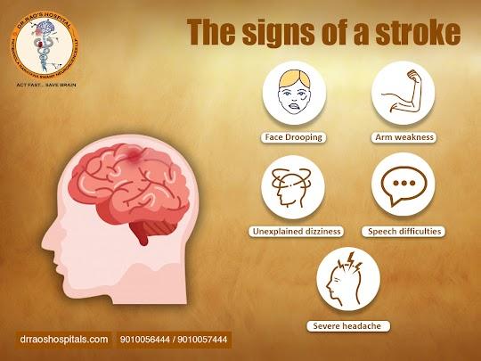
  1. Stroke: A stroke is a life-threatening, debilitating health event that occurs when the vital blood supply to the brain is disrupted, resulting in permanent damage to the brain tissue. There are two leading causes of stroke – blockages in the blood vessels, known as ischemic stroke, or bleeding in the brain, known as hemorrhagic stroke. Symptoms of stroke can vary depending on the area of the brain that has been affected and can include paralysis, difficulty speaking, and loss of vision. It is absolutely vital to be aware of the various signs and symptoms of stroke as quickly as you can, as the earlier treatment is started, the more probable it is that the outcome will be positive. The signs to look out for include sudden numbness of the face, arm, or leg, blurred vision, confusion, difficulty speaking, and severe headaches. If any of these are experienced, it is important to dial 9010056444 or 90100574444 immediately and seek prompt medical assistance.
  2. Epilepsy: Epilepsy is a serious neurological disorder that is characterized by recurrent and unpredictable seizures. Seizures are episodes of abnormal electrical activity in the brain that can be accompanied by convulsions, loss of consciousness, and strange changes in behavior. Seizures can last for only a few seconds or for several minutes, and can range from mild to severe in intensity. People living with epilepsy may experience seizures multiple times a day, or just once or twice a year. Epilepsy can be a debilitating condition, with devastating effects on a person’s physical and mental health. Treatment of the epilepsy typically involves a combination of medication and lifestyle modifications. Medications used to treat epilepsy may include anticonvulsants, antiepileptic drugs, or sedatives. In some cases, surgical intervention may be necessary to control the epileptic activity. In addition, lifestyle modifications such as avoiding triggers that can cause seizures, eating a healthy diet, and exercising regularly may help to reduce the frequency and severity of seizures.
  3. Multiple sclerosis (MS): Multiple Sclerosis (MS) is a chronic and often debilitating autoimmune disorder that affects the central nervous system. It is characterized by inflammation and damage to the myelin sheath, a protective insulating layer surrounding nerve fibers in the brain and spinal cord. This damage to the myelin sheath disrupts the normal transmission of nerve impulses, resulting in a wide range of possible symptoms, ranging from mild to severe. Common MS symptoms include vision problems, numbness, muscle weakness, and difficulty with coordination, balance, and speech. The cause of MS is unknown, but is thought to involve a combination of genetic and environmental factors. Diagnosis of MS involves a physical exam, neurological exam, MRI, and laboratory tests. Treatment of MS is aimed at reducingsymptoms and slowing the progression of the disease. Medications such as interferon beta, gling disease-modifying therapies, immunosuppressants, and corticosteroids may being the activity of the immune system. Other treatments may include physical therapy, occupational therapy, speech therapy , and cognitive behavioral therapy. In some cases, surgery may be recommended to help improve mobility and reduce pain. Medications such as nonsteroidal anti-inflammatory drugs, antispastic medicationss and muscle relaxants may be prescribed to help reduce pain and improve mobility. Physical therapy exercises may be “and stretching may be recommended to help strengthen muscles. Other treatments such as massage, acupuncture, and heat or cold therapy may be used to help reduce inflammation. In some cases, surgery may be necessary to correct the underlying cause of the mobility issue. Physical therapy can also help to improve balance, strengthen muscles, and increase range of motion. Exercises may be prescribed to help improve posture and improve coordination. Other treatments may include the use of assistive devices such as canes, walkers and wheelchairs, as well as physical therapy modalities such as heat, cold, ultrasound, and electrical stimulation. Occupational therapy may also be recommended to help with activities of daily living, such as dressing, bathing, eating, and grooming. Speech therapy may be recommended to help with communication and swallowing Speech therapists may also work on cognitive skills, such as memory, problem solving, and organization. Physical therapists may be recommended to help with mobility and balance. Occupational therapists may be recommended to help with activities of daily living, such as dressing, bathing, and eating. Speech-language pathologists may be recommended to help with communication and swallowing. Mental health professionals may be recommended to help with cognitive and behavioral issues. Occupational therapists may be recommended to help with activities of daily living, such as dressing and bathing, as well as to help with fine motor skills. Physical therapists may be recommended to help with strengthening and coordination. Speech therapists may be recommended to help with communication and swallowing difficulties.
  4. Parkinson’s disease: Parkinson’s disease is a progressive neurological disorder that affects movement, causing many symptoms, such as tremors, stiffness, slow movement, and balance issues. It is caused by the loss of nerve cells in the brain that produce a chemical called dopamine, which helps regulate movement and is essential for coordination and communication between the brain and muscles. People with Parkinson’s disease may experience a range of symptoms, including tremors, slowed movement, impaired balance, rigidity, and difficulty initiating movement. Over time, Parkinson’s disease can also cause changes in cognition, behavior, and emotion. Treatment for a variety of ailments and conditions usually involves a comprehensive approach, including a combination of medications, physical therapy, and lifestyle changes. Medications may include both Over-the-counter and prescription drugs are important medicines that are used to treat various ailments and health conditions. OTC drugs, such as those for pain relief, cold and flu symptoms, and allergies, can be purchased without a prescription at any drugstore, grocery store, or convenience store. Prescription drugs, on the other hand, are usually more potent medicines that require a doctor’s prescription. These medicines can help with chronic conditions like diabetes and high blood pressure, and can also be used to treat infections or serious illnesses such as cancer. Both types of drugs can be incredibly helpful in treating and managing health issues, but they should always be taken as prescribed by a medical professional., while physical therapy can include activities such as stretching, strength training, and aerobic exercises. Additionally, lifestyle changes, such as quitting smoking, reducing stress, and eating a healthy diet, can help improve overall health and well-being.
  5. Alzheimer’s disease: Alzheimer’s disease is a debilitating and progressive neurological disorder that affects memory, thinking, behavior, and day-to-day functioning in older adults. It is the most common form of dementia, a general term for memory loss and other cognitive abilities serious enough to interfere with daily life. Though the precise cause of this incurable illness remains unknown, age, genetics, and lifestyle choices have been identified as potential contributing factors. Diagnosis of Alzheimer’s disease typically involves a physical exam, a review of medical history, and a variety of cognitive and neurological tests to assess memory, thinking, language, judgment, and other mental functions. Imaging tests such as computed tomography (CT) or magnetic resonance imaging (MRI) may also be used to rule out other conditions that could be causing the symptoms. Blood tests may be used to check for certain metabolic or infectious diseases that could be causing the symptoms. Other tests such as electroencephalography (EEG) or electromyography (EMG) may be used to check for nerve or muscle disorders. Treatment of AD involves medications to help manage symptoms, lifestyle changes, and therapies to help improve cognitive and behavioral functioning.
  6. Headaches: Headaches are a pervasive neurological disorder that can be caused by a variety of underlying factors, such as muscle tension, sinus problems, eyestrain, and food sensitivities. In some cases, headaches can be indicative of a more serious underlying medical issue, so it’s important to consult a physician if the symptoms become severe or persistent. Common symptoms of a headache include a throbbing or pounding sensation in the head, sensitivity to light and noise, nausea, fatigue, and difficulty focusing. Fortunately, there are a variety of treatments and lifestyle modifications that can help alleviate symptoms and reduce the frequency of headaches. Some of these include relaxation techniques, proper hydration, avoidance of trigger foods, and regular exercise. Additionally, medications and supplements such as ibuprofen, aspirin, and magnesium can help reduce the severity and frequency of headaches.
  7. Traumatic brain injury (TBI): Traumatic Brain Injury (TBI) is an injury to the brain caused by a sudden, violent impact or blow to the head, usually occurring during a car accident, a fall, a sports injury, or an act of violence. The severity of the injury can range from mild to severe and can cause a range of symptoms, including changes in consciousness, confusion, memory problems, difficulty with concentration or comprehension, and difficulty with movement or coordination. TBI can also lead to long-term complications such as headaches, sleep disturbances, speech or language difficulties, depression, and even personality changes. Diagnosis of Traumatic Brain Injury (TBI) typically involves a comprehensive physical examination conducted by a qualified medical professional. During this exam, the doctor will ask questions about the patient’s medical history, recent head injury and current symptoms. The physical exam may also include a physical evaluation of the patient’s cognitive abilities and motor functioning, as well as neurological tests. In addition, imaging tests such as CT scans or MRI scans may be used to help diagnose TBI. Treatment of the TBI involves either conservative or Surgical management.

These are just a few of the many topics that fall under the umbrella of neurology. Other areas of focus within the field include sleep disorders, movement disorders, nerve and muscle disorders, and developmental disorders.

sensation-of-smell-the-best-treatment-at-dr-raos-guntur

Sensation of smell – the best treatment at Dr Raos, Guntur

Introduction

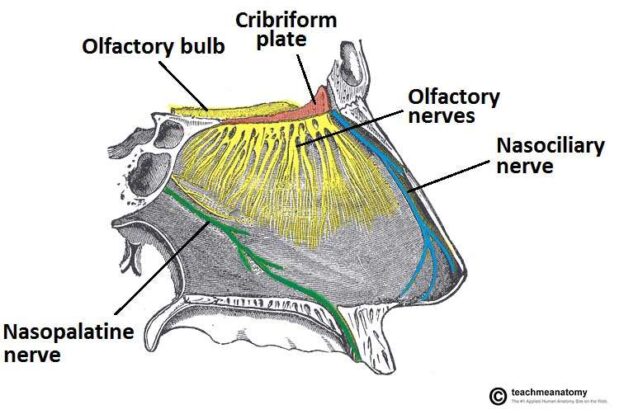
The human sense of smell is mediated by the olfactory system, which consists of the nose and the olfactory nerve. The olfactory nerve is a cranial nerve that transmits information about smells from the nose to the brain. The sense of smell is important for many aspects of daily life, including the ability to enjoy food and drink, to detect danger, and to communicate with others. Decreased sensation of smell can be caused by many different factors, including disease, injury, or aging. This condition can have a significant impact on quality of life, as it can make everyday activities such as eating and drinking less enjoyable, and can also lead to social isolation. If you are experiencing a decreased sense of smell, it is important to see a doctor so that the cause can be identified and appropriate treatment can be started. Looking for the best neurosurgery hospital or Best neurosurgeon or India’s best neurosurgeon or top ten best neurosurgeons, look no further than Dr Rao at Dr Rao’s hospital, the best spine surgery or best neurosurgery or best neurology hospital in Guntur, Andhra Pradesh, India. Call us at 9010056444 or 9010057444 for the best neurosurgeon or spine surgeon or neurologist in India. Dr Raos hospital is best for the vertigo / numbness / weakness /tingling treatment in Guntur and India.

causes

There are many potential causes of a decreased sense of smell, including: -Aging: As we age, our sense of smell can gradually decline. -Illness: Many illnesses can cause a decrease in sense of smell, including colds, sinus infections, the flu, and more serious conditions such as Alzheimer’s disease and Parkinson’s disease. -Nasal obstruction: Anything that blocks the airflow through your nose can reduce your sense of smell. This includes nasal polyps, a deviated septum, or allergies. -Medications: Some medications can cause a decrease in sense of smell, including blood pressure medications and antidepressants. -Radiation therapy: Radiation therapy to the head and neck can damage the olfactory nerve and cause a decrease in sense of smell. -Smoking: Smoking cigarettes can damage your sense of smell.

Associated symptoms

There are a few associated symptoms that can be experienced when someone has a decreased sense of smell. For example, they may have a hard time tasting food or distinguishing between different smells. Additionally, they may also experience a loss of appetite, fatigue, and difficulty breathing.

diagnsois

The diagnosis of decreased sense of smell can be made by a physician through a medical history and physical examination. However, if the cause is unknown, further testing may be needed to rule out other causes. These tests may include a CT scan or MRI of the head, sinus x-rays, or allergy testing.

treatment

The most common treatment for anosmia is a nasal corticosteroid spray. This type of medication can help to reduce inflammation in the nose and improve airflow. In some cases, a decongestant may also be recommended. If an infection is the underlying cause of the condition, antibiotics may be prescribed. In rare cases, surgery may be necessary to correct a structural problem in the nose that is causing anosmia.

prognosis

The prognosis for patients with a decreased sense of smell depends on the underlying cause. If the cause is a viral infection, the prognosis is generally good and the sense of smell usually returns to normal within a few weeks. However, if the cause is damage to the olfactory nerve, the prognosis is more uncertain. In some cases, the sense of smell may improve over time, but in other cases it may be permanently impaired.

Conclusion

In conclusion, a decreased sense of smell can be caused by a variety of factors, ranging from the common cold to more serious conditions such as Alzheimer’s disease. However, in most cases, a decreased sense of smell is not a cause for concern and will resolve itself over time. If you are experiencing a decreased sense of smell, consult your doctor to rule out any underlying medical conditions. Looking for the best neurosurgery hospital or Best neurosurgeon or India’s best neurosurgeon or top ten best neurosurgeons, look no further than Dr Rao at Dr Rao’s hospital, the best spine surgery or best neurosurgery or best neurology hospital in Guntur, Andhra Pradesh, India. Call us at 9010056444 or 9010057444 for the best neurosurgeon or spine surgeon or neurologist in India. Dr Raos hospital is best for the Loss of sensation of smell/ vertigo / numbness / weakness /tingling treatment in Guntur and India.

chiari-malformations-the-best-treatment-is-at-dr-raos-guntur

Chiari malformations – the best treatment is at Dr Raos, Guntur

Introduction

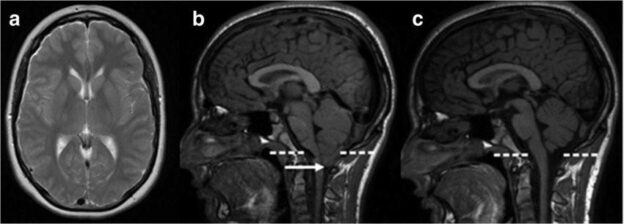
A Chiari malformation is a condition where the brain tissue extends into the spinal canal. It can cause a number of symptoms, including headaches, neck pain, and problems with balance and coordination. Chiari malformations are usually diagnosed using imaging tests, such as MRI. Treatment typically involves surgery to remove the excess brain tissue. Looking for the best neurosurgery hospital or Best neurosurgeon or India’s best neurosurgeon or top ten best neurosurgeons in India, look no further than Dr Rao at Dr Rao’s hospital, the best spine surgery or best neurosurgery or best neurology hospital in Guntur, Andhra Pradesh, India. Call us at 9010056444 or 9010057444 for the best neurosurgeon or spine surgeon or neurologist in India. Dr Raos hospital is best for the Chiari malformations/ numbness / weakness /tingling treatment in Guntur and India.

classification

There are four main types of Chiari malformations, which are classified according to the severity of the condition.

Type I is the most common and mildest form, and is often diagnosed during childhood.

Type II is more severe, and is typically diagnosed in infancy or early childhood.

congenital defects l Dr Raos hospital

Type III is the most severe form, and is usually diagnosed in utero.

Type IV is a rare form that is associated with certain genetic disorders.

Symptoms

The symptoms of Chiari malformations can vary depending on the severity of the condition. In some cases, there may be no symptoms at all. In other cases, symptoms may include:

-Headaches

-Neck pain

-Dizziness

-Nausea

-Vomiting

-Loss of balance

-Difficulty swallowing

-Weakness in the arms and legs

-Numbness or tingling in the extremities

-Problems with coordination and fine motor skills

-Blurred vision or double vision

-Hearing loss or ringing in the ears (tinnitus)

diagnosis

A diagnosis of Chiari malformation is made based on the symptoms present and imaging studies of the brain and spine. The most common imaging study used to diagnose Chiari malformation is magnetic resonance imaging (MRI). MRI can show the size and location of the herniated portion of the brain and any resulting compression of nearby structures.

treatment

There are a few different ways that Chiari malformations can be treated. One way is through decompression surgery. This type of surgery involves making an incision in the back of the head and removing a small portion of the bone at the base of the skull. This helps to relieve pressure on the brain and spinal cord. Another way to treat Chiari malformations is through a shunt. A shunt is a small tube that is inserted into the brain to help drain excess fluid. This helps to relieve pressure on the brain and can also help to improve symptoms. In some cases, medication may be used to help manage symptoms. Pain medication may be prescribed to help with headaches. Muscle relaxants may be prescribed to help with muscle spasms. And, in some cases, steroids may be prescribed to help reduce inflammation. The decision on which treatment is best for you will be made by you and your doctor based on the severity of your condition and your symptoms.

Living with it

Living with a Chiari malformation can be difficult. The symptoms can be debilitating and make it hard to live a normal life. There are, however, treatments available that can help ease the symptoms and improve quality of life. It is important to stay positive and work closely with your medical team to find the best treatment plan for you. There is no cure for Chiari, but with proper treatment, many people are able to lead happy and healthy lives.

Conclusion

The Chiari malformation is a condition that can be debilitating and even life-threatening. It is important to be aware of the symptoms and to seek medical help if you suspect you or your child may have the condition. There is no cure for Chiari, but treatment can improve quality of life. Looking for the best neurosurgery hospital or Best neurosurgeon or India’s best neurosurgeon or top ten best neurosurgeons in India, look no further than Dr Rao at Dr Rao’s hospital, the best spine surgery or best neurosurgery or best neurology hospital in Guntur, Andhra Pradesh, India. Call us at 9010056444 or 9010057444 for the best neurosurgeon or spine surgeon or neurologist in India. Dr Raos hospital is best for the Chiari malformations/ numbness / weakness /tingling treatment in Guntur and India.


పరిచయం

చియారీ వైకల్యం అనేది మెదడు కణజాలం వెన్నెముక కాలువలోకి విస్తరించే పరిస్థితి. ఇది తలనొప్పి, మెడ నొప్పి మరియు సమతుల్యత మరియు సమన్వయ సమస్యలతో సహా అనేక లక్షణాలను కలిగిస్తుంది. చియారీ వైకల్యాలు సాధారణంగా MRI వంటి ఇమేజింగ్ పరీక్షలను ఉపయోగించి నిర్ధారణ చేయబడతాయి. చికిత్సలో సాధారణంగా అదనపు మెదడు కణజాలాన్ని తొలగించడానికి శస్త్రచికిత్స ఉంటుంది.

వర్గీకరణ

చియారీ వైకల్యాల్లో నాలుగు ప్రధాన రకాలు ఉన్నాయి, ఇవి పరిస్థితి యొక్క తీవ్రతను బట్టి వర్గీకరించబడతాయి. టైప్ I అనేది అత్యంత సాధారణ మరియు తేలికపాటి రూపం, మరియు ఇది తరచుగా బాల్యంలో నిర్ధారణ అవుతుంది. రకం II మరింత తీవ్రమైనది, మరియు సాధారణంగా బాల్యంలో లేదా బాల్యంలోనే నిర్ధారణ అవుతుంది. రకం III అత్యంత తీవ్రమైన రూపం, మరియు సాధారణంగా గర్భాశయంలో నిర్ధారణ చేయబడుతుంది. టైప్ IV అనేది కొన్ని జన్యుపరమైన రుగ్మతలతో సంబంధం ఉన్న అరుదైన రూపం.

లక్షణాలు

చియారీ వైకల్యాల లక్షణాలు పరిస్థితి యొక్క తీవ్రతను బట్టి మారవచ్చు. కొన్ని సందర్భాల్లో, ఎటువంటి లక్షణాలు ఉండకపోవచ్చు. ఇతర సందర్భాల్లో, లక్షణాలు ఉండవచ్చు:

  • తలనొప్పి
  • మెడ నొప్పి
  • మైకము
    -వికారం
  • వాంతులు
  • సంతులనం కోల్పోవడం
    -మింగడం కష్టం
    -చేతులు మరియు కాళ్లలో బలహీనత
  • అంత్య భాగాలలో తిమ్మిరి లేదా జలదరింపు
  • సమన్వయం మరియు చక్కటి మోటార్ నైపుణ్యాలతో సమస్యలు
  • అస్పష్టమైన దృష్టి లేదా డబుల్ దృష్టి
  • వినికిడి లోపం లేదా చెవులలో రింగింగ్ (టిన్నిటస్)

నిర్ధారణ

చియారీ వైకల్యం యొక్క నిర్ధారణ ప్రస్తుతం ఉన్న లక్షణాలు మరియు మెదడు మరియు వెన్నెముక యొక్క ఇమేజింగ్ అధ్యయనాల ఆధారంగా తయారు చేయబడుతుంది. చియారీ వైకల్యాన్ని నిర్ధారించడానికి ఉపయోగించే అత్యంత సాధారణ ఇమేజింగ్ అధ్యయనం మాగ్నెటిక్ రెసొనెన్స్ ఇమేజింగ్ (MRI). MRI మెదడు యొక్క హెర్నియేటెడ్ భాగం యొక్క పరిమాణం మరియు స్థానాన్ని మరియు సమీపంలోని నిర్మాణాల యొక్క ఏదైనా కుదింపును చూపుతుంది.

చికిత్స

చియారీ వైకల్యాలకు చికిత్స చేయడానికి కొన్ని విభిన్న మార్గాలు ఉన్నాయి. డికంప్రెషన్ సర్జరీ ద్వారా ఒక మార్గం. ఈ రకమైన శస్త్రచికిత్సలో తల వెనుక భాగంలో ఒక కోత మరియు పుర్రె యొక్క బేస్ వద్ద ఉన్న ఎముక యొక్క చిన్న భాగాన్ని తొలగించడం జరుగుతుంది. ఇది మెదడు మరియు వెన్నుపాముపై ఒత్తిడిని తగ్గించడానికి సహాయపడుతుంది.

చియారీ వైకల్యాలకు చికిత్స చేయడానికి మరొక మార్గం షంట్ ద్వారా. షంట్ అనేది ఒక చిన్న గొట్టం, ఇది అదనపు ద్రవాన్ని హరించడంలో సహాయపడటానికి మెదడులోకి చొప్పించబడుతుంది. ఇది మెదడుపై ఒత్తిడిని తగ్గించడంలో సహాయపడుతుంది మరియు లక్షణాలను మెరుగుపరచడంలో కూడా సహాయపడుతుంది.

కొన్ని సందర్భాల్లో, లక్షణాలను నిర్వహించడంలో సహాయపడటానికి మందులు వాడవచ్చు. తలనొప్పికి సహాయపడటానికి నొప్పి మందులను సూచించవచ్చు. కండరాల నొప్పులతో సహాయం చేయడానికి కండరాల సడలింపులను సూచించవచ్చు. మరియు, కొన్ని సందర్భాల్లో, వాపును తగ్గించడంలో సహాయపడటానికి స్టెరాయిడ్స్ సూచించబడవచ్చు.

మీ పరిస్థితి మరియు మీ లక్షణాల తీవ్రత ఆధారంగా మీకు మరియు మీ వైద్యునిచే మీకు ఏ చికిత్స ఉత్తమమో నిర్ణయించబడుతుంది.

దానితో జీవించడం

చియారీ వైకల్యంతో జీవించడం కష్టం. లక్షణాలు బలహీనపరుస్తాయి మరియు సాధారణ జీవితాన్ని గడపడం కష్టతరం చేస్తాయి. అయితే, లక్షణాలను తగ్గించడానికి మరియు జీవన నాణ్యతను మెరుగుపరచడంలో సహాయపడే చికిత్సలు అందుబాటులో ఉన్నాయి.

మీ కోసం ఉత్తమమైన చికిత్స ప్రణాళికను కనుగొనడానికి సానుకూలంగా ఉండటం మరియు మీ వైద్య బృందంతో సన్నిహితంగా పని చేయడం ముఖ్యం. చియారీకి ఎటువంటి నివారణ లేదు, కానీ సరైన చికిత్సతో, చాలా మంది ప్రజలు సంతోషంగా మరియు ఆరోగ్యకరమైన జీవితాన్ని గడపగలుగుతారు.

ముగింపు

చియారీ వైకల్యం అనేది బలహీనపరిచే మరియు ప్రాణాపాయం కలిగించే పరిస్థితి. లక్షణాల గురించి తెలుసుకోవడం మరియు మీకు లేదా మీ బిడ్డకు ఈ పరిస్థితి ఉందని మీరు అనుమానించినట్లయితే వైద్య సహాయం పొందడం చాలా ముఖ్యం. చియారీకి చికిత్స లేదు, కానీ చికిత్స జీవన నాణ్యతను మెరుగుపరుస్తుంది.

vision-problems-the-best-treatment-is-at-dr-raos-guntur

Vision Problems – the best treatment is at Dr Raos, Guntur

Introduction

The optic nerve is a bundle of nerve fibers that transmits visual information from the eye to the brain. The optic nerve is also known as cranial nerve II. The optic nerve is important for vision. If the optic nerve is damaged, it can cause decreased vision or even blindness. Looking for the best neurosurgery hospital or Best neurosurgeon or India’s best neurosurgeon or top ten best neurosurgeons, look no further than Dr Rao at Dr Rao’s hospital, the best spine surgery or best neurosurgery or best neurology hospital in Guntur, Andhra Pradesh, India. Call us at 9010056444 or 9010057444 for the best neurosurgeon or spine surgeon or neurologist in India. Dr Raos hospital is best for the visual blurring/ vertigo / numbness / weakness /tingling treatment in Guntur and India.

causes

There are many potential causes of decreased vision. Some causes are more common than others, but all should be taken seriously. The most common cause of decreased vision is refractive error, which occurs when the eye does not focus light properly. This can be corrected with glasses or contact lenses. Other common causes include cataracts, glaucoma, and age-related macular degeneration. These conditions can often be treated with surgery or medication. Less common causes of decreased vision include diabetic retinopathy, stroke, and brain tumors. These conditions may require more aggressive treatment, such as surgery or radiation therapy.

Associated symptoms

There are several symptoms that may be associated with decreased vision. These include: -Blurry vision – Difficulty seeing at night – Difficulty reading – Eye fatigue – Headaches – Squinting

diagnsois

The first step in diagnosing decreased vision is to schedule an appointment with an eye doctor. During the appointment, the doctor will ask about your medical history and symptoms. They will also conduct a thorough eye exam. The eye exam will assess your vision and check for any underlying conditions that could be causing your decreased vision. The doctor may use a lighted instrument to examine your eyes and look for any damage to your optic nerve. They may also order tests, such as an MRI or CT scan, to get a better look at the structure of your eyes. Once the cause of your decreased vision has been determined, the doctor will develop a treatment plan. Treatment options vary depending on the underlying condition causing the decreased vision. In some cases, glasses or contact lenses can help improve vision. Other treatments may include surgery, medication, or lifestyle changes.

treatment

The most common treatment for decreased vision is to use corrective lenses. If the cause of the decreased vision is due to a refractive error, such as nearsightedness, farsightedness, or astigmatism, then glasses or contact lenses can be prescribed to help correct the problem. If the cause of the decreased vision is due to a more serious condition, such as cataracts, glaucoma, or macular degeneration, then surgery may be required. In some cases, medications may be used to treat the underlying condition causing the decreased vision.

prognosis

The prognosis for decreased vision caused by optic nerve damage is generally poor. However, if the damage is caught early and treated promptly, the chances for a better outcome are increased. In some cases, complete recovery is possible.

Conclusion

The optic nerve is an important part of the visual system, and any damage to it can lead to decreased vision. There are many possible causes of optic nerve damage, and associated symptoms can vary depending on the cause. Optic nerve damage can be diagnosed through a variety of methods, and treatment will vary depending on the underlying cause. The prognosis for patients with optic nerve damage depends on the severity of the damage and the underlying cause. Looking for the best neurosurgery hospital or Best neurosurgeon or India’s best neurosurgeon or top ten best neurosurgeons, look no further than Dr Rao at Dr Rao’s hospital, the best spine surgery or best neurosurgery or best neurology hospital in Guntur, Andhra Pradesh, India. Call us at 9010056444 or 9010057444 for the best neurosurgeon or spine surgeon or neurologist in India. Dr Raos hospital is best for the vertigo / numbness / weakness /tingling treatment in Guntur and India.

concussion-the-best-treatment-is-at-dr-raos-guntur

Concussion – the best treatment is at Dr Raos, Guntur

Introduction

A concussion is a type of traumatic brain injury (TBI) that is caused by a blow to the head or a jolt to the body. Concussions can occur in any type of accident, but they are most common in sports injuries and car accidents. A concussion is a serious medical condition that can cause long-term problems, so it is important to seek medical care as soon as possible after sustaining one. There are several different types of symptoms that can occur after a concussion, and they can range from mild to severe. The most common symptoms include headache, dizziness, nausea, vomiting, fatigue, difficulty concentrating, and difficulty remembering. Some people may also experience changes in mood, such as irritability or depression. In some cases, concussions can also cause seizures or coma. Most people who suffer from a concussion will recover within a few weeks with rest and proper medical care. However, some people may experience long-term effects from a concussion, such as problems with memory and concentration, headaches, dizziness, and sleep disorders. If you have any concerns about your health after sustaining a concussion, you should speak with your doctor. Looking for the best neurosurgery hospital or Best neurosurgeon or India’s best neurosurgeon for concussion treatment? Look no further than Dr Rao at Dr Raos hospital, Guntur.

When to seek medical care

If you think you or your child has a concussion, seek medical care right away. A concussion is a serious brain injury that can have lasting effects. Symptoms of a concussion can include:

• headache

• confusion

• memory problems

• nausea or vomiting

• dizziness or balance problems

• double or blurry vision

• sensitivity to light or noise

• feeling sluggish, hazy, foggy, or groggy

• concentration or memory problems

• changes in mood, such as feeling more irritable, sad, or anxious

Head Injury Prevention Tips

There are a number of things you can do to help prevent head injuries.

First, always wear a seatbelt when riding in a car. This will help protect your head and neck in the event of an accident.

Second, wear a helmet when participating in activities such as biking, skateboarding, or rollerblading. Helmets can help protect your head from serious injuries if you fall or are hit by another object.

Third, make sure the playing surface is safe before participating in any sport. If you see any potential hazards, such as loose gravel or potholes, avoid them.

Fourth, be aware of your surroundings at all times.

If you are walking near traffic, make sure to look both ways before crossing the street. By being aware of your surroundings, you can help avoid accidents that could lead to head injuries. Head injuries can happen anywhere, at any time.

By taking some simple precautions, you can help reduce your risk of sustaining a head injury.

Warning Signs of a Serious Brain Injury

If you or someone you know experiences any of the following symptoms after a head injury, it is important to seek medical attention immediately:

-Loss of consciousness

-Headache that gets worse or does not go away

-Repeated vomiting or nausea

-Slurred speech

-Drowsiness or inability to wake up

-Confusion

-Convulsions or seizures

-Weakness in one arm or leg

-Loss of coordination

Managing Concussions: The NFL Protocol

The National Football League (NFL) has a concussion protocol in place to protect players and ensure they receive the proper care and treatment if they suffer a head injury. The protocol is designed to standardize the evaluation and management of concussions, and it includes both sideline and locker room evaluations by team medical staff. If a player is suspected of having a concussion, they are removed from the game and cannot return until they are cleared by an independent medical consultant. The NFL’s concussion protocol has come under scrutiny in recent years, as some have criticized it for being too lenient and not doing enough to protect players. However, the league has made several changes to the protocol in recent years in an effort to improve player safety.

NCAA Concussion Update

The NCAA has updated its concussion protocol to require that any player who shows signs of a concussion must be removed from the game and cannot return until they are cleared by a medical professional. This is a major change from the previous protocol, which allowed players to stay in the game if they showed no symptoms of a concussion. The new protocol also requires that any player who is suspected of having a concussion must be evaluated by a medical professional at halftime or during the game. If the player is diagnosed with a concussion, they must be removed from the game and cannot return until they are cleared by a medical professional. The NCAA’s new concussion protocol is a major step forward in protecting athletes from serious brain injuries. This update will help to ensure that any player who suffers a concussion will receive the proper medical care and will not be allowed to return to play until it is safe for them to do so.

Conclusion

In conclusion, concussion is a serious medical condition that can have lasting effects. It is important to seek medical care if you think you or someone you know has suffered a concussion. There are also steps that can be taken to prevent head injuries. Be sure to watch for the warning signs of a serious brain injury and follow the appropriate protocol for managing concussions. Looking for the best neurosurgery hospital or Best neurosurgeon or India’s best neurosurgeon or top ten best neurosurgeons, look no further than Dr Rao at Dr Rao’s hospital, the best spine surgery or best neurosurgery or best neurology hospital in Guntur, Andhra Pradesh, India. Call us at 9010056444 or 9010057444 for the best neurosurgeon or spine surgeon or neurologist in India. Dr Raos hospital is best for the numbness / weakness /tingling treatment in Guntur and India.

కంకషన్

పరిచయం

ఒక కంకషన్ అనేది ఒక రకమైన బాధాకరమైన మెదడు గాయం (TBI), ఇది తలపై దెబ్బ లేదా శరీరానికి కుదుపు వల్ల వస్తుంది. ఏ రకమైన ప్రమాదంలోనైనా కంకషన్లు సంభవించవచ్చు, కానీ అవి స్పోర్ట్స్ గాయాలు మరియు కారు ప్రమాదాలలో సర్వసాధారణం. కంకషన్ అనేది తీవ్రమైన వైద్య పరిస్థితి, ఇది దీర్ఘకాలిక సమస్యలను కలిగిస్తుంది, కాబట్టి ఒకదానిని కొనసాగించిన తర్వాత వీలైనంత త్వరగా వైద్య సంరక్షణను పొందడం చాలా ముఖ్యం.

కంకషన్ తర్వాత సంభవించే అనేక రకాలైన లక్షణాలు ఉన్నాయి మరియు అవి తేలికపాటి నుండి తీవ్రమైన వరకు ఉంటాయి. అత్యంత సాధారణ లక్షణాలు తలనొప్పి, తల తిరగడం, వికారం, వాంతులు, అలసట, ఏకాగ్రత కష్టం మరియు గుర్తుంచుకోవడం కష్టం. కొంతమంది వ్యక్తులు చిరాకు లేదా నిరాశ వంటి మూడ్‌లో మార్పులను కూడా అనుభవించవచ్చు. కొన్ని సందర్భాల్లో, కంకషన్లు మూర్ఛలు లేదా కోమాకు కూడా కారణమవుతాయి.

కంకషన్‌తో బాధపడుతున్న చాలా మంది వ్యక్తులు విశ్రాంతి మరియు సరైన వైద్య సంరక్షణతో కొన్ని వారాలలో కోలుకుంటారు. అయినప్పటికీ, జ్ఞాపకశక్తి మరియు ఏకాగ్రత, తలనొప్పి, మైకము మరియు నిద్ర రుగ్మతలు వంటి సమస్యలు వంటి కంకషన్ నుండి కొంతమంది దీర్ఘకాలిక ప్రభావాలను అనుభవించవచ్చు. ఒక కంకషన్ తర్వాత మీ ఆరోగ్యం గురించి మీకు ఏవైనా ఆందోళనలు ఉంటే, మీరు మీ డాక్టర్తో మాట్లాడాలి.

ఎప్పుడు వైద్య సంరక్షణ పొందాలి

మీకు లేదా మీ బిడ్డకు కంకషన్ ఉందని మీరు భావిస్తే, వెంటనే వైద్య సంరక్షణ పొందండి. కంకషన్ అనేది తీవ్రమైన మెదడు గాయం, ఇది శాశ్వత ప్రభావాలను కలిగి ఉంటుంది.

కంకషన్ యొక్క లక్షణాలు వీటిని కలిగి ఉండవచ్చు:

• తలనొప్పి
• గందరగోళం
• జ్ఞాపకశక్తి సమస్యలు
• వికారం లేదా వాంతులు
• మైకము లేదా బ్యాలెన్స్ సమస్యలు
• డబుల్ లేదా అస్పష్టమైన దృష్టి
• కాంతి లేదా శబ్దానికి సున్నితత్వం
• నిదానంగా, మబ్బుగా, పొగమంచుగా లేదా గాఢంగా అనిపించడం
• ఏకాగ్రత లేదా జ్ఞాపకశక్తి సమస్యలు
• మరింత చిరాకుగా, విచారంగా లేదా ఆత్రుతగా అనిపించడం వంటి మానసిక స్థితి మార్పులు

తల గాయం నివారణ చిట్కాలు

తల గాయాలను నివారించడంలో మీరు సహాయం చేయగల అనేక అంశాలు ఉన్నాయి. మొదట, కారులో ప్రయాణించేటప్పుడు ఎల్లప్పుడూ సీటుబెల్ట్ ధరించండి. ప్రమాదం జరిగినప్పుడు ఇది మీ తల మరియు మెడను రక్షించడంలో సహాయపడుతుంది. రెండవది, బైకింగ్, స్కేట్‌బోర్డింగ్ లేదా రోలర్‌బ్లేడింగ్ వంటి కార్యకలాపాలలో పాల్గొనేటప్పుడు హెల్మెట్ ధరించండి. హెల్మెట్‌లు మీరు పడిపోతే లేదా మరొక వస్తువుతో కొట్టబడినప్పుడు మీ తలపై తీవ్రమైన గాయాల నుండి రక్షించడంలో సహాయపడతాయి. మూడవది, ఏదైనా క్రీడలో పాల్గొనే ముందు ఆడే ఉపరితలం సురక్షితంగా ఉందని నిర్ధారించుకోండి. మీరు వదులుగా ఉన్న కంకర లేదా గుంతలు వంటి ఏవైనా సంభావ్య ప్రమాదాలను చూసినట్లయితే, వాటిని నివారించండి. నాల్గవది, మీ పరిసరాల గురించి ఎల్లప్పుడూ తెలుసుకోండి. మీరు ట్రాఫిక్ దగ్గర నడుస్తుంటే, వీధిని దాటే ముందు రెండు వైపులా చూసుకోండి. మీ పరిసరాల గురించి తెలుసుకోవడం ద్వారా, తలకు గాయాలయ్యే ప్రమాదాలను నివారించడంలో మీరు సహాయపడవచ్చు.

తలకు గాయాలు ఎక్కడైనా, ఎప్పుడైనా జరగవచ్చు. కొన్ని సాధారణ జాగ్రత్తలు తీసుకోవడం ద్వారా, మీరు తలకు గాయం అయ్యే ప్రమాదాన్ని తగ్గించుకోవచ్చు.

తీవ్రమైన మెదడు గాయం యొక్క హెచ్చరిక సంకేతాలు

మీకు లేదా మీకు తెలిసిన ఎవరైనా తలకు గాయమైన తర్వాత క్రింది లక్షణాలలో దేనినైనా అనుభవిస్తే, వెంటనే వైద్య సంరక్షణను పొందడం చాలా ముఖ్యం:

-స్పృహ కోల్పోవడం
-అధ్వాన్నంగా లేదా తగ్గని తలనొప్పి

  • పదేపదే వాంతులు లేదా వికారం
  • అస్పష్టమైన ప్రసంగం
  • మగత లేదా మేల్కొలపడానికి అసమర్థత
  • గందరగోళం
  • మూర్ఛలు లేదా మూర్ఛలు
    -ఒక చేయి లేదా కాలులో బలహీనత
  • సమన్వయం కోల్పోవడం

కన్‌కషన్‌లను నిర్వహించడం: NFL ప్రోటోకాల్

నేషనల్ ఫుట్‌బాల్ లీగ్ (NFL) ఆటగాళ్లను రక్షించడానికి మరియు తలకు గాయం అయినట్లయితే వారికి సరైన సంరక్షణ మరియు చికిత్స అందుతుందని నిర్ధారించడానికి ఒక కంకషన్ ప్రోటోకాల్‌ను కలిగి ఉంది. ప్రోటోకాల్ కంకషన్ల మూల్యాంకనం మరియు నిర్వహణను ప్రామాణీకరించడానికి రూపొందించబడింది మరియు ఇది జట్టు వైద్య సిబ్బందిచే సైడ్‌లైన్ మరియు లాకర్ రూమ్ మూల్యాంకనాలను కలిగి ఉంటుంది. ఒక ఆటగాడికి కంకషన్ ఉన్నట్లు అనుమానించబడినట్లయితే, వారు ఆట నుండి తీసివేయబడతారు మరియు స్వతంత్ర వైద్య సలహాదారు ద్వారా క్లియర్ చేయబడే వరకు తిరిగి రాలేరు.

NFL యొక్క కంకషన్ ప్రోటోకాల్ ఇటీవలి సంవత్సరాలలో పరిశీలనలో ఉంది, కొంతమంది ఇది చాలా తేలికగా ఉందని మరియు ఆటగాళ్లను రక్షించడానికి తగినంతగా చేయలేదని విమర్శించారు. అయినప్పటికీ, ఆటగాళ్ల భద్రతను మెరుగుపరిచే ప్రయత్నంలో లీగ్ ఇటీవలి సంవత్సరాలలో ప్రోటోకాల్‌లో అనేక మార్పులు చేసింది.

NCAA కంకషన్ అప్‌డేట్

కన్‌కషన్ సంకేతాలను చూపించే ఏ ఆటగాడైనా తప్పనిసరిగా గేమ్ నుండి తీసివేయబడాలని మరియు వైద్య నిపుణుడిచే క్లియర్ చేయబడే వరకు తిరిగి రాకూడదని NCAA దాని కంకషన్ ప్రోటోకాల్‌ను నవీకరించింది. ఇది మునుపటి ప్రోటోకాల్ నుండి పెద్ద మార్పు, ఇది ఆటగాళ్ళు కంకషన్ లక్షణాలను చూపించకపోతే గేమ్‌లో ఉండటానికి అనుమతించింది.

కొత్త ప్రోటోకాల్ ప్రకారం ఏదైనా ఆటగాడు కంకషన్ ఉన్నట్లు అనుమానించబడినట్లయితే, హాఫ్‌టైమ్‌లో లేదా గేమ్ సమయంలో తప్పనిసరిగా వైద్య నిపుణులచే మూల్యాంకనం చేయబడాలి. ఆటగాడికి కంకషన్ ఉన్నట్లు నిర్ధారణ అయినట్లయితే, అతను తప్పనిసరిగా గేమ్ నుండి తీసివేయబడాలి మరియు వైద్య నిపుణులచే క్లియర్ చేయబడే వరకు తిరిగి రాకూడదు.

తీవ్రమైన మెదడు గాయాల నుండి అథ్లెట్లను రక్షించడంలో NCAA యొక్క కొత్త కంకషన్ ప్రోటోకాల్ ఒక ప్రధాన ముందడుగు. ఈ అప్‌డేట్ కంకషన్‌తో బాధపడే ఏ ఆటగాడికి సరైన వైద్య సంరక్షణ అందుతుందని నిర్ధారించడానికి సహాయం చేస్తుంది మరియు వారు అలా చేయడం సురక్షితంగా ఉండే వరకు తిరిగి ఆడేందుకు అనుమతించబడరు.

ముగింపు

ముగింపులో, కంకషన్ అనేది తీవ్రమైన వైద్య పరిస్థితి, ఇది శాశ్వత ప్రభావాలను కలిగి ఉంటుంది. మీరు లేదా మీకు తెలిసిన వారు కంకషన్‌కు గురయ్యారని మీరు భావిస్తే వైద్య సంరక్షణను పొందడం చాలా ముఖ్యం. తల గాయాలను నివారించడానికి కూడా తీసుకోగల చర్యలు ఉన్నాయి. తీవ్రమైన మెదడు గాయం యొక్క హెచ్చరిక సంకేతాల కోసం తప్పకుండా చూడండి మరియు కంకషన్లను నిర్వహించడానికి తగిన ప్రోటోకాల్‌ను అనుసరించండి.

the-best-minimally-invasive-neurosurgery-at-dr-raos

The best Minimally invasive neurosurgery at Dr Raos

Introduction

Minimally invasive neurosurgery (MIS) is a type of surgery that uses small incisions and specialized instruments to access the brain and spine. MIS procedures are typically performed using an endoscope, which is a long, thin tube with a light and camera attached to its end. This allows the surgeon to see inside the body without making a large incision. MIS procedures have many potential benefits over traditional open surgery, including less blood loss, shorter hospital stays, and quicker recovery times. In addition, MIS procedures are often less invasive and cause less damage to surrounding tissue. As a result, patients who undergo minimally invasive neurosurgery often experience fewer complications and a lower risk of infection. Despite these potential benefits, minimally invasive neurosurgery is not appropriate for all patients or all conditions. In some cases, open surgery may be the best option. Your surgeon will discuss all of your treatment options with you and help you decide if minimally invasive neurosurgery is right for you.

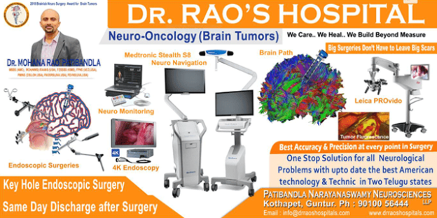
At Dr. Rao’s Hospital, one of the best neurology hospitals in Guntur, the endoscopic neurosurgery unit is equipped with state-of-the-art medical infrastructure and cutting-edge technology. Our medical head, Dr. Mohana Rao Patibandla, one of the best neurosurgeons in Guntur, holds years of experience in performing endoscopic neurosurgery in Guntur and had training from the USA. Dr. Rao’s hospital is known for the keyhole brain and spine surgeries.

Moreover, the hospital comprises a team of the best neurosurgeons and the best neurologists in Guntur. They are skilled in various advanced techniques for treating complex problems such as brain aneurysms, and spine and brain tumors that are difficult to reach with greater precision.

Conditions Treated Using minimally invasive neurosurgery Procedures

Minimally invasive neurosurgery can be used to treat a number of conditions affecting the brain and spine. These include:

-Brain tumors

-Meningiomas

-Pituitary tumors

-Trigeminal neuralgia

-Arteriovenous malformations (AVMs)

-Craniosynostosis

-Epidermoid cysts

-Hemangiomas

-Hydrocephalus

-Intracranial aneurysms

– Spine conditions

  • Degenerative disc disease
  • Herniated disc
  • Lumbar spinal stenosis
  • Spinal deformities such as scoliosis
  • Spinal infections
  • Spinal instability including spondylolisthesis
  • Vertebral compression fractures
  • Spinal tumors

How Minimally Invasive Surgery Works

Minimally invasive neurosurgery (MIS) is a type of surgery that uses smaller incisions and less tissue disruption than traditional open surgery. MIS techniques can be used to treat a variety of conditions affecting the brain and spine. MIS procedures are typically performed using an endoscope, which is a long, thin tube equipped with a light and camera. The endoscope is inserted through a small incision in the skull or spine, and the surgeon uses the camera to guide the endoscope to the site of the surgery. However, percutaneous (meaning “through the skin”) placement typically involves inserting rods and screws through relatively small skin incisions without cutting or dissecting the underlying muscle. With the aid of x-ray images, guidewires are placed through the skin and into the spinal vertebrae along the desired paths for the screws. Then, screws are placed over the guidewires and follow the path of the wires. These screws have temporary extenders that extend outside of the skin and are subsequently removed after helping to guide passage of rods to connect and secure the screws. With the use of spinal navigation and robots, spinal instrumentation is being placed more safely and accurately.

the best spine surgery hospital in Guntur, the best minimally invasive spine surgery hospital in Guntur
Minimally invasive spine surgery

During MIS surgery, the surgeon will make small incisions in the skin and muscle tissue to access the bones of the skull or spine. Once the endoscope is in place, the surgeon will use special instruments to remove or repair damaged tissue. MIS surgery is less invasive than traditional open surgery, and as a result, patients typically experience less pain and scarring. In addition, MIS surgery can often be performed on an outpatient basis, which means that patients can go home the same day as their procedure.

Common minimally invasive neurosurgery Surgery Treatment Options

There are many different types of minimally invasive neurosurgery procedures, each of which is designed to treat a specific condition. Some of the most common minimally invasive neurosurgery procedures include:

• Spinal cord stimulation: This procedure involves implanting a small device near the spinal cord that sends electrical impulses to the nerves in order to relieve pain.

• Deep brain stimulation: This procedure involves implanting a small device in the brain that sends electrical impulses to specific areas in order to relieve symptoms of conditions like Parkinson’s disease or dystonia.

• Intrathecal drug delivery: This procedure involves implantation of a small pump in the lower back that delivers medication directly to the spinal fluid. This is often used to treat pain or spasticity that is not responding to other forms of treatment.

• Peripheral nerve stimulation: This procedure involves implanting a small device near a peripheral nerve that sends electrical impulses to the nerve in order to relieve pain.

A number of specific techniques have been deployed for MIS surgery. Though the field continues to develop, the list below highlights some of the most common options.

Discectomy: Spinal discs are essentially elastic rings with soft material inside that serve as cushions between the vertebral bones. If the elastic ring becomes weakened, the soft tissue inside can extrude — or herniate — outside of the elastic ring. The herniated disc material can compress the nerves passing by, thus causing pain. If surgical treatment is recommended to trim or remove the herniated disc, it may be possible to perform this procedure with MIS surgery using tubular dilators and a microscope or endoscope.

Spinal decompression:Spinal stenosis, which is a narrowing of the vertebral canal, is a common condition that can result in compression of the nerves. This can produce a variety of symptoms, including pain, numbness and muscle weakness. If surgery is recommended, it may be possible to remove the bone and soft tissues causing the nerve compression through an MIS approach using tubular dilators and a microscope or endoscope. The more common decompressive procedures include laminectomy and foraminotomy.

Transforaminal Lumbar Interbody Fusion (TLIF): This is a MIS technique that is performed for patients with refractory mechanical low back and radicular pain associated with spondylolisthesis, degenerative disc disease and recurrent disc herniation. The procedure is performed from the back (posterior) with the patient on his or her stomach. Utilizing two small incisions, screws and rods are placed between two or more vertebral levels. The intervertebral disc is removed and a cage filled with bone is placed in that void with the goal of stabilizing the levels affected.

  • Minimally Invasive Lateral Interbody Fusion
  • Minimally Invasive Posterior Lumbar Interbody Fusion (PLIF)
  • Minimally Invasive Transforaminal Lumbar Interbody Fusion (TLIF)
  • Minimally Invasive Posterior Thoracic Fusion

Candidates for minimally invasive neurosurgery Surgery

Candidates for minimally invasive neurosurgery surgery are typically those who have conditions that require surgery but are not well suited for traditional, open surgery. These conditions can include tumors, aneurysms, and other conditions that require access to the brain or spine. In order to be a candidate for minimally invasive neurosurgery, you must first be evaluated by a neurosurgeon. During this evaluation, the neurosurgeon will determine if your condition is appropriate for this type of surgery. They will also take into account your overall health and any other factors that may impact your ability to tolerate the surgery.

minimally invasive neurosurgery Device Technology

One of the most important aspects of minimally invasive neurosurgery is the technology used to perform the procedures. This technology has evolved significantly over the past few years and has made these procedures much safer and more effective. Some of the most common devices used in minimally invasive neurosurgery include:

-Microscopes: These are used to provide the surgeon with a magnified view of the area being operated on.

-Endoscopes: These are long, thin tubes that are inserted into the body to allow the surgeon to see inside without making a large incision.

-Lasers: Lasers are often used to remove tissue or to cauterize blood vessels.

-Ultrasonic devices: These devices use sound waves to break up tissue so it can be removed more easily.

Conclusion

Minimally invasive neurosurgery is a type of surgery that uses small incisions and specialized instruments to treat conditions of the brain and spine. This type of surgery can be used to treat a variety of conditions, including tumors, herniated discs, and spinal stenosis. Minimally invasive neurosurgery is often used as an alternative to traditional open surgery, which can require large incisions and a longer recovery period. Candidates for minimally invasive neurosurgery include those who are seeking an alternative to traditional open surgery, have a condition that can be treated using minimally invasive techniques, and are in generally good health. The decision to undergo minimally invasive neurosurgery should be made in consultation with a qualified surgeon. Minimally invasive neurosurgery is a safe and effective treatment option for many conditions of the brain and spine. This type of surgery can offer patients a number of benefits, including a shorter recovery period, less pain, and a lower risk of complications. Taking every point into consideration, Dr. Rao is the best minimally invasive brain or spine surgeon in India as he has abroad experience, is a minimally invasive surgeon, fewer complication rate, compassionate to the patient, has the highest google rating for any neurosurgeon has in India, and is Best cutting edge technology at his Dr. Raos hospital/patibandla Narayana Swamy Neurosciences institute.

Top Ten Best Brain Tumor Neurosurgeons in India who are shaping Neurosurgery in India

Brain Tumors – All you need to know

Tumors may be benign or malignant and depend on their growth behavior. The brain is made up of neurons and supportive cells. Every cell has its particular function. Cells typically divide in a specific specified way and regenerate their successors. If a cell loses control over the division, the tumors will develop.

  • Benign brain tumors will not have cancer cells; the complete removal of the tumor will usually cease the tumor recurrence. They cause symptoms by pressing the surrounding structures.
  • Malignant brain tumors are aggressive and contain cancer cells. They are life-threatening. Malignant brain tumors grow rapidly and invade the tissue surrounding them. Malignant brain tumors may be contained or may be proliferating invasive. 

These brain tumors are graded as low grades (grades I and II) to high grades (grades III and IV). The grading is based on the microscopic cell pattern, corresponds to the malignancy potential, and grows faster. 

Brain Tumors Causes

The causes of brain tumors are unknown. If we know the brain tumor’s origin, targeted tumor therapies will be available. Brain tumors’ source and form in a person are mysterious but are not contagious.  

Risk factors for brain tumors
Brain tumor origin is unknown but some risk factors are known

Brain tumors commonly show bimodal age patterns involving children 3 to 12 years old and adults 40 to 70, but they can occur in any age group.

What are the Risk factors for developing brain tumors?

Oil refining, rubber manufacturing, and drug manufacturing

Chemists and embalmers 

Exposure to viruses is a possible cause. 

Heredity is a cause

In most cases no apparent risk factors. 

Tumors are the result of several factors acting together.

Types of Brain Tumors

Facts of the brain tumors - Dr Raos hospital

Brain tumors can be primary or secondary.

Primary Brain Tumors

Tumors originate within brain tissue. The type of cell origin categorizes primary brain tumors. The most prevalent brain tumors are gliomas arising from supportive tissue glial cells. Gliomas divided into

  • Astrocytomas arise from astrocytes. They may present in the brain or spinal cord. In children, they are often found in the brain stem, the cerebrum, and the cerebellum. In adults, they often arise in the cerebrum. A grade III astrocytoma is called anaplastic astrocytoma. A grade IV astrocytoma called GBM (glioblastoma multiforme).
  • Oligodendrogliomas arise from the cells that produce myelin, the fatty insulating covering that protects nerves. They commonly occur in the cerebrum. They grow slowly and are usually distinct from the brain tissue.
  • Ependymomas form from the lining of the ventricles. They occur in both the brain and the spinal cord. They occur commonly in childhood and adolescence.

Other tumors arise from the different tissues broadly:

  • Meningiomas grow from the coverings of the brain called meninges, usually benign. As these tumors grow slowly, the brain adapts to their presence so that meningiomas grow significantly when symptomatic. They occur primarily around 30 to 50 years of age and in women.
  • Schwannomas are benign tumors that arise from the counterparts of oligodendrogliomas in the peripheral nervous system, which produce the myelin that insulates peripheral nerves. Acoustic neuromas, V nerve, and XII nerve will commonly grow schwannoma, and spinal nerves will also have this kind of tumor, primarily in adults. This tumor harbors women twice as often as men.
  • Craniopharyngiomas develop from Rathke’s pouch remnants near the pituitary gland, usually benign; They are dangerous because they compress the hypothalamus. It follows a bimodal age pattern and occurs mainly in children and after 40 years.
  • Germ cell tumors arise from primitive totipotent cells or germ cells. Germinoma is a common type of germ cell tumor in the brain.
  • Pineal region tumors originate from the pineal gland. They are either benign, like pineocytoma, or malignant, like pineoblastoma.

Secondary Brain Tumors

Secondary brain tumors will have the source origin somewhere in the body. These are called metastasis. Based on the origin tissue, they will call it. For example, if lung cancer metastasizes to the brain, the tumor is called metastatic lung cancer. Treatment for these tumors depends on the systemic burden and other factors like general health, the patient’s age, and response to previous therapy.

Symptoms of Brain Tumor

Symptoms of brain tumor commonly involves the location and type of the brain tumor

The most frequent symptoms and signs of brain tumors include:

  • Confusion or personality changes,
  • Headaches that tend to be worse in the early morning and ease during the day and are associated with vomiting,
  • Nausea or vomiting,
  • Seizures (convulsions),
  • Stumbling in walking (ataxic gait),
  • Weakness in the arms or legs,
  • Changes in speech
  • Abnormal eye movements or changes in vision,
  • Drowsiness

Diagnosis of Brain Tumors

Diagnosis depends on clinical history and Imaging tests plus biopsy

Clinical history is essential as the symptoms sometimes guide us to the tissue of origin, like the brain stem, cerebrum, cerebellum, etc.

Based on the physical and neurologic examinations, we may ask for the following imaging tests:

  • CT scan – for bony details and screening 
  • MRI (magnetic resonance imaging) – for anatomical details

The doctor may also request other tests such as:

  • A skull x-ray – is rare nowadays.
  • An angiogram, or arteriogram, for highly vascular tumors.
  • Myelogram in those patients, we may not be able to do MRI or 3D purposes.
  •  

Collecting a biopsy tissue –  either through open surgery, minimally invasive surgery, or through the needle called a stereotactic biopsy.

  • A stereotactic needle biopsy is an image-guided surgery in which we remove the tissue from the deep-seated parts. We also use a brain path for the same and can remove the entire tumor minimally invasively. The biopsy collected was first seen under a microscope, then adding different markers to know the genetic mutations in cancer. Based on these sophisticated tests, you may get the tumor’s prognosis and the cancer response.

Treatment for brain tumors

Brain tumor can be removed using best imaging, navigation, best optics, best resection techniques and technology along with access retractors.
Key hole surgeries are the main surgeries all over the world nowadays and is now at Dr Raos hospitals

Treatment depends on the size, type, location, overall health of the patient, and preferences.

Brain Tumors Surgery:

Indications are brain tumors causing excessive swelling and are an imminent danger to the patient life

Biopsy

Accessible area

Reduce tumor burden

Best for adjuvant therapy like chemo or radiation therapy by reducing the tumor tissue

improve the quality of life along with 

Brain Tumors Radiation therapy

Side effects of radiation therapy depend on the dose and type of radiation received. Radiation therapy has several types – External beam radiation, Brachytherapy, and whole-brain radiation. External beam radiation therapy for benign tumors is around 54 Gy and malignant for tumors 60 Gy in 30 divided factors. Common side effects immediately following radiation include fatigue, headaches, vomiting, memory loss, scalp irritation, and hair loss.

Brain Tumors Stereotactic Radiosurgery

Stereotactic radiosurgery is also called knifeless surgery. Instead of a knife, it uses high-dose radiation to the confined area to control the tumor’s growth. You may get it from Gamma Knife or Linear accelerator, but the results are almost the same, with no difference. Radiosurgery is typically a daycare procedure.

Brain Tumors Chemotherapy: 

Chemotherapy uses drugs to kill tumor cells. Temozolomide is the commonly used drug either for concurrent or individual cycles. 

Chemotherapy can cause side effects depending on the type and dosage of medications. Chemotherapy will be given based on your biopsy results.

Brain Tumors Targeted drug therapy

Targeted drug treatments focus on specific genetic mutations present within cancer cells. By knocking these abnormalities, these targeted drugs can cause cancer cells to die.

Living with a Brain tumor

  • Physical therapy 
  • Speech therapy 
  • Occupational therapy 
  • Tutoring for school-age children 
the-best-cerebral-palsy-treatment-is-at-dr-raos-hospital

The best cerebral palsy treatment is at Dr Raos hospital


Introduction

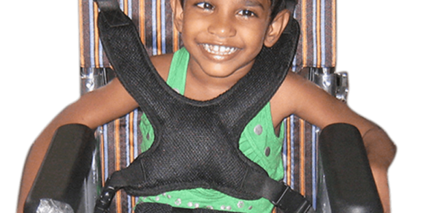
Cerebral palsy is a neurological disorder that affects muscle movement and coordination. It is caused by damage to the brain that occurs before, during, or after birth. Cerebral palsy can range from mild to severe, and it can lead to other health problems such as seizures, hearing loss, and vision problems. There is no cure for cerebral palsy, but there are treatments available to help improve muscle function and coordination. People with cerebral palsy can live long, healthy lives with the right support and care. Looking for the best cerebral palsy management or cerebral palsy treatment or cerebral palsy care in Guntur and Andhra Pradesh, look no further than Dr Raos hospital, the best neurology, best neurosurgery or best neurosurgery hospital in Guntur and Andhra Pradesh.


What is Cerebral Palsy?

Cerebral palsy is a neurological disorder that affects muscle movement and coordination. It is caused by damage to the brain that occurs before, during, or after birth. Cerebral palsy can range from mild to severe, and it can lead to other health problems such as seizures, hearing loss, and vision problems. There is no cure for cerebral palsy, but there are treatments available to help improve muscle function and coordination. People with cerebral palsy can live long, healthy lives with the right support and care.


What Causes Cerebral Palsy?

Cerebral palsy is caused by damage to the brain that occurs before, during, or after birth. The most common cause of cerebral palsy is damage to the brain that occurs during pregnancy. Other causes of cerebral palsy include damage to the brain that occurs during or after childbirth, and damage to the brain that occurs after birth.


What are the Symptoms of Cerebral Palsy?

The symptoms of cerebral palsy vary from person to person. Some common symptoms include muscle weakness, muscle stiffness, poor coordination, and tremors. Cerebral palsy can also lead to other health problems such as seizures, hearing loss, and vision problems.

What Diseases are Associated with Cerebral Palsy?

Cerebral palsy is often associated with other health problems such as seizures, hearing loss, and vision problems. Seizures are a common symptom of cerebral palsy, and they can be controlled with medication. Hearing loss and vision problems can also be caused by cerebral palsy, but they can be treated with surgery or other interventions.

How is Cerebral Palsy Diagnosed?

Cerebral palsy is usually diagnosed based on a medical history and a physical examination. A doctor will ask about the symptoms and when they started. A physical examination will be done to check for muscle weakness, muscle stiffness, and poor coordination. Imaging tests such as MRI or CT scans may also be done to check for damage to the brain.

What is the Treatment for Cerebral Palsy?

There is no cure for cerebral palsy, but there are treatments available to help improve muscle function and coordination. Physical therapy, occupational therapy, and speech therapy can all help people with cerebral palsy. Surgery may also be an option for some people with cerebral palsy. Medications can be used to control seizures and other symptoms.

How Can I Live with Cerebral Palsy?

People with cerebral palsy can live long, healthy lives with the right support and care. There are many resources available to help people with cerebral palsy and their families. Physical therapy, occupational therapy, and speech therapy can all help people with cerebral palsy. Surgery may also be an option for some people with cerebral palsy. Medications can be used to control seizures and other symptoms.

Conclusion

Cerebral palsy is a neurological disorder that affects muscle movement and coordination. It is caused by damage to the brain that occurs before, during, or after birth. Cerebral palsy can range from mild to severe, and it can lead to other health problems such as seizures, hearing loss, and vision problems. There is no cure for cerebral palsy, but there are treatments available to help improve muscle function and coordination. People with cerebral palsy can live long, healthy lives with the right support and care. Looking for the best cerebral palsy management or cerebral palsy treatment or cerebral palsy care in Guntur and Andhra Pradesh, look no further than Dr Raos hospital, the best neurology, best neurosurgery or best neurosurgery hospital in Guntur and Andhra Pradesh. Dr Rao is the best neurosurgeon or spine surgeon or neurologist in Guntur, Andhra Pradesh and India. Contact Dr Raos hospital @ 9010056444 or 9010057444 for further queries.

Dr. Mohana Rao Patibandla is one of the best neurosurgeons in Guntur, Andhra Pradesh, and India. He is an expert in brain tumors, epilepsy, spine surgery, minimally invasive spine surgery, keyhole brain surgery, aneurysm, stroke mechanical thrombectomy, and hemorrhage. He has over 25 years of experience in neurosurgery and has performed over 10,000 surgeries. He is also a member of the American Association of Neurological Surgeons and the World Federation of Neurological Surgeons.

dr-raos-hospital-is-the-best-for-epilepsy-surgery

Dr Raos hospital is the best for epilepsy Surgery

Welcome to Dr Raos hospital neuro department. Epilepsy is a neurological disorder that affects people of all ages. It is characterized by recurrent seizures that can vary in severity. Epilepsy can be caused by a variety of factors, including head injuries, brain tumors, stroke, and genetic predisposition. Although there is no definitive cure for all epilepsy diseases, it can be effectively managed with medication and other treatments like surgery. Dr Raos hospital is the best in India for the epilepsy diagnosis or medical or surgical treatment. They have the most experienced staff and the latest technology. This makes them the perfect choice for anyone looking for the best possible care.

National epilepsy month is observed in November in many countries around the world. This month is dedicated to raising awareness about epilepsy and its impact on people’s lives. National epilepsy day is celebrated on November 17th in India. This day is observed to create awareness about the condition and to dispel common myths and misconceptions about it.

There are many myths and misconceptions about epilepsy. One common myth is that people with epilepsy are possessed by demons or evil spirits. This is not true. Epilepsy is a medical condition that has nothing to do with demonic possession. Another myth is that people with epilepsy are not intelligent. This is also not true. Epilepsy does not affect a person’s intelligence. In fact, many people with epilepsy are highly intelligent and successful.

benzinga news - Dr Mohana Rao Patibandla, a 2022 ThreeBestRated Award-winning neurosurgeon in Guntur, Andhra Pradesh, Shares simple selfcare practices for epilepsy patients
benzinga news – Dr Mohana Rao Patibandla, a 2022 ThreeBestRated Award-winning neurosurgeon in Guntur, Andhra Pradesh, Shares simple selfcare practices for epilepsy patients

Risk Factors

There are a variety of factors that can increase the risk of developing epilepsy, including:

· Family history: Having a family member with epilepsy increases the risk of developing the condition.

· Head injuries: Head injuries, especially those that result in a concussion, can increase the risk of developing epilepsy.

· Brain tumors: Brain tumors can cause seizures and increase the risk of developing epilepsy.

· Stroke: Stroke can damage the brain and lead to epilepsy.

· Infections: Infections of the brain, such as meningitis, can increase the risk of developing epilepsy.

Symptoms

The most common symptom of epilepsy is seizures. Seizures can vary in severity from brief periods of unconsciousness to convulsions and loss of muscle control. Other symptoms of epilepsy may include:

· Auras: A feeling of déjà vu or an intense sense of fear or foreboding

· Alterations in behavior

· Loss of consciousness

· Confusion

· Memory loss

· Staring spells

Diagnosis

Epilepsy is typically diagnosed after a person has had two or more seizures that were not caused by a known medical condition. A variety of tests may be used to diagnose epilepsy, including:

· Neurological exam: A physical and neurological examination will be conducted to look for signs of epilepsy.

· Blood tests: Blood tests may be ordered to look for signs of infection or other conditions that could be causing the seizures.

· Imaging tests: An MRI or CT scan may be ordered to look for abnormalities in the brain that could be causing the seizures.

· EEG: An EEG may be ordered to record the electrical activity of the brain. This can help to diagnose epilepsy or rule out other conditions.

Treatment:

Treatment for epilepsy can vary depending on the individual’s needs. Medication is the most common form of treatment. Surgery may also be an option for some people with epilepsy especially for those having resistant epilepsy. The goal of treatment is to control seizures and improve the person’s quality of life.

Conclusion:

Epilepsy is a serious condition that should not be taken lightly. Epilepsy is a neurological disorder that affects the nervous system and is characterized by recurrent seizures. Epilepsy can be caused by a variety of factors and there is cure for the condition in some of the diseases. However, it can be managed with medication and lifestyle changes. However, with proper treatment, people with epilepsy can lead happy and fulfilling lives. Dr Raos hospital is the best in India for the epilepsy diagnosis or medical or surgical treatment. They have the most experienced staff and the latest technology. This makes them the perfect choice for anyone looking for the best possible care.

say_its_a_stroke dr-raos-hospital-is-best-in-stroke-awareness-in-guntur

The best Awake neurosurgery hospital in Guntur Dr. Raos

The best Awake neurosurgery hospital in Guntur, Dr. Raos

Dr. Raos hospital routinely uses local, regional and spinal anesthesia when performing most of the minimally invasive brain and spinal surgeries. There will be less post-operative pain and a shorter duration of surgery with the awake local and spinal anesthesia procedures compared to general anesthesia. 

“This is a real swift in neurosurgery following awake surgeries,” says Dr. Rao, a renowned neurosurgeon from Guntur in India. “Our studies and experience show that spinal and regional anesthesia are safe and offer real patient benefits.”

Dr. Raos Hospital’s regional anesthesia and spinal anesthesia protocols are used for selected individuals undergoing minimally invasive spinal surgeries like laminectomy, discectomy, or transforaminal lumbar interbody fusion (MIS-TLIF). These procedures are completed within 2 to 3 hours, the typical duration of spinal anesthesia or regional anesthesia.

There have been many myths about awake surgery regarding pain and not being able to stay still for the whole time, but those are not true in selected patients. 

Documenting the benefits for patients with awake surgery

General anesthesia has several disadvantages for individuals undergoing brain or spinal surgery, including nausea, vomiting, memory loss, and temporary impairment of motor and sensory functions. “General anesthesia might affect cognition in older individuals, which is one of the main concerns,” Dr. Rao says.

The specific advantages of regional or spinal anesthesia include the following:

· total operating room time will be decreased by 25%

· Lower overall and maximum pain in the first six hours after surgery

· Ambulation on an average 8 hours earlier than general anesthesia

· Shorter hospital stays of at least one day

Awake brain and spinal surgery is a multidisciplinary effort. “It requires mutual understanding of neurosurgeons and anesthesiologists who are experts in respective fields and should work together,” Dr. Rao says.

The minimally invasive spine surgeries are performed with patients in either a supine or prone position. Dr. Raos Hospital avoids spinal anesthesia for patients with a BMI over 35 or with obstructive sleep apnea due to carbon dioxide retention in the body.

Generally, we use awake brain surgery in tumor cases, DBS, and all kinds of minimally invasive brain surgeries on cooperative patients. The procedure we employ in the spine in almost all short, minimally invasive spine surgeries. We used motorized screws to enhance the speed of the procedure.

During all minimally invasive brain and spinal surgeries, patients wear listening devices like earphones and listen to their favorite music. “we are in constant communication with the patient, and we make sure that they are comfortable and doing well throughout the procedure,” Dr. Rao says.

“The dominance of general anesthesia is mostly due to a lack of exposure to regional scalp block and spinal anesthesia,” Dr. Rao says. “At Dr. Raos hospital, we are committed to innovating our approaches in ways that help patients; we always strive to get the best to our patients.”

If you are looking for awake neurosurgery with fewer disadvantages for your minimally invasive brain or spine surgery, please consult Dr Raos Hospital, the best neurosurgery hospital in India with the best minimally invasive keyhole neurosurgeon. Contact us @ 9010056444 or 9010057444