Tag Archives: Minimally Invasive Spine Surgery

The advancements in the best Brain tumor surgery

The advancements in the best Brain tumor surgery


Discover the advancements in brain surgery made possible by cutting-edge technologies at Dr. Rao’s Hospital, the best neurosurgery hospital in India. Dr. Rao, the best neurosurgeon in India, utilizes innovative techniques to provide exceptional care and improve patient outcomes.

In neurosurgery, technological advancements have revolutionized brain surgeries, leading to improved outcomes and enhanced patient care. At Dr. Rao’s Hospital, renowned as the best neurosurgery hospital in India, Dr. Rao, the best neurosurgeon in India, spearheads the utilization of cutting-edge technologies to deliver exceptional neurosurgical care.

The Evolving Landscape of Brain Surgery:

Brain surgery is a complex and delicate procedure that requires precision, skill, and advanced techniques. Over the years, neurosurgical innovations have transformed the field, enabling surgeons to approach brain surgeries with greater accuracy, minimal invasiveness, and improved patient safety. These innovations have revolutionized how brain tumors are removed, vascular abnormalities are treated, and neurological conditions are managed.

Dr. Rao: Pioneering Neurosurgeon in India:

Dr. Rao is an eminent neurosurgeon who stands at the forefront of neurosurgical advancements in India. With a wealth of experience and expertise, Dr. Rao has earned a reputation as the best neurosurgeon in India. He continually pushes the boundaries of neurosurgery, embracing the latest technologies and techniques to deliver the highest standard of care to his patients.

Cutting-Edge Technologies in Brain Surgery:

Dr. Rao’s Hospital is equipped with state-of-the-art technologies that have revolutionized the field of neurosurgery. These innovative tools and techniques enhance surgical precision, minimize invasiveness, and improve patient outcomes. Let’s explore some of the cutting-edge technologies utilized at Dr. Rao’s Hospital:

Neuro-navigation Systems:

These computer-assisted systems integrate preoperative imaging data with real-time intraoperative images, enabling surgeons to navigate the brain’s intricate anatomy precisely. They help guide surgeons during surgery, ensuring optimal outcomes while minimizing damage to healthy brain tissue.

Minimally Invasive Techniques:

Minimally invasive approaches, such as keyhole craniotomies and endoscopic procedures, involve smaller incisions, reduced tissue disruption, and faster recovery times. These techniques offer significant benefits, including shorter hospital stays, decreased postoperative pain, and a minimized risk of complications.

Intraoperative Imaging:

Advanced imaging technologies, such as intraoperative MRI (fMRI) and intraoperative CT (iCT), provide real-time visualization of the brain during surgery. This allows surgeons to assess the procedure’s progress and make immediate adjustments, improving surgical precision.

Stereotactic Radiosurgery:

Stereotactic radiosurgery delivers high-dose radiation precisely to the target area within the brain without requiring surgical incisions. It treats brain tumors, arteriovenous malformations (AVMs), and other neurological conditions, offering a non-invasive alternative to traditional surgery.

Neurostimulation:

Deep brain stimulation (DBS) and transcranial magnetic stimulation (TMS) are neuromodulation techniques used to treat neurological conditions such as Parkinson’s, epilepsy, and depression. These procedures involve the implantation or application of devices that deliver electrical or magnetic stimulation to specific brain areas, regulating abnormal neural activity.

Robotics-Assisted Surgery:

Robotics-assisted neurosurgery combines the precision of robotic systems with the surgeon’s skills. Robotic assistance provides enhanced dexterity, stability, and range of motion, enabling surgeons to perform complex procedures with improved accuracy and efficiency.

Advanced Imaging Techniques:

High-resolution MRI and diffusion tensor imaging (DTI) allow for detailed visualization of brain structures and pathways. These advanced imaging techniques aid in surgical planning, allowing surgeons to navigate critical areas and avoid damage to vital structures during brain surgery.

Intraoperative Monitoring:

Intraoperative monitoring techniques, such as electroencephalography (EEG), motor evoked potentials (MEPs), and somatosensory evoked potentials (SSEPs), assess brain function in real-time during surgery. This helps surgeons preserve vital functions, monitor neural activity, and minimize the risk of complications.

These cutting-edge technologies have significantly advanced the field of neurosurgery, improving patient outcomes, reducing risks, and expanding the possibilities for treating complex brain conditions. Dr. Rao and his team at Dr. Rao’s Hospital stay at the forefront of these innovations, incorporating them into their practice to provide the best possible care for their patients.

The Best Neurosurgery Hospital in India: Dr. Rao’s Hospital:

Dr. Rao’s Hospital is a beacon of excellence in neurosurgery in India. The hospital’s commitment to providing the highest level of care is reflected in its state-of-the-art infrastructure, advanced technologies, and a team of highly skilled healthcare professionals. Dr. Rao’s expertise, combined with the cutting-edge resources available at the hospital, ensures that patients receive world-class neurosurgical care.


Hashtags: #NeurosurgicalInnovations #CuttingEdgeTechnologies #BrainSurgery #BestNeurosurgeon #DrRao #NeurosurgeryHospital #DrRaosHospital

Neurosurgery in the Elderly: Addressing Unique Challenges and Optimizing Outcomes

Neurosurgery in the Elderly: Addressing Unique Challenges and Optimizing Outcomes

Discover the specialized care that Dr. Rao, the best functional neurosurgeon, and his staff offer at Dr. Rao’s Hospital, the top functional neurosurgery hospital in India. Learn how neurosurgery addresses the unique challenges faced by elderly patients and optimizes outcomes through personalized treatment plans, minimally invasive techniques, and comprehensive rehabilitation.

Our bodies undergo natural changes as we age, and the same applies to the brain and nervous system. Neurological conditions and disorders become more prevalent among the elderly, requiring specialized care and expertise. In this context, neurosurgery addresses older patients’ unique challenges and optimizes their outcomes. At Dr. Rao’s Hospital, recognized as the best functional neurosurgery hospital in India, Dr. Rao, the renowned and highly skilled functional neurosurgeon, provides comprehensive and compassionate care to elderly patients.

Unique Challenges in Neurosurgery for the Elderly:

Age-Related Factors:

Aging brings about physiological changes that impact surgical outcomes. The elderly often face compromised healing abilities, decreased immune function, and increased infection vulnerability. These factors need to be carefully considered when planning and performing neurosurgical interventions.

Comorbidities:

Older patients often have preexisting medical conditions like cardiovascular diseases, diabetes, and hypertension. These comorbidities can affect surgical risks, anesthesia management, and postoperative recovery. Dr. Rao and his team conduct thorough preoperative assessments to evaluate patients’ overall health and develop individualized treatment plans.

Frailty and Functional Decline:

Elderly individuals may experience frailty and a decline in functional status, impacting their ability to tolerate surgery and recover. Dr. Rao and the healthcare team at Dr. Rao’s Hospital implement multidisciplinary approaches to optimize perioperative care, including physical therapy, nutrition counseling, and rehabilitation services.

Optimizing Outcomes in Neurosurgery for the Elderly:

Comprehensive Evaluation

Dr. Rao and his team employ a comprehensive evaluation process to assess the elderly patient’s neurological condition, overall health, and functional status. This helps determine the most suitable treatment options and sets realistic expectations for the patient and their family.

Minimally Invasive Techniques

Whenever possible, minimally invasive neurosurgical approaches are preferred for elderly patients. These techniques involve smaller incisions, reduced tissue trauma, and faster recovery times, promoting improved outcomes and minimizing surgical risks.

Advanced Anesthesia Management

Anesthesia management is carefully tailored to the elderly patient’s needs, considering their age, comorbidities, and medication use. Dr. Rao collaborates closely with experienced anesthesiologists to ensure patient safety and comfort throughout the surgical procedure.

Geriatric-Focused Care:

The geriatric-focused care approach at Dr. Rao’s Hospital emphasizes individualized care plans, considering older patients’ unique needs and challenges. This may include specialized nursing care, medication management, and maintaining cognitive and functional abilities.

Rehabilitation and Support:

Postoperative rehabilitation plays a crucial role in optimizing outcomes for elderly patients. Dr. Rao’s team works closely with rehabilitation specialists to develop personalized rehabilitation programs that help regain mobility, function, and independence.

Dr. Rao’s Expertise in Functional Neurosurgery:

Dr. Rao, the best functional neurosurgeon in India, brings exceptional expertise and experience to neurosurgery for the elderly. His extensive knowledge of the aging brain and its specific requirements enables him to tailor treatment plans that address the unique needs and challenges older patients face. Dr. Rao’s dedication to patient-centered care, surgical precision, and innovation commitment ensure the best possible outcomes for elderly patients undergoing functional neurosurgical procedures.

Conclusion

In conclusion, neurosurgery for the elderly requires a specialized and multidisciplinary approach to address unique challenges and optimize outcomes. At Dr. Rao’s Hospital, the best functional neurosurgery hospital in India, Dr. Rao and his team provide comprehensive care to elderly patients, considering their physical and cognitive needs. With a focus on personalized treatment plans, minimally invasive techniques, advanced anesthesia management, and comprehensive rehabilitation, Dr. Rao and his team aim to restore neurological function, improve quality of life, and enhance independence in elderly patients.

A patient-centric approach is paramount at Dr. Rao’s Hospital, the best functional neurosurgery hospital in India. The hospital has a state-of-the-art infrastructure, cutting-edge technology, and a dedicated team of healthcare professionals specializing in geriatric neurosurgery. From the initial evaluation to the postoperative care and rehabilitation process, every step is carefully tailored to meet the unique needs of elderly patients.

The benefits of seeking neurosurgical care from Dr. Rao and his team at Dr. Rao’s Hospital extend beyond the expertise and advanced techniques. The compassionate and holistic approach to patient care ensures that elderly individuals receive the best surgical treatment and the necessary support and guidance throughout their journey.

In conclusion, neurosurgery for the elderly requires a specialized and comprehensive approach that considers the unique challenges and considerations associated with aging. At Dr. Rao’s Hospital, the best functional neurosurgery hospital in India, Dr. Rao, the leading functional neurosurgeon, combines his expertise, innovation, and patient-centered care to optimize outcomes for elderly patients. By addressing the specific needs of older individuals, providing minimally invasive techniques, and focusing on rehabilitation and support, Dr. Rao and his team are dedicated to restoring function, enhancing quality of life, and empowering elderly patients in their neurosurgical journey.


Deep Brain Stimulation: Revolutionizing the Treatment of Movement Disorders

Deep Brain Stimulation: Revolutionizing the Treatment of Movement Disorders

Discover the groundbreaking treatment of deep brain stimulation for movement disorders and the expertise of Dr. Rao, the best functional neurosurgeon, at Dr. Rao’s Hospital, the top neurosurgery hospital. Learn how DBS offers remarkable outcomes, improves quality of life, and restores independence for patients worldwide.

Movement Disorders

Movement disorders such as Parkinson’s disease, essential tremor, and dystonia can significantly impact a person’s quality of life. While medication and therapy are often the first line of treatment, some patients may experience limited benefits or intolerable side effects. In such cases, deep brain stimulation (DBS) has emerged as a revolutionary approach in neurosurgery, providing remarkable outcomes and improving patients’ lives worldwide.

Dr. Rao, the best functional neurosurgeon in the field

Dr. Rao, the best functional neurosurgeon in the field, is at the forefront of this groundbreaking treatment. With his exceptional expertise and innovative techniques, he has transformed the lives of individuals suffering from movement disorders. Dr. Rao’s Hospital, recognized as the best neurosurgery hospital, is at the forefront of deep brain stimulation, offering comprehensive care to patients seeking relief from the debilitating symptoms of movement disorders.

Deep brain stimulation

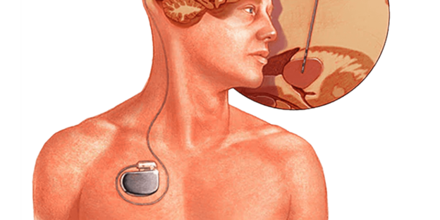
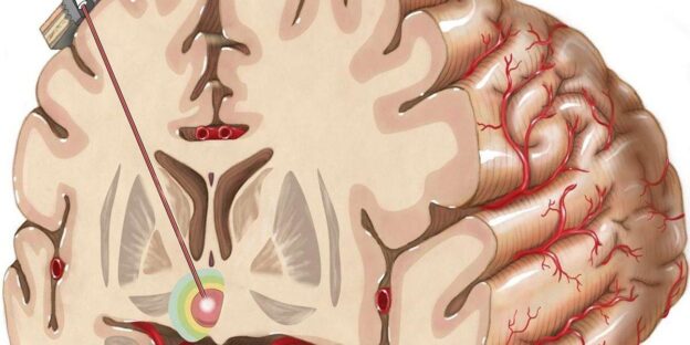
Deep brain stimulation involves the implantation of a small device, similar to a pacemaker, in the brain. This device delivers electrical impulses to specific areas, modulating abnormal neural activity and restoring normal brain function. The procedure is performed under the guidance of advanced imaging technology, ensuring precise electrode placement.

One of the critical advantages of deep brain stimulation is its adjustable nature. Following the surgery, the device can be fine-tuned and customized to each patient’s needs. This personalized approach allows for optimal symptom control and minimizes side effects.

The transformative impact of deep brain stimulation extends beyond symptom management. Patients often experience a significant improvement in motor function, reduced medication requirements, enhanced quality of life, and increased independence. Regaining control over daily activities and participating in social interactions provides newfound hope and freedom for individuals with movement disorders.

Dr. Rao the best Neurosurgeon in INDIA

Being the best functional neurosurgeon, Dr. Rao emphasizes a multidisciplinary approach to deep brain stimulation. Collaboration with neurologists, neuropsychologists, and physical therapists ensures comprehensive preoperative evaluation, precise electrode placement, and diligent postoperative care. This team approach contributes to the successful outcomes and long-term management of patients undergoing DBS.

Dr. Rao’s Hospital is the best neurosurgery hospital in INDIA

As the best neurosurgery hospital, Dr. Rao’s Hospital is equipped with cutting-edge technology, state-of-the-art operating theaters, and a compassionate team of healthcare professionals. The hospital provides a supportive and patient-centered environment, ensuring that each individual receives the highest standard of care throughout their journey.

Conclusion

In conclusion, deep brain stimulation has revolutionized the treatment of movement disorders, offering hope for patients who have exhausted traditional therapies. With the expertise of Dr. Rao, the best functional neurosurgeon, and the advanced facilities at Dr. Rao’s Hospital, patients can access world-class care and experience the transformative benefits of deep brain stimulation. The future of neurosurgery shines bright as this innovative treatment continues to enhance the lives of individuals with movement disorders.


Combating Sepsis on World Sepsis Day: Dr. Rao’s Hospital Leading the Way

Combating Sepsis on World Sepsis Day: Dr. Rao’s Hospital Leading the Way


Introduction

Every year, on September 13th, World Sepsis Day is a global reminder of the critical importance of recognizing and combating sepsis, a life-threatening condition affecting millions worldwide. Dr. Rao’s Hospital, renowned for its commitment to excellence in healthcare, is at the forefront of sepsis prevention and management. On this World Sepsis Day, let’s delve into what sepsis is, how it’s diagnosed, and how Dr. Rao’s Hospital’s superior care is making a difference.

Understanding Sepsis

A severe medical condition called sepsis, also known as septicemia, results from the body’s reaction to an infection. While the infection can start anywhere in the body, it often originates in the lungs, urinary tract, abdomen, or skin. What makes sepsis so perilous is its potential to cause widespread inflammation.

Etiology of Sepsis

The primary cause of sepsis is a bacterial, viral, or fungal infection. Common infections that can lead to sepsis include pneumonia, urinary tract infections, and skin infections. The invading pathogens release toxins into the bloodstream, triggering an alarmingly aggressive immune response.

Recognizing the Symptoms

Sepsis can manifest with various symptoms, including fever, increased heart rate, rapid breathing, confusion, extreme fatigue, and organ dysfunction. Early recognition is vital, as prompt treatment significantly improves outcomes.

Diagnosis and Assessment

Diagnosing sepsis involves a thorough clinical evaluation, blood tests, and imaging studies. The Sequential Organ Failure Assessment (SOFA) score helps gauge the severity of organ dysfunction. Timely diagnosis is a hallmark of Dr. Rao’s Hospital’s approach to sepsis management.

Managing Sepsis

Sepsis management demands a multidisciplinary approach. Dr. Rao’s Hospital excels in offering the following components of care:

  1. Source Control: Identifying and treating the source of infection, often through surgery or drainage, is crucial.
  2. Antibiotics and Antifungals: Administering appropriate medications to target the causative pathogens.
  3. Supportive Care: Respiratory support, fluid resuscitation, and medications to stabilize blood pressure.
  4. Nutritional Support: Ensuring patients receive adequate nutrition to support their immune system.
  5. Early Rehabilitation: Initiating physical therapy and rehabilitation as soon as possible for a smoother recovery.

Prognosis

Early intervention is pivotal for a favorable prognosis in sepsis cases. Dr. Rao’s Hospital’s commitment to swift and effective treatment significantly improves survival rates and reduces long-term complications.

Why Dr. Rao’s Hospital Shines in Sepsis Care

  1. Expertise: Dr. Rao’s Hospital boasts a team of highly skilled and experienced physicians, nurses, and specialists dedicated to sepsis management.
  2. Advanced Technology: The hospital is equipped with state-of-the-art diagnostic and therapeutic tools to aid in rapidly diagnosing and treating sepsis.
  3. Timely Care: Recognizing the urgency of sepsis, Dr. Rao’s Hospital prioritizes rapid intervention to halt its progression.
  4. Patient-Centered Approach: Dr. Rao’s Hospital understands the emotional and physical toll sepsis can take. Their patient-centered care focuses on both treatment and patient well-being.

Conclusion

On this World Sepsis Day, we’re reminded of the importance of sepsis awareness, early detection, and effective treatment. Dr. Rao’s Hospital’s unwavering commitment to sepsis care saves lives and improves outcomes. Sepsis may be a global health challenge, but with dedicated institutions like Dr. Rao’s Hospital leading the way, hope for a brighter, healthier future exists.

The Best Neurosurgery Advances in Trauma Care: Improving Outcomes for Critical Brain and Spinal Injuries.

The Best Neurosurgery Advances in Trauma Care: Improving Outcomes for Critical Brain and Spinal Injuries.

“Discover how Dr. Rao’s Hospital in Guntur, India, utilizes cutting-edge neurosurgical techniques to improve outcomes for critical brain and spinal injuries. Explore the advancements in trauma care and the expertise of Dr. Rao, the best neurosurgeon in India.”

Introduction

Traumatic brain and spinal injuries can have devastating consequences, impacting a person’s physical, cognitive, and emotional well-being. In recent years, significant advancements in neurosurgical techniques and technologies have revolutionized the management of trauma cases. Dr. Rao’s Hospital in Guntur, India, stands at the forefront of neurotrauma care, offering patients state-of-the-art treatments and comprehensive care. Led by Dr. Rao, the best neurosurgeon in India, the hospital combines expertise, cutting-edge technology, and a multidisciplinary approach to achieve optimal outcomes for individuals with critical brain and spinal injuries.

Understanding Traumatic Brain and Spinal Injuries

Traumatic brain injuries (TBIs) occur when external forces cause damage to the brain tissue. They can result from various incidents, such as falls, motor vehicle accidents, or sports-related accidents. On the other hand, spinal injuries involve damage to the spinal cord, leading to impaired motor and sensory function. Both conditions require prompt and specialized neurosurgical intervention to prevent further damage and enhance the chances of recovery.

Neurosurgical Advances in Trauma Care

Dr. Rao’s Hospital, renowned for its expertise in neurotrauma care, utilizes cutting-edge technologies and techniques to provide the best possible treatment for patients with critical brain and spinal injuries. These advancements have significantly improved outcomes, including:

Minimally invasive neurosurgery

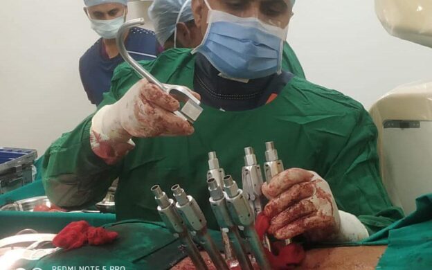
Minimally invasive neurosurgery has revolutionized the field of neurotrauma by offering a less invasive approach to treating critical brain and spinal injuries. This advanced surgical technique utilizes smaller incisions, specialized instruments, and advanced imaging technology to minimize tissue damage, reduce complications, and expedite patient recovery. In neurotrauma cases, minimally invasive neurosurgery significantly addresses traumatic brain and spinal injuries while minimizing the disruption of healthy tissues. This approach offers several advantages over traditional open surgery, including reduced postoperative pain, shorter hospital stays, faster recovery times, and improved cosmetic outcomes. One of the critical aspects of minimally invasive neurosurgery in neurotrauma is the use of advanced imaging techniques such as CT scans, MRIs, or fluoroscopy. These imaging modalities provide real-time visualization of the injured area, allowing the surgeon to locate the trauma and plan the surgical intervention precisely. This imaging guidance ensures accurate placement of surgical instruments and minimizes the risk of damaging healthy surrounding structures. During the surgical procedure, specialized instruments are utilized to access the injured site through small incisions. These instruments may include endoscopes, microscopes, or robotic-assisted tools, enabling surgeons to navigate delicate structures with enhanced precision. Minimally invasive techniques can be employed for a range of neurotrauma procedures, including hematoma evacuation, brain or spinal cord injury decompression, and the repair of skull fractures. The benefits of minimally invasive neurosurgery in neurotrauma are significant. Minimizing tissue disruption and trauma reduces the risk of complications such as infections, excessive bleeding, and postoperative scarring. Patients experience less pain and discomfort, resulting in faster recovery and earlier return to normal activities. Dr. Rao’s Hospital, renowned as the best neurosurgery center in India, is at the forefront of performing minimally invasive neurosurgical procedures for neurotrauma cases. The hospital uses cutting-edge methods and cutting-edge technology to give patients with severe brain and spinal injuries the best care possible under the direction of Dr. Rao, the top neurosurgeon in India. In conclusion, minimally invasive neurosurgery has transformed the field of neurotrauma by offering a less invasive and highly effective approach to treating critical brain and spinal injuries. Through advanced imaging guidance and specialized instruments, this technique reduces tissue damage, improves patient outcomes, and accelerates recovery. With its expertise in minimally invasive neurosurgery, Dr. Rao’s Hospital ensures that patients receive the best possible care, making it a premier destination for individuals seeking advanced neurotrauma treatment.

Image-guided surgery

Image-guided surgery has revolutionized the field of neurotrauma by providing neurosurgeons with real-time, precise navigation during surgical procedures. This advanced technology combines the power of imaging techniques with surgical instruments, enabling surgeons to target and treat traumatic brain and spinal injuries accurately. In neurotrauma cases, image-guided surgery allows for precise localization and visualization of the injury site, helping the surgeon plan and execute surgical interventions with enhanced accuracy. The process typically involves three key components: imaging, registration, and navigation. Imaging plays a vital role in image-guided surgery for neurotrauma. Various imaging modalities, such as computed tomography (CT), magnetic resonance imaging (MRI), and angiography, capture detailed images of the injured area. These images provide valuable information about the trauma’s location, extent, and nature, helping the surgeon understand the injury better. The next step is registration, which involves aligning the patient’s preoperative images with their actual anatomy during surgery. This is achieved using fiducial markers or surface landmarks to establish a coordinate system. The registered images are then integrated into a computer workstation, allowing the surgeon to visualize the patient’s anatomy in real time during the procedure. Once the registration is complete, the navigation component comes into play. The surgeon uses a tracking system that monitors the position and movements of surgical instruments in the patient’s anatomy. This information is displayed on a navigation system, providing a virtual map of the patient’s anatomy and the precise location of the surgical instruments in real time. During neurotrauma surgeries, image-guided navigation assists the surgeon in various ways. It helps locate and access the injury site precisely, ensuring minimal damage to healthy surrounding tissues. The surgeon can visualize critical structures, such as blood vessels or eloquent brain areas, to avoid complications during the procedure. Furthermore, image-guided navigation enables the surgeon to accurately place implants or perform complex reconstructive procedures with improved precision. The benefits of image-guided surgery in neurotrauma are numerous. It enhances surgical accuracy, reduces the risk of complications, and improves patient outcomes. By allowing surgeons to navigate through intricate anatomical structures, image-guided surgery minimizes the invasiveness of procedures, resulting in shorter operating times, reduced blood loss, and a faster recovery. Dr. Rao’s Hospital, a leading neurosurgery center in India, embraces the use of image-guided surgery to manage neurotrauma cases. With advanced imaging technology, state-of-the-art navigation systems, and the expertise of Dr. Rao, the best neurosurgeon in India, the hospital offers cutting-edge solutions for patients with traumatic brain and spinal injuries. In conclusion, image-guided surgery has significantly advanced the field of neurotrauma by providing surgeons with real-time, accurate navigation during surgical procedures. It enhances surgical precision, reduces complications, and ultimately improves patient outcomes. With its commitment to incorporating innovative technologies, Dr. Rao’s Hospital ensures that patients receive the best neurosurgical care, making it a premier destination for individuals seeking effective treatment for neurotrauma.

Neurocritical Care

Neurocritical care plays a crucial role in managing trauma-related brain and spinal injuries. The neurocritical care team at Dr. Rao’s Hospital is dedicated to providing specialized care for patients with severe traumatic brain injury (TBI) and spinal cord injury (SCI). These injuries can have devastating consequences and require immediate and intensive attention. In the case of traumatic brain injury, neurocritical care involves closely monitoring the patient’s neurological status, intracranial pressure, and cerebral blood flow. Advanced monitoring techniques, such as intracranial pressure monitoring and continuous electroencephalography (EEG), help the medical team assess the patient’s condition and guide treatment decisions. The team also ensures optimal oxygenation and ventilation to support brain function. For spinal cord injuries, neurocritical care focuses on preventing further damage and promoting recovery. This includes maintaining spinal alignment, managing spinal cord edema, and preventing complications like pressure sores and infections. The team works closely with physical, occupational, and rehabilitation specialists to develop a comprehensive treatment plan that maximizes the patient’s functional recovery. Dr. Rao’s Hospital has state-of-the-art facilities and cutting-edge technologies to provide advanced neurocritical care. The hospital has dedicated neurointensive care units staffed by highly skilled physicians, nurses, and critical care specialists. This multidisciplinary team collaborates to deliver personalized care and optimize outcomes for patients with traumatic brain and spinal injuries. In addition to the immediate management of trauma-related injuries, neurocritical care also focuses on preventing secondary complications and promoting long-term recovery. The team monitors and manages factors such as infection, fluid balance, blood pressure, and nutrition to create an optimal healing environment for the injured brain and spinal cord. The neurocritical care team at Dr. Rao’s Hospital is committed to providing compassionate care and ensuring the best possible outcomes for patients with traumatic brain and spinal injuries. Their expertise in neurosurgical interventions and comprehensive critical care management helps optimize recovery and improve the quality of life for patients and their families. Dr. Rao’s Hospital is a leader in neurocritical care for trauma patients by integrating advanced neurosurgical techniques, cutting-edge technology, and specialized expertise. The hospital’s commitment to excellence and innovation makes it the top choice for individuals seeking the best neurotrauma care in India.

Rehabilitation and neurorehabilitation

Rehabilitation and neurorehabilitation play a crucial role in the comprehensive care and recovery of individuals who have experienced neurotrauma. These specialized programs aim to optimize physical, cognitive, and emotional functioning, promote independence, and enhance patients’ overall quality of life. Neurotrauma can result in various impairments and disabilities, including motor deficits, sensory loss, cognitive challenges, and emotional disturbances. Rehabilitation addresses these specific needs through a multidisciplinary approach involving a team of healthcare professionals, including physiatrists, physical therapists, occupational therapists, speech therapists, psychologists, and social workers. The rehabilitation process begins soon after the acute phase of neurotrauma, once the patient’s condition has stabilized. The specific rehabilitation plan is tailored to the individual’s unique needs and goals, considering the severity of the injury, the affected areas, and the patient’s overall health. The primary objectives of rehabilitation in neurotrauma include

Physical Rehabilitation

Physical therapy aims to improve mobility, strength, balance, and coordination. Therapists employ various techniques, exercises, and assistive devices to help patients regain motor skills, increase their range of motion, and restore functional independence.

Cognitive Rehabilitation

Neurotrauma can affect cognitive abilities, including memory, attention, problem-solving, and language. Cognitive rehabilitation programs use specialized techniques and exercises to improve cognitive function, enhance attention and memory skills, and promote overall cognitive well-being.

Speech and Language

For patients who experience difficulties in speech and language following neurotrauma, speech therapy focuses on improving communication skills, articulation, voice control, and swallowing abilities.

Psychosocial Support

Neurotrauma can significantly impact emotional well-being, leading to anxiety, depression, and other psychological challenges. Psychologists and social workers provide counseling, support, and coping strategies to help patients and their families navigate the emotional aspects of recovery.

Functional Independence Training

Rehabilitation programs also focus on improving daily living activities and functional independence. This includes self-care, mobility, and community integration training to help patients regain independence and reintegrate into their daily lives. Neurorehabilitation is a long-term process that requires patience, dedication, and consistent effort from both the healthcare team and the patient. Setting realistic goals and milestones is essential, as progress may vary for each individual. The duration and intensity of rehabilitation depend on the severity of the neurotrauma and the progress made during the recovery process. Dr. Rao’s Hospital, recognized as the best neurosurgery center in India, provides comprehensive rehabilitation and neurorehabilitation services for patients with neurotrauma. A group of skilled healthcare professionals working under Dr. Rao’s direction closely collaborate with patients to create individualized rehabilitation programs that maximize recovery and enhance functional outcomes. In conclusion, rehabilitation and neurorehabilitation are essential components of the comprehensive care and recovery process for individuals with neurotrauma. These programs address patients’ specific physical, cognitive, and emotional needs, promoting functional independence and improving overall quality of life. Dr. Rao’s Hospital, with its expertise in neurotrauma rehabilitation, offers comprehensive and individualized programs to help patients regain their optimal level of functioning and reintegrate into their daily lives with confidence and independence.

The Role of Dr. Rao, the Best Neurosurgeon in India

Dr. Rao, a renowned neurosurgeon with extensive experience in neurotrauma care, leads the team at Dr. Rao’s Hospital. His expertise in intricate neurosurgical procedures and his compassionate approach to patient care have earned him recognition as the best neurosurgeon in India. Dr. Rao‘s commitment to staying at the forefront of medical advancements allows him to offer patients the latest and most effective treatment options.

Conclusion

Neurosurgical advances in trauma care have transformed how critical brain and spinal injuries are managed, offering new hope to patients and their families. Dr. Rao’s Hospital, recognized as the best neurotrauma hospital in India, combines expertise, state-of-the-art technology, and a patient-centered approach to deliver exceptional care. Under the leadership of Dr. Rao, the hospital continues to push boundaries and improve outcomes for individuals with traumatic brain and spinal injuries.

If you or a loved one has experienced a critical brain or spinal injury, seeking immediate medical attention from a specialized neurosurgical center like Dr. Rao’s Hospital can make a significant difference in the recovery journey.



Deep Brain Stimulation for Epilepsy: A Promising Treatment Approach

Deep Brain Stimulation for Epilepsy: A Promising Treatment Approach

Discover how deep brain stimulation (DBS) revolutionizes epilepsy treatment, offering improved seizure control and enhanced quality of life. Dr. Rao’s Hospital, a leading neurosurgery center, provides cutting-edge DBS procedures for epilepsy patients.

Introduction: Epilepsy is a neurological disorder characterized by recurrent seizures that can significantly impact a person’s quality of life. While medication is the primary treatment option, some individuals may experience inadequate seizure control or intolerable side effects. Deep brain stimulation (DBS) has emerged as a promising therapeutic approach in such cases. Dr. Rao’s Hospital, known for its excellence in neurosurgery, offers cutting-edge DBS procedures to provide relief and improve the lives of epilepsy patients.

Understanding Deep Brain Stimulation:

Deep brain stimulation is a surgical procedure involving the implantation of electrodes in specific brain regions. These electrodes deliver controlled electrical impulses to modulate abnormal brain activity and reduce the frequency and severity of seizures. DBS targets the areas responsible for generating epileptic activity and regulating abnormal neural pathways.

The Benefits of Deep Brain Stimulation for Epilepsy:

  1. Seizure Control: DBS has shown significant efficacy in reducing seizure frequency and intensity in individuals with epilepsy who are unresponsive to medication. Precisely targeting brain regions involved in seizure generation allows personalized and effective seizure management.
  2. Improved Quality of Life: By providing better seizure control, DBS enhances individuals’ overall quality of life with epilepsy. It allows them to engage in daily activities, pursue education and employment, and enjoy social interactions with reduced fear of seizures and their associated limitations.
  3. Medication Reduction: Deep brain stimulation can decrease reliance on anti-seizure medications, often with side effects and long-term complications. By reducing medication dosages, patients may experience improved cognitive function and a lower risk of adverse drug reactions.

The Deep Brain Stimulation Procedure:

  1. Electrode Placement: During the DBS procedure, electrodes are surgically implanted into specific brain regions implicated in seizure activity. Advanced imaging techniques and neurophysiological mapping guide the precise placement of the electrodes to ensure optimal targeting.
  2. Neurostimulator Implantation: A neurostimulator, similar to a pacemaker, is implanted under the skin, typically in the chest or abdominal area. This device generates electrical impulses and is programmed to deliver the appropriate stimulation parameters tailored to the individual’s needs.
  3. Programming and Adjustment: Following the surgical implantation, the medical team programs the neurostimulator to deliver electrical impulses customized to the patient’s seizure patterns. Regular follow-up visits allow for fine-tuning and adjusting the stimulation parameters for optimal seizure control.

Collaborative Care Approach:

Dr. Rao’s Hospital adopts a collaborative approach to DBS for epilepsy, involving a multidisciplinary team of neurologists, neurosurgeons, neuropsychologists, and neurostimulation specialists. This collaborative effort ensures comprehensive evaluation, accurate patient selection, meticulous surgical planning, and ongoing post-operative care.

Ongoing Monitoring and Support: Patients who undergo DBS for epilepsy receive continuous monitoring and support from the medical team at Dr. Rao’s Hospital. Regular follow-up visits allow for assessment of seizure control, adjustment of stimulation parameters, medication management, and addressing any concerns or side effects that may arise.

Conclusion:

Deep brain stimulation has emerged as a promising treatment approach for individuals with epilepsy who have not achieved adequate seizure control with medications. By precisely targeting brain regions involved in seizure generation, DBS offers the potential for improved seizure management and enhanced quality of life. With its expertise in neurosurgery and advanced neurostimulation techniques, Dr. Rao’s Hospital is at the forefront of providing comprehensive care and innovative treatments for epilepsy patients.


Hashtags:

  1. #EpilepsyTreatment
  2. #DeepBrainStimulation
  3. #SeizureControl
  4. #NeurosurgeryInnovation
  5. #QualityofLife
  6. #DrRaosHospital
  7. #NeurologicalDisorders
  8. #InnovativeTreatments
  9. #EpilepsyAwareness
  10. #NeuroscienceAdvancements
The best treatment for the Lumbar Canal Stenosis

The best treatment for the Lumbar Canal Stenosis

Creating a Concept Map for Lumbar Canal Stenosis:

Context:

  • Dr. Rao’s Hospital, located in Guntur, Andhra Pradesh, India, is a renowned neurosurgery and spine care center.
  • Dr. Rao, a distinguished neurosurgeon, leads the hospital’s expert medical team.
  • Lumbar Canal Stenosis (LCS) is frequently treated at Dr. Rao’s Hospital due to its expertise in spine-related disorders.
  • LCS is a common spinal condition, often affecting older adults and significantly impacting patients’ quality of life.

Disease Overview:

  • Definition: Lumbar Canal Stenosis refers to the narrowing of the spinal canal in the lumbar (lower back) region, leading to spinal cord compression or nerve damage.
  • Key clinical and presenting features:
    • Chronic lower back pain
    • Radiating leg pain (neurogenic claudication)
    • Leg weakness and numbness
    • Gait disturbances
  • Relevant anatomical and physiological features:
    • The lumbar spinal canal houses the spinal cord and nerve roots.
    • Stenosis can occur due to bone spurs, herniated discs, or thickening of ligaments.

Differential Diagnoses:

  • Herniated Disc: Disc material pressing on nerves.
  • Peripheral Vascular Disease: Leg pain due to poor blood flow.
  • Osteoarthritis: Joint pain and stiffness in the lower back.

Patient History:

  • Important positive aspects:
    • Gradual onset of symptoms.
    • Relief when bending forward.
    • Age-related degeneration.
  • Important negative aspects:
    • Absence of bowel or bladder dysfunction.
    • No trauma history
    • No systemic symptoms

Clinical Signs:

  • Abnormal clinical signs:
    • Reduced lumbar range of motion.
    • Positive straight-leg raise test.
    • Muscle weakness in the affected legs

Investigations:

  • MRI: To visualize the spinal canal and nerve compression.
  • X-ray: To assess for bone spurs or instability.
  • CT Myelogram: If MRI is contraindicated.

Management Plan:

  • Lifestyle Modifications:
    • Physical therapy and exercises
    • Weight management.
    • Use of assistive devices if needed.
  • Pharmacological Interventions:
    • Pain management with NSAIDs
    • Neuropathic pain medications
  • Surgical Interventions:
    • Decompressive laminectomy.
    • Spinal fusion (if instability is present).
  • Other Interventions:
    • Epidural steroid injections for pain relief

Disease Prevention/Health Promotion:

  • Maintain a healthy weight.
  • Engage in regular physical activity.
  • Use proper body mechanics when lifting heavy objects.

Current Guidelines:

  • Refer to the latest guidelines from reputable sources such as the American Association of Neurological Surgeons (AANS).
  • Key recommendations include conservative management as the first-line treatment and surgical intervention for severe cases or those unresponsive to conservative treatments.

Dr. Rao and Dr. Rao’s Hospital:

This concept map integrates the expertise of Dr. Rao and the exceptional medical services offered at Dr. Rao’s Hospital in the context of Lumbar Canal Stenosis.

Neurosurgery and Epilepsy: Innovative Treatments for Seizure Control and Quality of Life

Neurosurgery and Epilepsy: Innovative Treatments for Seizure Control and Quality of Life

Explore innovative neurosurgical treatments for epilepsy that provide effective seizure control and enhance the quality of life for patients. Dr. Rao’s Hospital, a renowned neurosurgery center, offers cutting-edge therapies to improve epilepsy management.

Introduction:

Epilepsy is a neurological disorder characterized by recurrent seizures. While medication is the primary treatment for epilepsy, some individuals may experience inadequate seizure control or adverse side effects from medication. In such cases, neurosurgery can be a viable option to improve seizure control and enhance the quality of life for those with epilepsy. Dr. Rao’s Hospital, renowned for its expertise in neurosurgery, offers innovative treatments that revolutionize epilepsy care and provide hope for patients seeking long-term seizure management.

Understanding Epilepsy:

Before delving into neurosurgical treatments, it’s essential to understand epilepsy and its impact on individuals’ lives. Convulsions, loss of consciousness, and sensory disturbances are just a few of the symptoms that result from abnormal electrical activity in the brain, which causes epileptic seizures. Epilepsy can significantly impact a person’s physical, cognitive, and emotional well-being, affecting their daily activities, relationships, and overall quality of life.

The Role of Neurosurgery in Epilepsy Management:

Neurosurgery offers alternative treatment options for individuals with epilepsy who have not achieved adequate seizure control with medications. These surgical interventions aim to identify and remove the epileptogenic focus—the area of the brain where seizures originate—to reduce seizure frequency and improve overall seizure management. Dr. Rao, an esteemed neurosurgeon, specializes in epilepsy surgery and deeply understands the intricacies involved in treating this complex condition.

Innovative Neurosurgical Treatments for Epilepsy:

Resective Surgery:

This procedure involves the removal of the epileptogenic brain tissue responsible for initiating seizures. Dr. Rao is an expert at using advanced brain mapping techniques, such as functional MRI and intraoperative electrocorticography, to find the epileptogenic focus and remove it safely while keeping important brain functions intact.

Laser Ablation:

This minimally invasive technique utilizes laser energy to destroy or ablate seizure-generating brain tissue. Advanced imaging technologies guide the insertion of a laser probe into the targeted area. Dr. Rao’s proficiency in laser ablation ensures accurate targeting and optimal patient outcomes.

Responsive Neurostimulation:

In some cases where the epileptogenic focus cannot be entirely removed, responsive neurostimulation devices are implanted to detect abnormal electrical activity and deliver targeted electrical stimulation to prevent seizures. Dr. Rao’s expertise in neuromodulation therapies allows for precise device placement and effective seizure control.

Improving Quality of Life:

Neurosurgical treatments aim to reduce seizure frequency and significantly improve individuals’ overall quality of life with epilepsy. Better seizure control allows patients to participate in daily activities, drive, pursue education and employment opportunities, and enjoy a more independent and fulfilling life. Dr. Rao’s Hospital provides comprehensive post-surgical care, including ongoing monitoring, medication adjustments, and supportive services to ensure optimal seizure management and long-term well-being.

Collaborative Approach and Individualized Care:

Dr. Rao’s Hospital adopts a collaborative and multidisciplinary approach to epilepsy care. A team of experts, including neurologists, neurosurgeons, neuropsychologists, and epilepsy nurses, work together to provide individualized treatment plans tailored to each patient’s unique needs. The collaboration ensures comprehensive evaluation, accurate diagnosis, and effective management of epilepsy, promoting the best possible outcomes for patients.

Conclusion:

Neurosurgery has emerged as a transformative field in the management of epilepsy. Through innovative techniques and advancements in technology, such as resective surgery, laser ablation, and responsive neurostimulation, individuals with epilepsy can experience improved seizure control and enhanced quality of life. Dr. Rao’s Hospital, with its expertise in neurosurgery, will improve the quality of epilepsy patients.


Hashtags:

  1. #Neurosurgery
  2. #EpilepsyTreatment
  3. #SeizureControl
  4. #QualityofLife
  5. #InnovativeTreatments
  6. #NeurologicalDisorders
  7. #DrRaosHospital
  8. #NeuroscienceAdvancements
  9. #EpilepsyAwareness
  10. #MedicalAdvancements
The best Neurosurgery for Trigeminal Pain at Dr Rao’s

The best Neurosurgery for Trigeminal Pain at Dr Rao’s


Introduction:

Facial pain can significantly impact an individual’s quality of life, challenging even the simplest daily activities. Finding effective treatment options becomes crucial, whether it’s trigeminal neuralgia or other facial conditions causing discomfort. This is where the expertise of Dr. Rao, the best neurosurgeon in Guntur and India, and the advanced facilities at Dr. Rao’s Hospital, the leading neurosurgery hospital in Guntur and India, come into play. This blog will explore neurosurgical approaches to facial pain, explicitly focusing on relieving trigeminal neuralgia and other facial conditions and how Dr. Rao and his team are transforming patients’ lives through their expertise and state-of-the-art treatments.

Understanding Facial Pain:

Facial pain refers to any discomfort or sensation of pain experienced in the face region. One of the most debilitating facial pain conditions is trigeminal neuralgia, characterized by sudden, intense, repetitive facial pain episodes. Trigeminal neuralgia affects the trigeminal nerve, which transmits sensory information from the face to the brain. Other facial conditions that can cause pain include glossopharyngeal neuralgia, hemifacial spasm, and geniculate neuralgia.

Neurosurgical Approaches to Facial Pain:

Microvascular decompression (MVD)

Microvascular decompression (MVD) is a highly specialized neurosurgical procedure used to alleviate facial pain caused by the compression of nerves by blood vessels. Patients with trigeminal neuralgia receive significant relief from this technique thanks to Dr. Rao’s extreme precision and expertise. The first step in MVD is using advanced imaging techniques, such as magnetic resonance imaging (MRI) and computed tomography (CT) scans, to locate the compressed nerve precisely. Dr. Rao and his team analyzed the images to identify the blood vessel responsible for the compression. Once the precise location of the compression is determined, Dr. Rao proceeds with the surgery. The procedure is typically performed under general anesthesia to ensure patient comfort. A small incision is made behind the ear on the affected side of the face. Using delicate surgical instruments and a microscope for enhanced visualization, Dr. Rao carefully exposes the area where the compressed nerve and blood vessel are in contact. He then places a small cushioning material, such as Teflon or a similar biocompatible material, between the blood vessel and the nerve. This cushioning material aims to create a barrier between the two structures, effectively alleviating the pressure and reducing the irritation on the nerve. The pain signals are interrupted by separating the nerve from the compressing blood vessel, leading to significant pain relief. During the procedure, Dr. Rao ensures meticulous attention to detail, carefully protecting surrounding structures and nerves. The goal is to achieve long-term pain relief while minimizing the risk of complications. Following the completion of the MVD procedure, the incision is closed with sutures, and the patient is carefully monitored in the recovery area. Dr. Rao’s comprehensive postoperative care plan includes pain management and close follow-up to ensure optimal healing and recovery. MVD has been demonstrated to be a successful treatment option for nerve compression-related facial pain. With Dr. Rao’s expertise in this specialized technique, patients can experience substantial relief from debilitating conditions like trigeminal neuralgia. The advanced imaging technology and meticulous surgical skills available at Dr. Rao’s Hospital further enhance the success and safety of the procedure. In conclusion, Microvascular Decompression (MVD) is a neurosurgical procedure that plays a vital role in relieving facial pain caused by the compression of nerves by blood vessels. Dr. Rao’s utilization of advanced imaging techniques, precise surgical skills, and a patient-centered approach ensures the best possible outcomes for patients seeking relief from facial pain. With Dr. Rao’s expertise and the advanced facilities at Dr. Rao’s Hospital, patients can rest assured that they receive the highest quality of care for their neurosurgical needs.

Stereotactic radiosurgery (SRS)

Stereotactic radiosurgery (SRS) is a remarkable non-invasive neurosurgical technique for treating facial pain and other neurological conditions. Dr. Rao and his experienced team at Dr. Rao’s Hospital utilize state-of-the-art technology to deliver highly focused radiation to the targeted area, effectively reducing pain signals and relieving patients. The critical advantage of SRS is its ability to precisely deliver radiation to the affected nerve without incisions or anesthesia. This makes it an excellent option for patients who may not be suitable candidates for conventional surgery due to factors such as their overall health or the location of the affected nerve. The procedure begins with thoroughly evaluating the patient’s condition and a detailed imaging study, such as magnetic resonance imaging (MRI) or computed tomography (CT) scans. These images are used to locate the area to treat and develop a customized treatment plan. During the SRS procedure, the patient lies comfortably on a treatment table, and a specialized device, such as a gamma knife or a linear accelerator, is used to deliver precise and highly focused radiation beams to the targeted area. The advanced technology in SRS allows for sub-millimeter accuracy, ensuring that the radiation is precisely delivered to the affected nerve while sparing surrounding healthy tissue. The focused radiation damages the targeted nerve, reducing its ability to transmit pain signals. Over time, the treated nerve becomes less active, leading to a significant reduction in facial pain. The exact mechanism by which SRS provides pain relief is not fully understood, but it is believed to involve a combination of nerve damage and the reduction of inflammation. One of the significant advantages of SRS is its minimal invasiveness. The procedure does not require incisions, and patients can typically return home on the same day without needing a hospital stay. The recovery time is relatively short, allowing patients to resume their daily activities with minimal disruption. SRS has been proven to be a safe and effective treatment option for various neurological conditions, including facial pain. Dr. Rao’s expertise in utilizing SRS technology ensures that patients receive the most accurate and precise treatment possible. The advanced technology and equipment available at Dr. Rao’s Hospital further enhance the effectiveness and safety of the procedure. Patients undergoing SRS for facial pain will benefit from a multidisciplinary approach, with close collaboration between Dr. Rao and a team of specialists, including radiation oncologists and neurologists. This comprehensive approach ensures that each patient’s unique needs are addressed and that the treatment plan is tailored to optimize outcomes and minimize side effects. In summary, stereotactic radiosurgery (SRS) is a non-invasive neurosurgical technique that delivers highly focused radiation to the targeted area, reducing pain signals and relieving patients with facial pain. Dr. Rao’s expertise in utilizing state-of-the-art SRS technology and the advanced facilities at Dr. Rao’s Hospital ensure that patients receive the highest level of care and optimal outcomes for their neurosurgical needs.

Peripheral Nerve Stimulation (PNS):

Peripheral nerve stimulation involves the placement of small electrodes near the affected nerves to deliver electrical impulses, effectively blocking pain signals. Dr. Rao and his team carefully implant the electrodes connected to a small device that delivers electrical stimulation. PNS provides long-term pain relief and can be adjusted according to individual needs.

Motor Cortex Stimulation (MCS):

Motor cortex stimulation involves the placement of electrodes in the motor cortex region of the brain, which helps modulate pain signals. By delivering electrical impulses to the motor cortex, Dr. Rao can reduce facial pain and improve the quality of life. MCS is typically recommended for patients who have not responded to other treatment options.

Dr. Rao’s Expertise and Dr. Rao’s Hospital’s Advanced Facilities:

Dr. Rao, renowned as the best neurosurgeon in Guntur and India, brings a wealth of experience and expertise in treating facial pain conditions. His deep understanding of neurosurgical techniques and meticulous approach ensure precise and effective treatment. Dr. Rao’s Hospital, equipped with advanced technology and a dedicated team of healthcare professionals, provides a supportive and compassionate environment for patients undergoing neurosurgical interventions. The hospital’s commitment to patient-centered care and continuous innovation makes it the go-to destination for individuals seeking relief from facial pain.

Conclusion:

In conclusion, neurosurgical approaches to facial pain, including microvascular decompression (MVD) and stereotactic radiosurgery (SRS), offer hope and relief for patients suffering from trigeminal neuralgia and other facial pain disorders. Dr. Rao, renowned as the best neurosurgeon in Guntur and India, along with the exceptional team at Dr. Rao’s Hospital, is committed to providing innovative and effective treatments to restore the quality of life for patients experiencing facial pain.

Microvascular decompression (MVD) is a precise surgical procedure that relieves nerve compression by placing cushioning material between blood vessels and affected nerves. This technique, performed with the utmost precision and using advanced imaging technology, provides long-term relief by addressing the root cause of the facial pain.

Stereotactic radiosurgery (SRS), on the other hand, offers a non-invasive alternative to conventional surgery. By delivering highly focused radiation to the targeted nerve, SRS disrupts pain signals and reduces inflammation, resulting in significant pain relief. This state-of-the-art technique, available at Dr. Rao’s Hospital, allows patients who may not be suitable candidates for traditional surgery to experience relief and an improved quality of life.

Both MVD and SRS exemplify the advancements in neurosurgery, emphasizing precision, minimal invasiveness, and enhanced outcomes for patients with facial pain. Dr. Rao’s expertise in utilizing these techniques, coupled with the cutting-edge technology and compassionate care provided at Dr. Rao’s Hospital, ensures that patients receive the highest standard of treatment and comprehensive support throughout their journey to recovery.

By choosing Dr. Rao and Dr. Rao’s Hospital, patients with facial pain can have confidence in the expertise and experience of the best neurosurgeon in Guntur and India. The multidisciplinary approach, collaborative teamwork, and patient-centered care provided by the hospital’s dedicated professionals ensure that each patient’s unique needs are met and that treatment plans are tailored to optimize outcomes and improve their overall quality of life.

If you or your loved ones are experiencing facial pain, it is essential to consult with Dr. Rao, an expert in neurosurgical approaches. With his vast knowledge and experience, along with the exceptional facilities and personalized care at Dr. Rao’s Hospital, you can embark on a path toward relief, restoration, and an improved quality of life.

Don’t let facial pain control your life. Contact Dr. Rao’s Hospital today and take the first step towards a pain-free future.

Exploring Pituitary Tumors by Dr. Rao, the best Endoscopic neurosurgeon

Exploring Pituitary Tumors by Dr. Rao, the best Endoscopic neurosurgeon

Exploring Pituitary Tumors: A Guide to Understanding and Treatment

Pituitary tumors are a complex medical condition requiring expert care and personalized treatment. These tumors can affect the small, pea-sized gland at the base of the brain – the pituitary gland – which plays a crucial role in regulating various hormonal functions. With a focus on delivering comprehensive care and groundbreaking solutions, Dr. Rao and Dr. Rao’s Hospital are at the forefront of addressing pituitary tumors. Join us on a journey to discover the intricacies of these tumors and the exceptional care available at our hospital.

I. General Questions About a Pituitary Tumor or Pituitary Adenoma

What causes a pituitary tumor or adenoma to develop?

Pituitary tumors, also known as pituitary adenomas, develop due to abnormal growth in the pituitary gland cells. The exact cause of these tumors is often unclear, but specific genetic mutations and changes in cell regulation are believed to contribute. Some pituitary adenomas are sporadic, while others can be linked to hereditary conditions such as multiple endocrine neoplasia type 1 (MEN1) or the Carney complex.

Is a pituitary tumor a brain tumor?

Yes, a pituitary tumor is a type of brain tumor. It originates explicitly from the pituitary gland at the brain’s base. However, pituitary adenomas are typically considered benign, unlike other types of brain tumors, meaning they are not cancerous and do not spread to other body parts.

Is a pituitary tumor cancerous?

No, the majority of pituitary tumors are not cancerous. Most pituitary adenomas are benign tumors that do not metastasize or invade surrounding tissues. However, a tiny percentage of pituitary tumors, known as pituitary carcinomas, can be malignant and exhibit aggressive behavior.

What are the symptoms of a pituitary tumor?

The symptoms of a pituitary tumor can vary depending on the size and type of tumor. Common symptoms may include headaches, vision problems (due to pressure on the optic nerve), hormonal imbalances (resulting in changes in menstrual cycles, growth, libido, etc.), fatigue, unexplained weight gain or loss, and, in some cases, nausea and vomiting.

What is the best treatment for a pituitary tumor?

The best treatment approach for a pituitary tumor depends on factors such as the tumor’s size, type, hormone production, and the patient’s overall health. Treatment options include observation (for small, non-secreting tumors), medication (to manage hormone imbalances), surgery (to remove the tumor), and radiation therapy (for residual tumors or cases where surgery is not feasible).

Why are regular visits, blood tests, and MRI scans necessary if a tumor has been successfully removed?

Regular follow-up visits, blood tests, and MRI scans are essential to monitor for tumor recurrence or residual tissue growth. Even if a tumor is successfully removed, there’s a possibility that small remnants could remain, and pituitary function and hormone levels need to be closely monitored to ensure optimal health.

Is radiation necessary for all patients? Who should have radiation treatment for the pituitary?

Radiation treatment is not necessary for all patients with pituitary tumors. When surgery is not an option, when the tumor returns after surgery, or when the tumor is secreting hormones that are difficult to control with medication alone, it is frequently considered. The decision for radiation is individualized based on factors like tumor type, size, location, and the patient’s overall health.

Are all types of pituitary radiation the same?

No, there are different types of radiation therapy for pituitary tumors, including conventional external beam radiation and more targeted approaches like stereotactic radiosurgery. Stereotactic radiosurgery, such as Gamma Knife or CyberKnife, delivers precise radiation doses to the tumor while minimizing damage to surrounding healthy tissue.

What are the side effects of radiation?

Radiation therapy for pituitary tumors can have side effects, including fatigue, headaches, hair loss (if the radiation targets the scalp), skin irritation, and, in some cases, changes in hormone levels or worsening of hormone-related symptoms. However, modern radiation techniques aim to minimize these effects.

Does a pituitary tumor shorten life expectancy?

In general, most benign pituitary tumors do not significantly affect life expectancy. However, the impact on life expectancy can vary based on factors such as tumor size, hormone production, and the effectiveness of treatment. Malignant pituitary carcinomas are sporadic and can be more aggressive, potentially impacting life expectancy, but they constitute a tiny portion of pituitary tumors.

https://youtu.be/W21Zo9ui-_I

II. Diagnosis of Pituitary Tumor

How is a pituitary tumor diagnosed?

Diagnosing a pituitary tumor is a multi-step process that combines medical history, physical examination, hormone testing, and imaging studies:

  • Medical History and Physical Examination: A healthcare provider begins by discussing the patient’s symptoms, medical history, and familial conditions. The doctor assesses neurological function, visual fields, and other relevant factors during a physical examination.
  • Hormone Testing: Pituitary tumors can cause hormonal imbalances due to excessive hormone production. Blood tests are performed to measure hormone levels in the body. Specific hormone tests may include prolactin, growth hormone (GH), insulin-like growth factor 1 (IGF-1), adrenocorticotropic hormone (ACTH), thyroid-stimulating hormone (TSH), and others. These tests help identify the type of tumor and its effects on hormone regulation.
  • Imaging Studies: The most common imaging technique for visualizing the pituitary gland and detecting tumors is magnetic resonance imaging (MRI) of the brain. An MRI provides detailed images that show the tumor’s size, location, and characteristics. It helps doctors assess if the tumor is pressing on nearby structures, such as the optic nerve, and plan appropriate treatment strategies.

Specific Types of Pituitary Tumors: How to Diagnose Different Types of Functional Tumors:

Prolactinomas

  • Prolactinomas are tumors that overproduce prolactin, leading to symptoms like irregular menstrual cycles, infertility, and milk production in non-pregnant individuals. The diagnosis involves measuring prolactin levels through blood tests. Elevated prolactin levels often indicate the presence of a prolactinoma. An MRI may be performed to visualize the tumor and assess its size and location within the pituitary gland.

Acromegaly and gigantism (GH-secreting tumors)

  • These tumors lead to excessive growth hormone (GH) production, causing abnormal growth of bones and tissues. Diagnosis includes measuring GH and insulin-like growth factor 1 (IGF-1) levels in the blood. Elevated levels of these hormones suggest the presence of an acromegaly-causing tumor. Oral glucose tolerance tests (OGTT) can also help confirm the diagnosis. In healthy individuals, glucose ingestion suppresses GH release, but in patients with acromegaly, GH remains elevated. Cushing’s Disease (ACTH-secreting tumors): Cushing’s disease is characterized by excessive cortisol production, resulting in weight gain, high blood pressure, and other symptoms. Diagnosis involves measuring cortisol levels through blood and urine tests. The dexamethasone suppression test helps differentiate between Cushing’s disease and other elevated cortisol causesImaging studies, such as MRI, locate the tumor and assess its size.

TSH-secreting tumors (TSHomas):

  • These rare tumors produce excess thyroid-stimulating hormone (TSH), leading to hyperthyroidism. The diagnosis includes measuring TSH and thyroid hormone levels in the blood. Elevated TSH levels and increased thyroid hormone levels suggest a TSH-secreting tumor.

Non-Functioning Pituitary Adenomas:

  • Non-functioning pituitary adenomas do not secrete excess hormones. They can still cause symptoms due to their size and pressure on surrounding structures. Diagnosis involves imaging studies like MRI to visualize the tumor’s location, size, and potential impact on adjacent tissues.

The diagnosis and subsequent treatment plan for pituitary tumors often involve a collaborative effort among endocrinologists, neurosurgeons, radiologists, and other medical professionals. The goal is to accurately identify the tumor type, assess its impact on hormone regulation and overall health, and determine the most suitable course of treatment for each patient.

https://youtu.be/O2r1LuVzzRw

III. Medical Treatment of Pituitary Tumors

Medical therapy for pituitary tumors aims to reduce hormone overproduction and, ideally, shrink tumor size to alleviate symptoms. Here’s information about medical treatment for prolactin-producing tumors (prolactinomas):

A. Prolactin-Producing Tumor (Prolactinoma):

What are the benefits and limitations of medical treatment?

In many cases, medical treatment can effectively normalize prolactin levels, alleviate symptoms, and reduce tumor size. However, effectiveness can vary between individuals. Limitations include the potential for side effects, variable responses, and the need for ongoing treatment.

How do the medications work?

Medications like cabergoline and bromocriptine belong to a class called dopamine agonists. They mimic the action of dopamine, a neurotransmitter that inhibits prolactin secretion. These medications reduce prolactin production and normalize levels by binding to dopamine receptors on tumor cells.

Why don’t these medications permanently reduce prolactin to normal levels?

Tumor size, patient tolerance, and individual variations in drug sensitivity are just a few examples of the variables that can affect how well a medication works. Some tumors may be more resistant to treatment.

Do these medications “cure” the tumor? Can I stop the medication later?

While these medications can significantly shrink tumors and normalize hormone levels, they do not permanently cure the underlying condition. Tapering or stopping medication can be considered if prolactin levels remain stable and tumor size decreases, but this should be done under medical supervision.

Is one medication more effective than another?

Cabergoline is often preferred due to its higher potency and less frequent dosing than bromocriptine. However, the choice of drugs depends on individual factors and a doctor’s recommendation.

What if I wish to become pregnant?

Both cabergoline and bromocriptine can be used during pregnancy under medical guidance. Cabergoline is generally preferred due to its safety profile.

How many times a day are these medications taken?

Bromocriptine is usually taken multiple times a day, while cabergoline is taken once or twice a week, making it more convenient for patients.

Are there any potential risks to taking cabergoline or bromocriptine?

Both medications can cause side effects such as nausea, dizziness, and headaches. Rarely, they may lead to more serious issues like low blood pressure and heart valve abnormalities.

What is the cost of these medications?

The cost varies depending on location, insurance coverage, and the specific drug. Cabergoline tends to be more expensive than bromocriptine.

If my prolactin level returns to normal, will I be able to get pregnant?

Normalized prolactin levels improve fertility. However, pregnancy should be discussed with a healthcare provider to ensure optimal management.

I don’t want to become pregnant now; can I take a birth control pill?

Birth control pills can be used alongside prolactinoma treatment. However, interactions between medications should be discussed with a doctor.

Will I have to take bromocriptine or cabergoline for the rest of my life?

The duration of treatment varies. Some patients can eventually taper off or discontinue medication if their condition remains stable, while others may need long-term treatment for ongoing symptom control.

Discussing all aspects of medical treatment with a qualified healthcare provider is essential. Treatment decisions should be personalized based on the patient’s condition, preferences, and medical history.

https://youtu.be/Z9FzO8KI6LQ

III. Medical Treatment of Pituitary Tumors

B. Growth Hormone Producing Tumor (Acromegaly):

Why should I have treatment after I’ve had surgery to remove the tumor and I feel better?

Some residual tumor tissue or hormone overproduction might remain even after successful surgery. Therapy helps ensure complete control of hormonal levels, prevents tumor regrowth, and reduces the risk of long-term complications.

My tumor makes growth hormone; what is the IGF-1 test, and why is it important?

Insulin-like growth factor 1 (IGF-1) is a hormone that reflects the overall production of growth hormone (GH). Elevated IGF-1 levels indicate excessive GH secretion and ongoing disease activity, even if GH levels appear normal. Monitoring IGF-1 helps assess the effectiveness of treatment.

What is the role of medical treatment for a growth hormone-producing tumor?

Medical treatment lowers GH and IGF-1 levels, even if surgical tumor removal is not wholly successful. It’s also considered in cases where surgery is not an option due to tumor size or location.

What medications are available for the treatment of Acromegaly?

Medications called somatostatin analogs (e.g., octreotide, lanreotide) are commonly used. They mimic somatostatin, a hormone that inhibits GH secretion. Dopamine agonists (e.g., cabergoline) can also be used. Pegvisomant, a GH receptor antagonist, directly blocks GH action and is another option.

How effective are medications?

Medications effectively control GH and IGF-1 levels in a significant percentage of patients. The response varies; some patients achieve expected levels, while others experience substantial reductions in hormone levels.

What are the side effects of these medications?

Side effects can include gastrointestinal symptoms (nausea, diarrhea), injection site reactions (for injectable forms), and, in rare cases, gallstones. Dopamine agonists can cause dizziness, nasal congestion, and digestive issues. Regular monitoring helps manage potential side effects.

Can medical treatment be used instead of surgery for Acromegaly?

Surgery is often the primary treatment. When surgery is not practical, when there are still tumors after surgery, or when surgery does not entirely control the tumor, medical therapy is an option.

Does medical treatment shrink the tumor?

Some medications can lead to tumor shrinkage, but the primary goal is to control hormone levels and symptoms. Tumor shrinkage might take time and may not occur in all cases.

What is the cost of medical treatment?

The cost of medical treatments varies depending on factors such as the specific medication, location, and insurance coverage. Discussing cost considerations with a healthcare provider or insurance representative is essential.

Patients with acromegaly should work closely with their healthcare team to determine the most appropriate treatment plan based on their circumstances, tumor characteristics, and treatment goals. Regular monitoring and communication with medical professionals are crucial for successful long-term management.

III. Medical Treatment of Pituitary Tumors

C. ACTH Producing Tumor (Cushing’s Disease):

Are there any medical treatments for ACTH-producing tumors?

Yes, medical treatments can be considered for Cushing’s Disease if surgery is not entirely successful or is not an option. Medications like ketoconazole, pasireotide, and cabergoline can lower cortisol levels and manage symptoms.

What are the side effects of ketoconazole?

Ketoconazole is an antifungal medication that can also suppress cortisol production. Side effects may include nausea, liver enzyme elevation, rash, and potential drug interactions due to its impact on liver enzymes.

What are the side effects of mitotane (Lysodren)?

Mitotane treats Cushing’s and can cause nausea, vomiting, diarrhea, rash, and neurological symptoms. Regular monitoring of adrenal function and other parameters is necessary due to its impact on the adrenal glands.


I had pituitary surgery for Cushing’s; why do I have to take steroid (cortisol) replacement (hydrocortisone (Cortef), prednisone, or dexamethasone)?

Pituitary surgery can disrupt the balance of cortisol production. Sometimes, the pituitary’s ability to signal the adrenal glands to produce cortisol might be compromised. As a result, patients may need cortisol replacement to prevent adrenal insufficiency.

I have been cured of my Cushing’s; why don’t I feel normal six months after my surgery?

Even after successful surgery to remove an ACTH-producing tumor, it can take time for the body to recover from the effects of excess cortisol. Patients might experience fatigue, weight changes, mood swings, and other symptoms as their body adjusts to normal cortisol levels.

Individual responses to medical treatments and recovery from Cushing’s Disease can vary. Patients must communicate openly with their healthcare provider to manage symptoms, monitor progress, and adjust treatment plans.

III. Medical Treatment of Pituitary Tumors

D. Non-Functioning Pituitary Tumor:


Is there any medical treatment for this type of tumor?

Since non-functioning pituitary tumors do not overproduce hormones, the tumor’s size, effects on nearby structures, and symptoms frequently determine whether medical treatment is necessary. In some cases, if the tumor is causing compression or symptoms, surgery may be the primary treatment option. However, medical management might be considered in specific situations.

Should I try medication such as bromocriptine before surgery?

Since non-functioning pituitary tumors do not produce excess hormones that these medications can target, they are typically not treated with medications like bromocriptine. The treatment approach usually involves surgical removal of the tumor if it’s causing symptoms or affecting nearby structures.

Will I need additional treatment after surgery?

After surgery to remove a non-functioning pituitary tumor, additional treatment is needed depending on the tumor’s size, invasiveness, and extent of removal. Some patients may require no further treatment if the tumor is fully resected and not causing ongoing issues. Others might need long-term monitoring to ensure the tumor does not regrow or cause problems in the future.

Treatment decisions for non-functioning pituitary tumors are highly individualized based on the patient’s specific situation, the tumor’s characteristics, and the medical team’s expertise. Regular follow-up and communication with healthcare providers are essential to ensuring the best possible outcomes.

III. Medical Treatment of Pituitary Tumors

E. Craniopharyngioma:

Is a craniopharyngioma a pituitary tumor?

A craniopharyngioma is not technically a pituitary tumor, although it is located near the pituitary gland in the brain. Craniopharyngiomas develop from remnants of tissue left over from fetal development and can affect the pituitary gland’s function due to their location.

Is there medical treatment for a craniopharyngioma?

Medical treatment for craniopharyngiomas typically involves a combination of surgery, radiation therapy, and sometimes chemotherapy. Complete surgical removal is often the preferred initial treatment. However, if the tumor cannot be entirely removed or if it recurs, radiation therapy (including proton therapy or stereotactic radiosurgery) can be considered to target residual tumor cells. Unlike pituitary tumors that respond to medical therapy to control hormone production, craniopharyngiomas are usually managed through surgical and radiation treatments due to their unique nature and location.

Treatment decisions for craniopharyngiomas depend on the tumor’s size, location, the patient’s age, overall health, and potential impact on pituitary function. Close collaboration with a multidisciplinary medical team is crucial to developing an appropriate treatment plan tailored to the individual patient’s needs.

V. Replacement Therapy for Hypopituitarism

What is hypopituitarism?

Hypopituitarism is when the pituitary gland fails to produce adequate amounts of one or more hormones. This can result in hormonal imbalances affecting the body’s overall function.

Why does hypopituitarism happen?

Hypopituitarism can occur due to pituitary tumors, surgical removal of the pituitary gland, radiation therapy to the pituitary area, traumatic brain injury, infections, autoimmune diseases, and other factors that damage the pituitary gland.

https://youtu.be/Wyb6VAovIgg

Is hypopituitarism the loss of all normal pituitary function?

Hypopituitarism can involve partial or complete loss of pituitary function, depending on the extent of damage to the gland. Some patients may have deficiencies in only a few hormones, while others may have more widespread hormonal deficits.

What are the pituitary hormones, and what do they do?

Pituitary hormones include adrenocorticotropic hormone (ACTH), thyroid-stimulating hormone (TSH), growth hormone (GH), luteinizing hormone (LH), follicle-stimulating hormone (FSH), prolactin, and antidiuretic hormone (ADH). They regulate various bodily functions such as stress response, thyroid function, growth, reproduction, and water balance.

Can all of these hormones be replaced?

Many pituitary hormones can be restored through hormone replacement therapy to address the hormonal deficiencies caused by hypopituitarism.

What does hormone replacement involve? What do I have to do?

Hormone replacement involves taking synthetic hormones through oral medications, injections, nasal sprays, or transdermal patches. The specific regimen depends on the deficient hormone and the patient’s needs.

How long do I have to take hormone replacement therapy?

Hormone replacement therapy is often lifelong for those with permanent hypopituitarism. The goal is to maintain hormonal balance and overall health.

Why should a woman take estrogen (female hormone) treatment?

Women with hypopituitarism may require estrogen replacement to manage menstrual irregularities, maintain bone health, and alleviate menopausal symptoms if their ovaries are not functioning adequately.

Why should a man take testosterone (male hormone) treatment?

Men with hypopituitarism may need testosterone replacement to address symptoms like fatigue, reduced libido, and muscle weakness caused by low testosterone levels.

Is fertility possible in a patient with a loss of pituitary function?

Fertility can be challenging for patients with hypopituitarism. Some may still have residual pituitary function, allowing fertility, while others may require medical assistance to conceive.

Should a man have his sperm frozen? If so, when?

Men with hypopituitarism who wish to preserve fertility should consider sperm freezing before starting testosterone replacement therapy. This should be discussed with a healthcare provider.

Is growth hormone important in adults?

Yes, growth hormone plays a role in adulthood by maintaining bone density, muscle mass, and overall body composition. Adults with growth hormone deficiency might benefit from hormone replacement to improve their health.

Individualized treatment plans for hypopituitarism should be developed in consultation with a knowledgeable endocrinologist or healthcare provider. The goal is to optimize each patient’s hormone levels and overall well-being.

CONCLUSION

Pituitary tumors present challenges and opportunities for effective treatment in intricate medical conditions. With their potential to affect hormone production and neurological functions, these tumors demand specialized care that combines expertise, innovation, and compassionate support. Dr. Mohana Rao Patibandla, widely recognized as one of the best neurosurgeons in the field, stands at the forefront of pituitary tumor management.

Dr. Rao’s extensive training, encompassing various neurosurgery subspecialties, and his commitment to patient-centric care make him a beacon of hope for those grappling with pituitary tumors. His journey, characterized by outstanding accomplishments and unwavering dedication, proves the transformative power of medical knowledge.

Within the halls of Dr. Rao’s Hospital, a haven of cutting-edge technology and advanced treatment options, the synergy of Dr. Rao’s proficiency and a state-of-the-art facility creates an environment where patients receive the highest quality of care. With a holistic approach that prioritizes patient well-being and recovery, Dr. Rao’s Hospital is a sanctuary for those seeking solutions for pituitary tumors.

Whether it’s a minimally invasive endoscopic approach, neuroendoscopic surgery, or a well-planned craniotomy, the treatment strategies offered here are tailored to each patient’s unique needs. The unwavering commitment to excellence, coupled with a compassionate touch, sets Dr. Rao’s Hospital apart as a center of excellence in pituitary tumor management.

If you or your loved ones are facing the challenges of pituitary tumors, Dr. Mohana Rao Patibandla, along with his expert team at Dr. Rao’s Hospital, is here to provide guidance, care, and hope. You can contact him at 9010056444 or visit https://drraoshospitals.com to learn more about the comprehensive services and personalized treatment plans available. Pituitary tumor recovery can result in medical success and a renewed sense of vitality and well-being with the proper knowledge and a supportive environment.