Category Archives: Neurology Services

best-hydrocephalus-treatment-in-guntur-best-neurosurgeon-in-guntur-best-neurosurgeon-in-india

Best Hydrocephalus treatment in Guntur

Hydrocephalus is a condition in which excess fluid accumulates in the brain. CSF has several vital functions in the brain, including a shock absorber for the brain and spinal cord, a vehicle for nutrients and removing wastes, and maintaining the constant flow between the skull and spine to regulate pressure. Hydrocephalus occurs due to an increase in the production of the fluid, a decreased rate of CSF absorption, or a block in normal flow through the ventricular system. Hydrocephalus can range from neonates to adults due to different conditions. Hydrocephalus can be caused by several factors, including genetic mutations, meningitis, brain tumors, injuries, etc.


Dr. Rao’s hospital is the best in the country for hydrocephalus treatment. He is also one of the best brain surgeons and neurosurgeons in Guntur. In addition to Dr. Mohana Rao Patibandla‘s excellent medical skills, he is compassionate and caring. He has helped many patients with hydrocephalus and is the best doctor for this condition in Guntur.


A. Causes of hydrocephalus:
1.Genetic mutations: Congenital Hydrocephalus
2.Infections – Communicating Hydrocephalus, Acquired Hydrocephalus
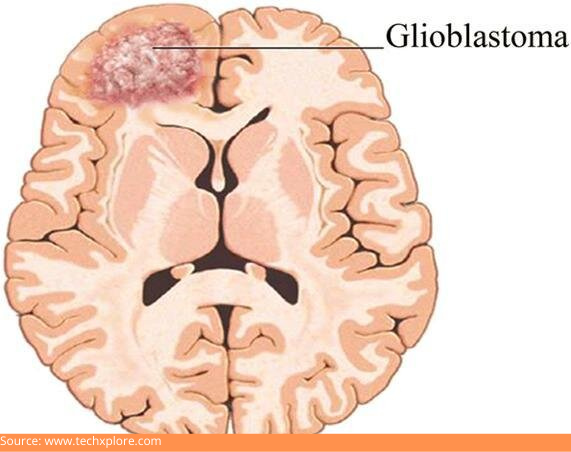
3.Injuries or tumors – Non-communication (Obstructive) Hydrocephalus
4.Normal Pressure Hydrocephalus


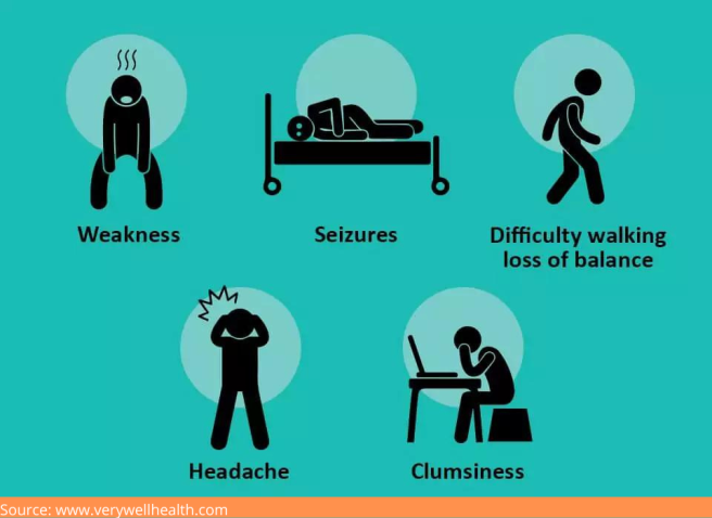
B. Symptoms of hydrocephalus: The symptoms vary from person to person and age. Infants and young children more often present with increased intracranial pressure like vomiting, and adults present with difficulty walking, peeing, or thinking.
1. Infants – increase in the size of the head, progressively increasing head circumference, Bulging fontanelle, prominent scalp veins, Sunset sign, Vomiting, Seizures, Sleepiness, Irritability
2. Children and adolescents: Nausea or vomiting, papilloedema, Blurred vision, double vision, Balance abnormalities, developmental delay, Changes in personality, concentration issues, Seizures, Poor appetite, incontinence
3. Adults: Headache, vomiting, gait apraxia, balance issues, incontinence, and vision issues, memory loss, dementia


C. Diagnosis of hydrocephalus:
1. Physical exam: We need to identify the cause and severity of the hydrocephalus. Thorough patient history, clinical examination, and diagnostic tests are essential in diagnosing hydrocephalus.
2. Imaging tests:
Lumbar puncture (spinal tap) is contraindicated in obstructive hydrocephalus.
Computed tomography scan (CT or CAT scan)
Magnetic resonance imaging (MRI)
ICP monitoring
3. Tests for genetic mutations


D. Treatment of hydrocephalus: Treatment of the cause of water retention is the priority. Removing the obstruction caused by the tumor or blood is a direct way of treating hydrocephalus. Indirect treatments for hydrocephalus are the most common mode of treatment, including shunt or endoscopic third ventriculostomy. The shunt will have a catheter and valve, and is flexible tube drains excessive CSF. The shunt stays for a lifetime and keeps the intracranial pressure within normal limits. Endoscopic third ventriculostomy is a new procedure that involves creating a new hole in the floor of the third ventricle as an alternative path for CSF flow. It avoids the presence of lifelong implants in the body.
1. Surgery: Shunting or Endoscopic third ventriculostomy or tumor removal.
2. Medication: Acetazolamide to decrease fluid production.


E. Follow-up
Hydrocephalus is a persistent condition, so continuous follow-up and Follow-up diagnostic tests, including MRIs, CT scans, and x-rays, need to be done to check the shunt function.
Alarming symptoms to see a doctor:

Tenderness, Redness, pain, or swelling of the skin at the incision or along the tube
Fever
Irritability
Nausea, vomiting,
Decreased vision
Headache
Double vision
Drowsiness
Abdominal pain
Return of preoperative neurological symptoms


F. Prognosis
The prognosis depends on the etiology, earlier diagnosis and treatment, and the extent of symptoms. Some patients show good improvement, while others do not. Elevated pressure symptoms will be relieved. In normal pressure hydrocephalus, dementia can be reversed.

The earlier the diagnosis and treatment of hydrocephalus will be, the better the outcome. Improvement of the condition varies significantly for each patient. 50% of the shunts fail at two years; the valve may be clogged and needs additional surgeries. Surgery is often necessary to replace the blocked or malfunctioning portion of the shunt system when a shunt malfunctions. Luckily, most of the complications can be dealt with successfully.

Conclusion 

Hydrocephalus is a condition in which excess fluid accumulates in the brain. Hydrocephalus can be caused by several factors, including genetic mutations, infections, trauma, tumor, and injuries. Know the signs and symptoms of hydrocephalus so that you can get them diagnosed and treated early.

Dr. Rao’s hospital is the best Neurosurgery hospital in Guntur, and Dr. Mohana Rao Patibandla is the best Neurosurgeon for hydrocephalus, brain surgery, and neurosurgery in Guntur. He is very experienced and skilled in these fields, and he is also very compassionate and caring. If you need treatment for hydrocephalus or surgery for your brain or spine, you should go to Dr. Mohana Rao Patibandla.

The Science of Brain-Computer Interfaces: How to Control Computers with Your Mind

Cauda Equina Syndrome (CES) – Dr Rao at Dr Rao’s Hospital

Cauda Equina Syndrome (CES) – The best treatment at Dr Rao’s Hospital

Get the best neurosurgeon in Guntur. Dr. Mohana Rao Patibandla is doing minimally invasive spine surgery with a fixation for a patient with cauda equine syndrome secondary to spine trauma.

Compression of the spinal cord results in Cauda Equina Syndrome, a severe neurological condition. Early diagnosis and treatment are essential to improving the chances of a good outcome. The cauda equina is a bundle of nerves at the base of the spinal cord that controls movement and sensation in the legs and lower body. When these nerves are compressed, it can cause pain, numbness, and weakness in the legs. Cauda Equina Syndrome is a medical emergency and requires immediate treatment. Guntur’s best spine surgery hospital offers round-the-clock emergency surgical services and care. In a recent India today article.

Causes:

Various factors, including a herniated disc, spinal tumors, or trauma to the spine, can cause this compression.

One of the most common causes of CES is a herniated disc. A herniated disc occurs when the outer layer of the disc ruptures, allowing the inner layer to bulge out and compress the nerves. This can happen due to various factors, including age-related wear and tear, injury, or lifting heavy objects.

Another common cause of CES is spinal tumors. Spinal tumors are growths that develop on or near the spine. They can be benign (non-cancerous) or malignant (cancerous). While benign tumors are not usually life-threatening, they can still compress the nerves and cause CES.

Symptoms

Symptoms of a herniated disc include pain, numbness, and weakness in the legs and lower body. These symptoms can vary depending on the location of the herniated disc. In some cases, a herniated disc can also cause urinary incontinence. Malignant tumors are more likely to cause CES because they grow quickly and can spread to other body parts. Spinal tumor symptoms include back pain, numbness, and weakness in the legs and lower body. In some cases, a spinal tumor can also cause paralysis.

Diagnosis and Treatment

Once the diagnosis of CES is made and the etiology is established through clinical history, physical examination, diagnostic tests like an MRI of the spine, CT myelogram, and emergent surgery are the treatments of choice. The goal of CES treatment is to free up the compressed nerve roots. if not treated in time, it will result in paralysis and incontinence. The duration of surgery symptoms is less than 48 hours, leading to a better prognosis and looking for the best neurosurgery or spine surgery hospital in Guntur. Look no further than Dr. Rao’s hospital! We’re the best in the business, and our neurosurgeon is the best in India!

Conclusion:

Cauda Equina Syndrome is a severe condition that can cause permanent nerve damage. The syndrome occurs when the cauda equina, a bundle of nerves at the base of the spine, is compressed. A herniated disc, spinal tumors, or spine trauma are just a few causes of this compression.

CES is a medical emergency and requires prompt treatment to prevent permanent nerve damage. Suppose you experience any of the symptoms of CES. In that case, it is essential to seek medical attention immediately and call Dr. Rao’s hospital at 9010056444 or 9010057444 for the best spine surgeon in India, Dr. Mohana Rao Patibandla.

First Biplane cath lab in Andhra Pradesh, India at Dr Rao's Hospital for the advanced interventional neurology procedures

The Best Neurosurgery Hospital in Guntur With The Latest State of the Art Technology

The Best Neurosurgery Hospital in Guntur With The Latest State of the Art Technology

A hybrid operating theater is a type of operating room that combines the features of a traditional operating room and a catheterization lab. It is typically used for procedures that require both surgical and interventional radiology (IR) techniques. check more about drraohospitals on india today.

First biplane cathlab in Andhra Pradesh and Telangana diagnostic-tests-in-neurosurgery-the-best-at-dr-raos-guntur

first biplane cath lab in Andhra Pradesh and Telangana diagnostic-tests-in-neurosurgery-the-best-at-dr-Rao’s hospital-Guntur


Hybrid operation theater @ Dr. Rao’s hospital.

Some of the benefits of using a hybrid operating theater include: 

1. Increased efficiency: procedures typically requiring two separate rooms can be completed in one hybrid room, saving time and money. 

2. Better patient outcomes—Having all of the necessary equipment and personnel in one room can help ensure that procedures are performed safely and effectively. 

3. Enhanced safety: having a dedicated hybrid operating room can help reduce the risk of infection and other complications. 

4. Improved patient comfort—Patients may feel more comfortable knowing that they will not have to be moved between two different rooms during their procedure. 

Our hospital’s Biplane Cath Lab Hybrid operation theater is equipped with the latest 4K endoscopy and neuronavigation technology. This state-of-the-art equipment lets us provide our patients with the best possible care. 

Our hospital’s Biplane Cath Lab Hybrid operation theater is equipped with the latest 4K endoscopy and neuronavigation technology. This state-of-the-art equipment lets us provide our patients with the best possible care. 4K endoscopy provides superior image quality, which helps our doctors diagnose and treat conditions more accurately. 

This advanced equipment helps us to safely and effectively perform procedures such as brain surgery. Neuronavigation technology allows us to precisely target areas in the brain, while neuromonitoring helps us to monitor the patient’s neurological status during surgery. This information is essential to ensure that the procedure is being performed safely and effectively. 

Conclusion: Our hospital’s Biplane Cath Lab Hybrid operation theater is equipped with the latest 4K endoscopy and neuronavigation technology. This state-of-the-art equipment lets us provide our patients with the best possible care. Neuronavigation and neuromonitoring technology help us to safely and effectively perform procedures such as brain surgery.


Dyskinesia - the best treatment at Dr Rao's hospital, Guntur

Dyskinesia: Signs, Symptoms, Causes, Diagnosis and the best treatment

Dyskinesia: Signs, Symptoms, Causes, Diagnosis and the best treatment

Dyskinesia is a chronic neurological disorder that causes involuntary body movements. It is characterized by uncontrollable movements of the joints and muscles, often with a simultaneous movement disorder.

Dyskinesia can affect individuals at all levels of society. Although it frequently manifests as a behavioral symptom, overexposure to toxic substances or diseases like Huntington’s disease and encephalitis can also be the cause.

This post aims to provide information about dyskinesia, how it’s diagnosed, how it’s treated, and why you should take this condition seriously.

Dr Rao Hospital is the best spine surgery hospital in India. It provides the best treatment and surgery for all types of spine diseases.

Diagnosis of Dyskinesia

The first impression of a patient with Dyskinesia is that of a behavioral problem. However, repeated episodes may contradict this impression. In most cases, patients with this disorder present an intractable condition to medical professionals.

A diagnostic test is used to detect the presence of Dyskinesia. This may include muscle tone and reflex tests, neurological motor tests, and coordination assessments.

An MRI is the best way to identify if there are any other types of disorders.

Parkinson’s or Huntington’s disease, as well as any brain lesions that could affect the neuromuscular system. Moreover, Dr Rao Hospital is the best spine surgery hospital in India, and it provides the best treatment and test facilities for MRI, CT scans, and more.

Causes of Dyskinesia

Although many medical experts think that damage to the basal ganglia causes dyskinesia, the etiology and cause are still under investigation.

The best Neurosurgeon in Guntur, Andhra Pradesh, suggests that the cause is neurological in nature and damage to the basal ganglia may be linked to other neurological disorders, such as Alzheimer’s disease or Parkinson’s disease.

Symptoms that tell about Dyskinesia

Depending on the underlying cause and severity of the condition, dyskinesia can have mild or severe symptoms. Some symptoms include sudden muscular movements, uncontrolled muscle jerking or spasms, and sudden movements repeated over a short period.

Best neurologist in Guntur say that the symptoms may appear purposeless, but in most cases, the movement is functional (referred to as functional dystonia).

Dyskinesia’s involuntary movements may range from mild tics (twitches) to severe spasticity. People with dyskinesia often experience pain and other symptoms related to their condition.

People with this disorder must receive a thorough evaluation by a medical specialist, such as a spine surgeon, who can determine what other causes of movement are present and take appropriate treatment steps.

Some signs of Dyskinesia

a. Rapid, jerky movements of part or all of the body
b. Muscle spasms
c. Abnormal movements that are repeated and appear purposeless
d. Spontaneous, uncontrolled muscle activity in one or both arms, legs, or face
e. Fluid-filled cavities within muscles
f. Uncontrollable tremor or shaking.

Conditions Linked to Dyskinesia

Treatment is tailored to the underlying cause and severity of the condition, which the evaluation of a medical specialist such as a spine surgeon in Guntur may determine.

Best spine surgeon in Guntur suggest that Dyskinesia can be linked to the following conditions:

  1. Dystonia: Dystonia is a term for movement disorders that affect voluntary muscle groups (usually the muscles of facial expression). It can cause a variety of symptoms, including muscle cramps, spasms, and unusual postures.
  2. Parkinson’s disease: Parkinson’s is a disorder that often affects the basal ganglia system, which is responsible for controlling coordinated movement. In individuals with Parkinson’s disease, movement coordination is lost, and involuntary movements occur (see dyskinesia).
  3. Tardive dyskinesia: Tardive dyskinesia is a rare, irreversible movement disorder that usually appears after the long-term use of antipsychotic medications. Symptoms include involuntary jerking and repetitive movements.
  4. Huntington’s disease: Huntington’s disease is a hereditary neurodegenerative disorder that causes involuntary movements (see dystonia).

Treatment Options for Dyskinesia

The goal of dyskinesia treatment is to eliminate or reduce the symptoms. Different treatments are used for different types of dyskinesias.

Most treatment options have limitations and should be discussed with a medical professional before implementation. The likelihood of success varies depending on the person with dyskinesia, the type of dyskinesia, and how well it is treated.

There are no effective treatments for all types of dyskinesias. However, if you experience dystonic movements, you may be able to take medication to prevent them.

From occurring without causing serious side effects, such as low blood pressure or sudden death (central nervous system toxicity).

Some steps to lower the chances of Dyskinesia and increase Recovery

  • Don’t take too much alcohol, especially if you are prone to having a seizure
  • Use a high-quality sleeping pill or herbal sleeping pill.
  • Good sleep hygiene is essential. If you drink too much coffee, don’t drink any at night for at least 24 hours.
  • Take a supplement that promotes the enzymatic breakdown of lipid compounds in human cells.
  • Do not take it rectally, if possible.
  • If you take it rectally, you will see that the supplement’s concentration is high and that you can absorb the amount needed.
  • Use a lot of fiber, antifungal fiber, etc., in combination with glutamine, especially for those with H. pylori or any other kind of gastric bacterial overgrowth in the stomach.

It is important to realize that dyskinesia does not have to be permanent and cannot be cured with surgery alone. Physical therapy and other behavioural therapies can sometimes avoid or reduce dyskinesia.

Best Neurosurgeon in India

Glioblastoma –Symptoms, Causes, Diagnosis, And The best Treatment

Glioblastoma –Symptoms, Causes, Diagnosis, And The best Treatment

It is pretty normal for people to have mood changes and irritability once in a while. We all have one of those days when we just want to be left alone. However, if you notice changes in behavior accompanied by severe headaches, failing memory, and blurred eyesight, amongst other things, it is time to sit up and take notice.

Dr. Mohana Rao, Founder of Dr. Rao’s Hospital – a world-class neurology hospital in Guntur, says that these may be symptoms of a serious condition or brain ailment called Glioblastoma.

If you or a loved one has been suddenly experiencing sensations and having episodes that are out of the ordinary, please do not take it lightly. Get in touch with Dr. Mohana Rao, a renowned and respected neurosurgeon in Andhra Pradesh, to avail of his expertise in comprehensive nerve, brain, and spine care. 

What Is Glioblastoma?

D:\Digicore\UNOSEARCH\Dr Mohana Rao Neurosurgeon Guntur\All About Glioblastoma\Source Gioblast.jpg

Glioblastoma or glioblastoma multiforme (GBM) is a condition that strikes unexpectedly, usually with a sudden seizure or some days of headaches that get worse each day. It is cancer that can develop in your brain or spinal cord and is very destructive. Glioblastoma originates from astrocytes, which are cells that support your nerve cells.

This cancer can strike anyone at any age, but it affects older people more frequently. It can induce headaches, seizures, nausea, and vomiting that worsen with time.

What Are The Symptoms Of Glioblastoma?

D:\Digicore\UNOSEARCH\Dr Mohana Rao Neurosurgeon Guntur\All About Glioblastoma\Source Symp.png

Symptoms of GBM usually appear suddenly. As the tumor grows, it exerts pressure on your brain, resulting in:

  • Hazy or blurry vision
  • Headaches, which become more and more frequent and severe
  • Appetite loss
  • Memory problems
  • Mood swings or changes in personality
  • Weakness in the muscle, and trouble walking
  • Nausea and vomiting
  • Seizures
  • Speech difficulties
  • Numbness or tingling

If you have noticed any of the above symptoms, please consult with Dr. Mohana Rao, among the best neurosurgeons in Andhra Pradesh, to find out what may be causing it.

What Can Cause Glioblastoma?

Despite various studies and research, experts do not know why some people develop malignant brain tumors, including glioblastoma.

However, some risk factors have been observed and understood.

Who Is At Risk Of Glioblastoma?

GBM most typically affects persons between the ages of 45 – 70. Although men are at a little higher risk than women, the disease affects people of all genders and ages.

These variables may put you at a higher risk:

  • Exposure to chemicals like petroleum, synthetic rubber, pesticides, and vinyl chloride
  • Genetic tumor-causing disorders like neurofibromatosis (tumors that form in your nervous system), Turcot syndrome, and Li-Fraumeni syndrome (both of which are rare genetic disorders increasing the risk of cancer in your brain and spine)
  • Radiation therapy to the head had previously been used

How Is Glioblastoma Diagnosed?

There are several ways to diagnose a brain tumor, including-

Neurological exam:

Your doctor will discuss what changes you have observed, and experienced. He might examine your eyesight, hearing, coordination, reflexes, balance, and strength, among other things. Problems in any of these areas could indicate which section of your brain is being impacted by the tumor.

Imaging tests:

Imaging tests can enable your doctor to figure out where your brain tumor is and how big it is. Brain tumors are frequently diagnosed with MRI, which may be combined with specialized MRI imaging, like functional MRI and MRS (magnetic resonance spectroscopy).

The specialist may also conduct other imaging tests like CT scan (computed tomography) and PET (positron emission tomography).

Biopsy:

Depending on your situation and the position of your tumor, a needle biopsy can be performed before or during the surgery to remove some glioblastoma tissue. A laboratory examines the sample of suspicious tissue to assess the sort of cells present and the level of their aggressiveness.

The specialist can conduct specialized tests of the extracted tumor cells to determine and understand their mutation types. The types of mutation the cells have developed can provide information about the severity of your condition and the best treatment option.

If you suspect that the recent changes you have been undergoing are related to a brain condition, please visit Dr. Rao’s Hospital. It is among the leading neurology hospitals in Guntur with cutting-edge technology and sophisticated equipment. 

What are the treatment options available?

Glioblastoma is a cancer that is very hard to treat and often incurable. Treatments may help to decrease the growth of cancer and alleviate symptoms.

The primary treatment is surgery during which your neurosurgeon will operate and work on removing as much of the cancerous tissues as possible. Glioblastoma cannot be completely removed because it develops into normal brain tissue. As a result, the majority of patients undergo further treatments following surgery to address the leftover cells.

However, some people cannot undergo surgery due to health issues or the location of the tumor. In such cases, radiation therapy and chemotherapy will be the primary treatment.

The treatments for GBM include:

Radiation Therapy:

Radiation therapy employs high-energy beams such as protons or X-rays to damage the cancer cells and stop their growth. 

IMRT (Intensity-modulated radiation therapy):

IMRT enables the targeting of radiation to the tumor while limiting the exposure to healthy brain tissue in the surrounding area.

Stereotactic radiosurgery:

Stereotactic radiosurgery ( or gamma knife radiosurgery) is an advanced form of radiation therapy. This therapy uses highly focused X-ray beams to precisely target the tumor, resulting in minimal harm to healthy tissues. Providers may apply this approach when GBM increases after going through initial IMRT.

D:\Digicore\UNOSEARCH\Dr Mohana Rao Neurosurgeon Guntur\All About Glioblastoma\Source stereo.jpg

Targeted Drug Therapy:

Targeted medications target the specific defects in cancer cells that allow them to develop and flourish. The medicines focus on those abnormalities and cause them to die.

Chemotherapy:

Chemotherapy is a treatment that uses medications to kill cancerous cells. Fine, circular wafers filled with chemotherapy medicine could be inserted into your brain during the surgery in some cases. The slowly dissolving wafers release the medication to kill the cancer cells.

TTF (Tumor treatment fields):

TTF entails putting adhesive pads on your scalp. The pads are wired to a handheld device that produces an electrical field that disrupts the ability of tumor cells from multiplying.TTF is used in conjunction with chemotherapy and can be administered following radiation therapy.


What Is The Outlook Or Prognosis For Glioblastoma Patients?

GBM is a malignancy that is aggressive and difficult to treat. Suitable treatments help to reduce symptoms, make you more comfortable, and help you live longer. The condition does not have a cure yet but many clinical trials are being conducted to develop newer glioblastoma therapies. 

The average life expectancy is 15 months, with about 25% of patients surviving after 2 years and 7% for 5 years.

Conclusion

D:\Digicore\UNOSEARCH\Dr Mohana Rao Neurosurgeon Guntur\All About Glioblastoma\Source Caring.jpg

It can be tough to discover that you have GBM. The disease spreads swiftly, making therapy difficult. GBM researchers are still looking for novel ways to treat it. For the time being, treatments can help you manage your symptoms and enhance the quality of your life. 

It is critical to understand that there was nothing you might have done or avoided that would have led to the development of a brain tumor in you or someone you love.

Do watch out for any sudden behavioral changes or discomforts that you feel, or are expressed by a loved one. The faster you address the symptoms, the faster the treatment can begin to alleviate discomfort. 

Please schedule an appointment and seek medical attention from Dr. Mohana Rao, a top-notch neurosurgeon in Guntur, Hyderabad. Known for his integrity, professionalism, and expertise, he is passionate about improving the quality of people’s lives.

Biplane Cath Lab - cerebral-angiography-the-best-at-dr-raos-guntur

Causes, Prevention and The Best Treatment of Cerebrovascular Disorders

Causes, Prevention and The Best Treatment of Cerebrovascular Disorders

A stroke, also known as a brain attack, occurs when the brain’s blood supply is cut off, depriving the brain of oxygen and causing brain cells to die. When cells die, the damaged part of the brain loses control of whatever part of the body it controls. It affects people of all ages, genders, and races. It is the leading cause of disability.

Although stroke is a brain disease, it can impact the entire body. The consequences of a stroke are determined by which section of the brain is affected and how serious the injury is. 

Some of the common disabling symptoms of a stroke are:

  • Paralysis or issues controlling movement. 
  • Sensory abnormalities, including pain. 
  • Problems using or comprehending language. 
  • Problems with thinking and memory. 
  • Emotional disturbances. 

Stroke can have fatal consequences. Thanks to innovative and sophisticated procedures and therapies, life-long impairment or death is no longer addressed.

At Dr. Rao’s Hospital, one of the best neurology hospital in Guntur, Andhra Pradesh, highly qualified and experienced neurosurgeons collaborate to ensure that patients receive the care they require, whether it’s urgent care, brain surgery, cutting-edge diagnostics and treatment, cutting-edge clinical trials, or rehabilitation.

Because time is of the essence when treating stroke, our emergency department teams are prepared to deliver immediate, high-quality, and best stroke and cerebrovascular treatment in Andhra Pradesh. We also have a rapid response team that assesses patients and initiates particular procedures to determine the etiology of the stroke.

Now, let’s know,

What Causes a Stroke?

High blood pressure (hypertension): 

Itis a primary cause of stroke.

Vascular diseases: 

Vascular illnesses, such as clogged arteries, heart valve malfunction, irregular heartbeat, heart infection, and heart abnormalities, can lead to strokes, especially in the elderly.

Smoking or chewing tobacco: 

Smoking can cause several health problems, including fatty deposits in the neck artery, high blood pressure, and blood thickening. Blood clots in the blood vessels can result due to this. Even exposure to second hand smoke can put you at risk for a stroke.

Obesity: 

Being obese or overweight increases the risk of having a stroke.

Diabetes: 

Obesity, high blood pressure, and diabetes are all variables that increase the chances of stroke.

Family history: 

Family genes can also increase the chance of stroke, heart attack, or other major risk factors such as diabetes or high blood pressure.

Now, let’s know more about,

Acute Treatment Strategies for Stroke

If someone has a stroke, they should seek emergency medical attention from a neurologist in Guntur. You can be treated for a stroke with treatments like mechanical thrombectomy.

Neurosurgeons in Guntur use the following equipment to perform mechanical thrombectomy to remove blood clots.

1. Stentriever Mechanical Thrombectomy

A stentriever is the most recent mechanical device. A self-expanding mesh tube is linked to a wire-guided through a catheter (thin tube). The surgeon places the catheter in a groin artery and guides it through numerous blood channels until it reaches the blood clot in the brain.

2. Aspiration Thrombectomy 

Manual cluster yearning or the use of catheter equipment to suck the coagulation out of the vessel are both used in aspiration thrombectomy.

Now, let’s see,

Procedure of Mechanical Thrombectomy

Mechanical thrombectomy is a procedure used to remove blood clots. Neurosurgeons remove blood clots using a vacuum or mechanical gear in mechanical thrombectomy. Therefore, the blood flow is restored, and the stroke is cured.

To achieve the finest results, it’s best to seek mechanical thrombectomy treatment from the best neurology hospital in Guntur.

What are the Advantages of Mechanical Thrombectomy?

  • Mechanical thrombectomy treatment allows patients to recover from a stroke within a 24-hour window following the commencement of the stroke. This will assist more individuals in recovering from stroke and avoiding the adverse effects of stroke. 
  • For patients who have had a stroke, mechanical thrombectomy offers a gleam of hope. Mechanical thrombectomy provides them hope and bravery in their battle against stroke. It resurrects their long-dead hopes and gives them the strength to battle stroke. It is a potent tool that aids patients in overcoming dangerous strokes.
  • Mechanical thrombectomy also speeds up the recovery of patients with stroke. Though mechanical thrombectomy is a valuable technique for treating stroke, it is only available at a few neurology hospitals in Guntur.

Let’s discuss,

Stroke Prevention Strategies

Regardless of family history or age, there are several steps one can take to reduce the risk of having a stroke:

1. Keeping an eye on the blood pressure

One of the leading causes of stroke is high blood pressure. To improve vascular health, an individual should regularly monitor blood pressure and treat it if it rises.

2. Keeping weight in check

Obesity is connected to comorbidities such as diabetes and hypertension, which increase the risk of a stroke. Reduce your calorie consumption and increase your physical activity. Consult your doctor to develop a personal weight-loss plan that works for you.

3. Regular exercise is essential

Daily exercise will help you stay in shape and better control your weight and blood pressure, minimizing your risk of stroke.

4. Avoiding smoking and consuming alcohol

Smoking increases plaque build-up and blood thickening in the neck arteries, which can lead to clotting. Drinking increases the risk of stroke by causing high blood pressure, diabetes, obesity, liver damage, and various other disorders. Reduce your risk of stroke by eating a healthy diet and quitting smoking. In addition, if you consume alcohol, do it in moderation.

5. Keeping an eye on the blood sugar level

Exercise, drugs, and a balanced diet can all help you keep track of your blood sugar levels. Diabetes can cause blood arteries to rupture and clot inside them over time.

Consult our expert neurosurgeons at Dr. Rao’s Hospital if you need cerebrovascular disease treatment or a stroke specialist in Andhra Pradesh.

Spinal-Cord-Tumors

Get The best Treatment For Spinal Cord Tumors

Get The Best Treatment For Spinal Cord Tumors at Dr Rao’s Hospital

Spinal Cord Tumors, on occasion, probably go unnoticed because they may no longer be unusual, and their signs resemble those of more significant, not uncommon, conditions. Therefore, your physician must recognize your clinical record and perform each fashionable bodily and neurological exam.

Dr. Mohan Rao Patibandla, one of Guntur’s most renowned and best neurosurgeons, has years of expertise in treating spinal tumors. You can consult with him and get all the suitable guidance regarding the process of treating a spinal tumor. In a recent  mid-day. 

If your physician suspects a spinal tumor, those checks can assist in verifying the prognosis and pinpoint the tumor’s location:

Spinal magnetic resonance imaging (MRI).

 MRI uses an effective magnetic area and radio waves to correctly photograph your spine, spinal cord, and nerves. MRI is usually the preferred way to diagnose spinal cord tumors and surrounding tissue tumors. An evaluation agent that ensures that the tissues and systems can be injected right into a vein on your hand or forearm throughout the take-a-look.

Computerized tomography (CT)

This test uses a thin beam of radiation to provide specific photographs of your spine. Sometimes, it is mixed with an injected contrast dye to make unusual modifications inside the spinal canal or spinal cord less challenging to see. A CT test is hardly ever used to assist in diagnosing spinal tumors.

Biopsy

The simplest way to determine the precise form of a spinal tumor is to study a small tissue sample (biopsy) under a microscope. The biopsy results will assist in deciding on treatment options.

Treatment

Ideally, a spinal tumor remedy aims to cure the tumor. However, this aim can be complex, using the threat of everlasting harm to the spinal cord tumors and surrounding nerves. Doctors additionally have to forget your age and normal health.

The sort of tumor and whether or not it arises from the systems of the backbone or spinal canal or has unfolded on your backbone from someplace else to your frame additionally have to be taken into consideration in figuring out a remedy plan.

Treatment alternatives for maximum spinal tumors include:

1. Monitoring

Some spinal tumors can be found earlier than they cause symptoms—frequently while you are being evaluated for every other condition. If small tumors are not developing or urgent on surrounding tissues, looking at them cautiously can be all that is needed.

2. Surgery

This is frequently the remedy of preference for tumors that may be eliminated with an appropriate chance of spinal cord tumors or nerve damage. Newer strategies and devices permit neurosurgeons to attain tumors that have been inaccessible as soon as taken into consideration. The high-powered microscopes utilized in microsurgery make differentiating tumors from wholesome tissue simpler.

Doctors can also screen the features of the spinal wire and different crucial nerves for the duration of the surgical operation, minimizing the threat of wounding them. In a few instances, very high-frequency sound waves are probably used during the surgical operation to interrupt tumors and do away with the fragments.

3. Radiation therapy.

This can be used to dispose of the remnants of tumors that continue to be after surgical procedure, to deal with inoperable tumors or to deal with the ones tumors in which surgical procedure is simply too risky.

 Medications may also assist in easing a number of the aspect results of radiation, which include nausea and vomiting.

4. Chemotherapy.

A fashionable remedy for plenty of varieties of most cancers, chemotherapy makes use of medicinal drugs to damage most cancer cells or forestall them from growing. Your health practitioner can decide whether or not chemotherapy is probably helpful for you, both on your own or in a mixture with radiation therapy.

 Side outcomes may also include fatigue, nausea, vomiting, an accelerated threat of infection, and hair loss.

5. Other drugs.

Because surgical procedures and radiation remedies, in addition to tumors themselves, can irritate the spinal cord, doctors, once in a while, prescribe corticosteroids to lessen the swelling, both after a surgical procedure or at some point in radiation treatments.

Although corticosteroids lessen irritation, they may be commonly used for quick intervals to avoid extreme aspects, such as muscle weakness, osteoporosis, excessive blood pressure, diabetes, and a multiplied susceptibility to infection.

Dr. Rao’s Contact Information:

the best Head-Injury treatment in andhra Pradesh by Dr Rao at Dr Rao's hospital

Head Injury Precautions and Treatments

Head Injury Precautions and Treatments

A knock on the head and a skull fracture are all examples of head injuries. Some severe head traumas might result in brain damage or even death.

Knowing the signs and symptoms of a concussion and some basic first aid can help individuals respond promptly to a head injury, decreasing the risk of consequences. In a recent India today article.

Dr. Rao’s hospital in Guntur has successfully performed many head injury surgeries. It is one of the most renowned and best neurology hospitals in Guntur, with expertise over the years.

Types

There are different types of brain injuries, each of which can be more or less devastating.

 Traumatic brain injury (TBI) occurs when the brain is damaged, usually as a result of an accident. TBI can occur when an object hits a person’s head or when an object enters the brain through the skull.

 A high-velocity ball that hits the head, a massive fall to the head, or a gunshot wound are examples.

Shaking the head vigorously can also cause SHT because the brain can jump or spin in the skull. Whiplash is a common cause of these injuries.

Traumatic brain injury (TBI) is damage to the brain that frequently results in blood clots or bruises. An intracranial hematoma (ICH) is a blood clot in the brain. Depending on its size and location in the brain, the ego can be moderate or heavy.

A concussion is a type of traumatic brain injury (TBI) in which the brain temporarily ceases to function normally. Concussion symptoms are usually not severe or long-lasting, but they can be bothersome.

The brain is protected by the skull, which is formed of thick, sturdy bone. A powerful strike, on the other hand, has the potential to crack or break the skull.

An open head wound, or a penetrating head wound, occurs when the skull is fractured and a bone or other item pierces the brain.

When should you see a doctor?

Both moderate and severe head injuries require medical attention right away. If the symptoms of a mild head injury persist for more than two weeks, get medical help.

The signs and symptoms of a concussion do not usually show right away. A person’s symptoms may appear days or weeks after an incident.

Always proceed with caution while dealing with a head injury. Even after a minor mishap, if someone is worried about their symptoms, they should see a doctor.

First aid and treatment

Mild head injuries are frequently treated at home. Swelling can be reduced by applying a cold pack to the affected region.

Tylenol can also be used, but non-steroidal anti-inflammatory medications (NSAIDs), such as ibuprofen and aspirin, should only be used if a doctor has prescribed them. Someone should check on a person periodically over the first 24 hours following a moderate head injury.

It is critical to seek medical attention if a person loses consciousness or exhibits indications of disorientation or memory loss.

After a brain injury, a person should avoid using drugs or alcohol, driving, or participating in contact sports. They may have to miss work or school.

Severe head injuries necessitate medical care right away. Anyone who encounters someone with severe head injury symptoms should dial 9010056444 or their local emergency number.

It is advisable not to move a person who has suffered a severe head injury to prevent aggravating their ailments. It’s also not a good idea to try to take someone’s helmet off if they’re wearing one.

Severe head injuries frequently require hospitalization, surgery, or long-term care.

Prevention

Although avoiding head injuries is not always feasible, specific actions may be taken to decrease the risk. These are some of them:

  • When in a car, wear a seatbelt
  • When cycling or riding a motorcycle, you should always wear a helmet.
  • when practicing contact sports, take safety precautions

Because a brain injury can have long-term repercussions, it’s critical to take measures wherever possible.

Dr. Rao’s Contact Information:

osteoporotic-spine-surgery

The Best osteoporotic spine surgery in Andhra Pradesh

The Best osteoporotic spine surgery in Andhra Pradesh

Osteoporosis is a bone disease that affects both men and women. Millions of people worldwide have it or are at greater risk of getting it.

A healthy bone has microscopic gaps on the inside, like a honeycomb. Osteoporosis enlarges these gaps, causing the bone to deteriorate in strength and density. Furthermore, the exterior of the bone becomes thinner and weaker.

This condition can strike anyone at any age, but it is more common in the elderly, particularly women. Osteoporosis makes people more prone to fractures, especially in the spine, known as vertebral compression fractures.

Early diagnosis and treatment can be beneficial as they prevent further compression and irreversible changes in the spine. You can consult Dr. Mohana Rao Patibandla, one of the best neurosurgeons in Guntur, at Dr. Rao’s Hospital for advanced surgical intervention.

What are the symptoms of spine fractures?

A compression fracture can result in back or neck pain and loss of height. It is a fracture in one of your neck or back vertebrae that is so weak that it breaks under normal spinal pressure.

The fracture can cause symptoms such as:

  1. Severe, sudden back pain
  2. Have problems twisting or bending your body?
  3. Pain that increases when you walk or stand
  4. Curved-shaped spine
  5. There is far more significant pain and discomfort in moving if the fracture is in the lower spine.
  6. Kyphosis of the spine, loss of height, and neuro deficiencies
  7. Neurological deficits include weakness of the limbs, tingling numbness, bed wetting, and stool incontinence or constipation.

What are the causes of osteoporosis?

Osteoporosis is a natural aging process in which our bones weaken as we get older. The vertebrae in the spine might narrow and flatten due to it. This can result in a hump or rounded back in older adults, giving them a bent-forward look.

The weaker vertebrae are particularly vulnerable to fracture. When too much pressure is applied to a weak vertebra, its front cracks and loses height, resulting in a vertebral compression fracture. They are most commonly caused by falls. However, people with osteoporosis can sustain a fracture by coughing, twisting, or sneezing.

How is osteoporosis diagnosed?

Your doctor will evaluate your medical history and do a physical exam to determine if you have osteoporosis. They may also conduct blood and urine tests to look for disorders that could cause bone loss. Tests to check the levels of calcium and vitamin D are also included.

To determine whether you have a spine fracture, your doctor will recommend an X-ray, MRI, or CT scan to examine your bones more closely.

Further, he will ask you to undergo a bone density test called bone densitometry, or dual-energy X-ray absorptiometry (DEXA).

Treatment for vertebral compression fracture

Nonsurgical treatment

Most people with a spinal compression fracture recover within three months without needing to have the fracture fixed. Simple measures, such as a brief period of rest and the use of pain medications when necessary, are all that are required. The doctor may ask the patients to wear a brace to limit their movement and allow the spinal compression fracture to heal.

The doctor may also recommend lifestyle changes and prescribe calcium, Vitamin D, bisphosphonates, and anabolic bone drugs.

Surgical Treatment

Surgery may be recommended if you have significant pain that does not respond to nonsurgical treatment. Patients with recent spinal compression fractures are the best candidates for surgery. Your doctor will discuss which operation is best for you based on the type of spinal compression fracture you have.

Dr. Rao’s Hospital is one of the best neurology hospitals in Guntur, where you are offered advanced minimally invasive vertebral augmentation procedures.

Kyphoplasty

During this procedure, the surgeon inserts a balloon tamp into the fractured vertebra. The balloon is then inflated, which helps restore the vertebra’s height and shape. The device leaves behind a small cavity that the doctor fills with special bone cement.

Vertebroplasty

Here, the surgeon injects the bone cement into the narrowed vertebra to keep it stable. It helps to alleviate discomfort and help avoid additional vertebral fractures and a bent spine.

Cemented augmented fenestrated screws

In patients with low bone quality due to osteoporosis, fenestrated screws for cement augmentation provided effective and long-lasting outcomes.

They are used for spine stabilization in the elderly, allowing for rapid functional recovery in fractures and deformities.

It also overcomes the problem of screws loosening in individuals with inadequate bone stock due to osteoporosis, causing morbidity.

Contact Dr. Mohana Rao Patibandla, one of the best neurosurgeons in Guntur, Andhra Pradesh, for superior treatment options for spinal compression fractures due to osteoporosis. According to mid-day

Dr. Rao’s Contact Information:

Deep-Brain-Stimulation

The best Deep Brain Stimulation (DBS): A Brain Pacemaker

Deep Brain Stimulation (DBS): A Brain Pacemaker—The best at Dr Rao’s Hospital

Deep brain stimulation (DBS) electrodes are implanted in particular regions of the brain. These electrodes generate electrical impulses, which control the aberrant impulses. Alternatively, electrical impulses in the brain can impact specific cells and substances.

Dr. Rao’s Hospital Guntur is one of Guntur’s most renowned and best neurology hospitals. They have years of expertise in implementing deep brain stimulation.

Under the skin of the upper chest, a pacemaker-like device controls deep brain stimulation. A cable that travels beneath your skin connects this device to electrodes in your brain.

Deep brain stimulation is frequently used to treat a variety of ailments, including:

  1. Parkinson’s disease is a neurological disorder that affects people.
  2. Essential tremor is a kind of tremor that affects
  3. Dystonia
  4. Epilepsy
  5. Obsessive-compulsive disorder (OCD) is a kind of obsessive-compulsive disorder.

Why is it done this way?

People with movement disorders, including essential tremors, Parkinson’s disease, and dystonia, as well as mental diseases like obsessive-compulsive disorder, can benefit from deep brain stimulation. The Food and Drug Administration has also authorized it for use in treating seizures in people with difficult-to-treat epilepsy.

This treatment is only for those who can’t control their symptoms with medicine.

Risks

Although deep brain stimulation is thought to be a low-risk procedure, any operation has the potential for consequences. Furthermore, brain stimulation might have adverse effects.

The dangers of surgery

Deep brain stimulation entails drilling small holes in the skull for the electrodes to be implanted into the brain tissue and surgery to implant the device with the batteries under the skin in the chest. Surgical complications might include:

  1. Leads are misplaced.
  2. There is bleeding in the brain.
  3. Stroke
  4. Breathing difficulties
  5. Nausea
  6. Problems with the heart
  7. Seizure

The device will be turned on a few weeks following surgery, and determining your optimal settings will begin. Some settings may have adverse effects, although these usually improve with more device changes.

The Food and Drug Administration suggests talking with your doctor and adopting water safety precautions before swimming. A few reports have suggested that DBS treatment impairs the movements needed for swimming.

How do you get ready?

Consider the advantages and disadvantages first.

Deep brain stimulation is a method that has the potential to be both serious and hazardous. Even if you believe you could be a candidate for deep brain stimulation, you and your doctors should carefully weigh the risks and benefits.

Prepare for surgery after that.

Before surgery, you’ll almost definitely need medical testing to confirm that deep brain stimulation is a safe and effective therapy option for you. Before the procedure, you may need brain imaging tests, such as an MRI. These studies help map the brain areas where electrodes will be implanted.

What you may anticipate

During the procedure,

Here’s how deep brain stimulation surgery works in general:

Brain Surgery: Your medical team will provide a unique head frame to keep your head still throughout the brain surgery procedure (stereotactic head frame). The experts will next map your brain using neuroimaging (brain MRI or CT) to decide where the electrodes should be put.

Surgery on the chest wall

During the second step of the surgery, the pulse generator (the part of the device that contains the batteries) is implanted under the skin in the chest near the collarbone. General anesthesia will be used during this procedure. The wires from the brain electrodes are routed under the skin to the battery-operated pulse generator. Then, the generator is set up to send electrical impulses to your brain in a loop. The generator is under your control, and you can turn it on or off with a unique remote control.

Following the surgery

The pulse generator in your chest will turn on in your doctor’s office a few weeks after surgery. With a unique remote control, the physician can easily configure his device from outside his body. The intensity of the stimulation is tailored to your needs, and it can take up to six months to achieve the proper balance.

Results

Although deep brain stimulation will not cure your condition, it can help alleviate some of the symptoms. If deep brain stimulation is successful, your symptoms will improve significantly, but they won’t completely disappear. However, certain conditions may require the use of medications.

Deep brain stimulation is not always practical. The effectiveness of deep brain stimulation depends on a variety of factors. Before surgery, talk to your doctor about the improvement you can expect from your condition.

Dr. Rao’s Contact Information: