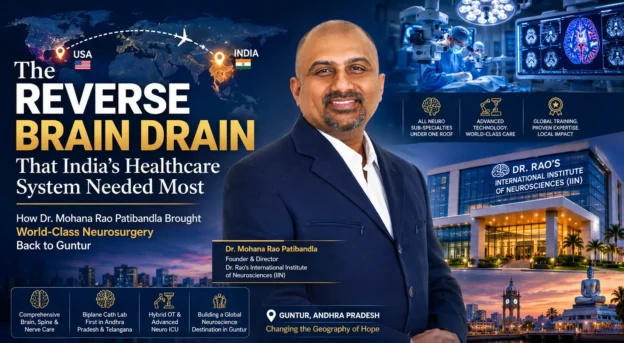
Year in Review 2025: Advancing Neurosurgical Excellence at Dr. Rao’s Hospital, Guntur
As 2025 concludes, it marks a year of structured growth, academic engagement, and continued advancement in neurosurgical care at Dr. Rao’s Hospital, Guntur. Under the leadership of Dr. Mohana Rao Patibandla, the hospital strengthened its position as a dedicated center for brain, spine, and nerve disorders in Andhra Pradesh.
This year was not defined by expansion alone, but by measurable refinement — refinement of surgical protocols, refinement of patient safety systems, and refinement of data-driven clinical practice.
Strengthening Brain Tumor and Skull Base Surgery
Brain tumor and skull base procedures remained a major focus in 2025. Complex cases involving deep-seated lesions, eloquent cortex tumors, posterior fossa tumors, and skull base meningiomas were managed using advanced operative microscopy, endoscopic assistance, and structured intraoperative neuromonitoring (IONM).
Minimally invasive approaches were emphasized wherever appropriate to reduce tissue disruption and enhance recovery. Structured volumetric follow-up for skull base meningiomas and postoperative MRI surveillance protocols were further standardized during the year.
The integration of stereotactic radiosurgery planning into comprehensive treatment pathways allowed selected patients to benefit from non-invasive options for small tumors and vascular malformations.
Spine Surgery: Precision and Preservation
Spine surgery services at Dr. Rao’s Hospital saw continued refinement in 2025, particularly in:
Minimally invasive lumbar decompression
Cervical spine stabilization
Complex thoracic procedures
Tumor-related spinal interventions
Instrumented fusion with neuromonitoring support
Intraoperative neuromonitoring protocols were applied more consistently during deformity correction and high-risk spine cases, reinforcing neurological preservation strategies.
Structured rehabilitation and early mobilization pathways were strengthened, improving postoperative recovery timelines and patient outcomes.
Stereotactic Radiosurgery and Vascular Neurosurgery
A significant academic and clinical highlight of 2025 was the continued emphasis on stereotactic radiosurgery (SRS) for complex intracranial pathologies.
Dr. Mohana Rao Patibandla’s contributions to international multicenter research on stereotactic radiosurgery for arteriovenous malformations (AVMs), skull base meningiomas, and dural arteriovenous fistulas continued to inform institutional protocols.
Published work in leading journals such as the Journal of Neurosurgery and Neurosurgery has provided outcome benchmarks for:
Spetzler-Martin Grade III–V AVMs
Posterior fossa meningiomas
Central skull base meningiomas
Foramen magnum meningiomas
Pediatric high-grade AVMs
In 2025, these evidence-based principles were further integrated into patient selection, treatment planning, and long-term volumetric monitoring at Dr. Rao’s Hospital.
Data-Driven Neurosurgical Practice
One of the defining themes of 2025 was structured data analysis. Outcome tracking systems were strengthened to ensure that every complex procedure contributed to institutional learning.
Key focus areas included:
Intraoperative neuromonitoring dataset analysis
Tumor volumetric response documentation
Complication rate tracking
Functional neurological outcome assessment
ICU protocol standardization
By maintaining structured records, the hospital reinforced its commitment to evidence-based neurosurgery rather than anecdotal practice.
Pediatric Neurosurgery and Functional Disorders
Pediatric brain tumors, hydrocephalus management, and selected epilepsy surgery evaluations continued to be managed with multidisciplinary input.
Functional neurosurgery and radiosurgical planning for endocrine-related conditions were also part of academic engagement during the year, reflecting Dr. Rao’s continued involvement in collaborative research initiatives.
Institutional Infrastructure and Technology Upgrades
2025 also saw continued infrastructure optimization at Dr. Rao’s Hospital. Upgrades included:
Enhanced high-resolution surgical microscopy
Advanced endoscopic systems
Improved neuro-navigation integration
Strengthened intraoperative neuromonitoring setup
Structured ICU workflow refinement
The focus remained on precision, reproducibility, and patient safety rather than expansion for visibility.
Academic Engagement and Global Visibility
During 2025, Dr. Mohana Rao Patibandla’s work was featured in multiple national and international editorial platforms. Participation in academic conferences and research collaborations reinforced the hospital’s connection to global neurosurgical discourse.
Such engagements contribute not only to professional visibility but to continuous learning and adaptation of best practices in patient care.
Commitment to Regional Healthcare Advancement
A defining institutional philosophy continues to be the strengthening of advanced neurological care within Andhra Pradesh. By maintaining comprehensive neurosurgical capability in Guntur, Dr. Rao’s Hospital reduces the need for patients to travel to distant metropolitan centers.
The hospital serves patients from:
Andhra Pradesh
Telangana
Coastal and Rayalaseema regions
Neighboring states
This regional strengthening of tertiary neurosurgical services remains central to long-term planning.
Continued participation in international multicenter research
Institutional consolidation and workflow refinement
Regional referral growth
Looking Ahead to 2026
As Dr. Rao’s Hospital moves into 2026, priorities include:
Further strengthening of minimally invasive spine surgery programs
Expanded radiosurgery planning integration
Advanced neuro-rehabilitation services
Structured outcome publication initiatives
Continued academic collaboration
The focus remains sustainable progress rather than rapid expansion.
Conclusion: A Year of Structured Progress
Reflecting on 2025, Dr. Rao’s Hospital describes the year as one of maturity and consolidation. Clinical excellence was reinforced through structured systems, research engagement, and technological refinement.
Under the leadership of Dr. Mohana Rao Patibandla, the hospital continues to advance as a dedicated center for neurology, neurosurgery, and spine surgery in Guntur, Andhra Pradesh — guided by precision, evidence, and patient-centered care.
Patients from across Andhra Pradesh seeking advanced brain and spine care often search for the best neurosurgeon in Guntur, reflecting the growing demand for specialized tertiary neurological services within the region.
CONTACT INFORMATION
Dr. Rao’s Hospital / Patibandla Narayana Swamy Neurosciences LLP
12-19-67, Old Bank Road, Kothapet, Besides AK Khan Biryani point, Guntur, Andhra Pradesh, India 522001
Stereotactic Radiosurgery for AVMs and Skull-Base Meningiomas: Evidence, Experience, and Practical Integration at Dr. Rao’s Hospital
Stereotactic radiosurgery (SRS) has become a cornerstone in the multidisciplinary management of complex intracranial vascular lesions and skull-base tumors. Over the last decade, international multicenter studies — many of which include contributions from Dr. Mohana Rao Patibandla — have refined our understanding of SRS for arteriovenous malformations (AVMs) and skull-base meningiomas, clarifying indications, outcomes, volumetric response patterns, and safety considerations. At Dr. Rao’s Hospital in Guntur, these evidence signals inform patient selection, technical planning, and follow-up pathways, enabling local patients to access world-class, data-driven radiosurgical care close to home.
This article summarizes the current evidence base, highlights key publications with Dr. Patibandla as a co-author, and explains how this body of work is operationalized at Dr. Rao’s Hospital for the benefit of patients and referral partners.
Why Stereotactic Radiosurgery?
SRS delivers a conformal, high-dose radiation target to a lesion in one or a few sessions with submillimeter accuracy. For selected AVMs and skull-base meningiomas, SRS offers:
A non-invasive option when open surgery carries prohibitive morbidity, or when lesions are surgically inaccessible (e.g., deep brain AVMs, central skull base meningiomas).
High local control rates for small- to medium-sized meningiomas while preserving adjacent cranial nerve function when careful dose planning and volumetric constraints are observed.
A treatment pathway that can be combined with staged microsurgery, fractionated radiotherapy, or observation depending on patient and lesion factors.
Evidence from international multicenter consortia has clarified which lesion subgroups benefit most from SRS, how radiation dose and target volume predict outcomes, and what long-term surveillance strategies are effective. These multicenter analyses allow repeated findings to be validated across institutions, equipment types, and patient populations — strengthening clinical confidence in SRS protocols.
Key Multicenter Evidence (Selected Publications)
The following selected publications — many authored or coauthored by Dr. Patibandla — are among the most influential studies shaping current practice. Links to the publications are provided so clinicians can review methodology and results in full.
SRS for Spetzler-Martin Grade III AVMs. An international multicenter study evaluated outcomes of SRS for Grade III AVMs and provided important data on obliteration rates and hemorrhage risk over time; this work is frequently cited when counseling patients about radiosurgical outcomes for intermediate-grade AVMs.
SRS for Spetzler-Martin Grade IV & V AVMs. Multicenter analysis of higher-grade AVMs (IV & V) provided pragmatic outcome data that help stratify which high-risk lesions might still be appropriate for radiosurgical strategies (for example, staged SRS or combined modality approaches) versus when conservative management may be preferable.
Posterior Fossa (WHO Grade I) Meningiomas — Volumetric Outcomes. Long-term volumetric evaluations of posterior fossa meningiomas treated with SRS indicate reasonably high local control with structured follow-up and underscore the importance of dose planning in preventing cranial nerve toxicity.
Central Skull Base Meningiomas — Volumetric Evaluation & Long-Term Outcomes. Central skull base meningiomas present particular challenges because of intimate cranial nerve and brainstem relationships; volumetric SRS series in the literature have demonstrated meaningful tumor control with acceptable cranial neuropathy rates when careful planning is performed.
Foramen Magnum and Posterior Fossa Meningiomas — Multicenter Experience. Additional multicenter studies provide outcome benchmarks specifically for foramen magnum lesions and posterior fossa meningiomas, informing margin selection and surveillance intervals.
Endocrine-Related Radiosurgery (Pituitary Targets). Dr. Patibandla has also contributed to international SRS literature on functional pituitary targets (Cushing’s disease, acromegaly), which illustrates multidisciplinary radiosurgery practice principles, dose constraints, and endocrine follow-up pathways that are informative for skull base work.
Collectively, these multicenter works move beyond single-center series: they provide reproducible metrics such as volumetric tumor reduction, obliteration curves for AVMs, cranial nerve preservation rates, and timeframes for expected radiographic responses. These are essential parameters for shared decision-making with patients.
Translating Evidence to Practice: Patient Selection & Workflows at Dr. Rao’s Hospital
At Dr. Rao’s Hospital, evidence from the multicenter literature underpins a systematic, multidisciplinary workflow for radiosurgical candidates. Key components include:
1. Multidisciplinary Case Review
Every potential SRS candidate — whether for AVM or skull base meningioma — is reviewed in a multidisciplinary forum involving neurosurgery, neuro-radiology, radiation oncology, neuro-anesthesia, and neurorehabilitation. This forum evaluates surgical risk, radiosurgical candidacy, and whether staged or multimodality therapy is preferable.
2. Indications & Contraindications
AVMs: SRS is considered for small to medium AVMs (<10–12 mL typically), deep and eloquent location AVMs where surgical morbidity is high, and certain higher-grade AVMs after careful multidisciplinary assessment (taking cues from published Spetzler-Martin multicenter outcomes). For very large AVMs, staged SRS or multimodality treatment may be proposed.
Skull base meningiomas: SRS is often recommended for small-to-medium sized tumors, residual/recurrent tumors following surgery, or for surgically inaccessible central skull base and posterior fossa meningiomas where cranial nerve preservation is paramount. Volumetric data help guide fractionation and dose.
3. Imaging & Targeting Protocols
High-resolution MRI with thin slices, contrast sequences, and fused CT angiography (for AVMs) provide the accurate structural and vascular maps required for contouring. For AVMs, digital subtraction angiography (DSA) remains the gold standard in many cases and is integrated into planning when needed. Institutional imaging protocols align with those used in major multicenter studies to allow comparable dosimetric decisions and outcome tracking.
4. Dose, Volume, and Fractionation Decisions
Radiation dose and fractionation are chosen based on lesion size, proximity to critical structures (brainstem, cranial nerves), and published volumetric outcome data. For example, smaller skull base meningiomas may be treated with a single-fraction high-dose SRS regimen if critical distances permit; where nerves or brainstem proximity is close, hypofractionated SRS (fractionated stereotactic radiotherapy) may be selected to decrease cranial neuropathy risk — an approach supported by multicenter experience.
5. Patient Counselling & Shared Decision Making
Patients receive structured counselling about expected radiographic response timelines (e.g., months to years for volumetric reduction), possibilities of transient radiation-induced swelling, and the small but real risk of delayed cranial neuropathy or radionecrosis. Prognostic metrics derived from multicenter series (obliteration curves for AVMs, actuarial local control for meningiomas) are used to provide evidence-based probabilities.
6. Follow-up & Outcome Monitoring
Standardized follow-up protocols include MRI at 6 months, 12 months, then annually for 3–5 years (adjusted by pathology and response). For AVMs, DSA is used selectively to confirm obliteration, particularly if MRI suggests complete response. All outcomes are logged in a structured database to permit continuous quality assessment and contribute to local and international research activities.
Practical Examples from the Literature
The international multicenter AVM studies led by collaborators including Dr. Patibandla provide real-world outcome rates used in clinical counseling. For instance, Grade III AVMs treated with SRS demonstrate meaningful obliteration rates over a multi-year timeline with variable hemorrhage risk during the latency period after radiosurgery — data that shape follow-up intensity and urgent care planning.
Large multicenter work on Grade IV–V AVMs demonstrates that while outcomes are more guarded, tailored strategies (staged SRS, combined embolization + SRS, or conservative management) can be considered within a framework of multidisciplinary risk assessment. These studies emphasize individualized care rather than one-size-fits-all algorithms.
For skull base meningiomas, volumetric analyses from World Neurosurgery and Journal of Neurosurgery cohorts show consistent long-term tumor control with low cranial nerve toxicity when volumetric limits and dose constraints are respected. These datasets are particularly useful in cases where resection would carry substantial morbidity.
Finally, the inclusion of pituitary radiosurgery studies (e.g., on Cushing’s disease and acromegaly), while distinct in therapeutic goals, contributes to a common knowledge base about skull base radiosurgical planning, endocrine follow-up, and multidisciplinary coordination. These works illustrate how radiosurgery teams coordinate with endocrinology, neurosurgery, and radiation oncology for best outcomes.
Outcomes, Safety, and Risk Management
SRS is not risk-free; responsible practice demands balancing benefit and harm. Key safety themes include:
Latency hemorrhage risk in AVMs: Patients treated with SRS face a latency period before obliteration; during this time, hemorrhage remains possible. Counselling and emergency plans are part of routine care. Multicenter data provide the best estimates of this risk by lesion grade and radiosurgical dose.
Cranial nerve toxicity in skull base SRS: Careful dose fractionation and strict dose constraints to nerves and brainstem minimize cranial neuropathies; multicenter volumetric evaluations provide thresholds that guide safe planning.
Radionecrosis and edema: Close imaging surveillance and low thresholds for corticosteroid management or surgical decompression (rare) ensure patient safety where symptomatic radiation effects occur.
At Dr. Rao’s Hospital, these risks are managed through evidence-informed protocols, real-time multidisciplinary decision making, and structured emergency pathways — the very elements proven in large multicenter cohorts.
Research, Collaboration, and the Way Forward
The global multicenter collaborations that Dr. Patibandla has been part of reflect the modern scientific approach to complex neurosurgical problems: pooling cases across centers to achieve statistical power, harmonizing outcome definitions, and producing practice-informing data. Dr. Rao’s Hospital maintains institutional data registries consistent with international reporting standards to enable local contributions to future multicenter efforts.
Priority areas for ongoing research include:
Prospective registries for AVM radiosurgery outcomes stratified by grade and prior embolization status.
Volumetric imaging markers that predict early tumor response versus pseudoprogression in skull base meningiomas.
Comparative studies of single-fraction versus hypofractionated SRS for central skull base lesions.
Long-term cranial nerve function tracking after skull base radiosurgery.
These initiatives will help continue the cycle of evidence generation and clinical refinement.
Conclusion: Evidence Informs Care
Stereotactic radiosurgery is a sophisticated, evidence-based tool in the neurosurgical armamentarium for AVMs and skull base meningiomas. Multicenter studies — to which Dr. Mohana Rao Patibandla has substantially contributed — provide the outcome benchmarks and safety thresholds that clinics like Dr. Rao’s Hospital use to offer precision, documented, and patient-centered radiosurgical care. Patients and referring physicians seeking expert evaluation in Andhra Pradesh can rely on a multidisciplinary approach that emphasizes selection, safety, and long-term follow-up — the pillars of modern radiosurgical practice.
Selected References & Links (Representative publications with Dr. Patibandla contributions)
Dr. Rao’s Hospital, Guntur — offers multidisciplinary radiosurgical evaluation and treatment planning for AVMs and skull base meningiomas. For consultation or referral:
Stereotactic radiosurgery (SRS) is a non-invasive treatment that delivers highly focused radiation to an arteriovenous malformation (AVM) in the brain. The radiation causes the abnormal blood vessels to gradually close over months to years, reducing the risk of future bleeding without open surgery.
How effective is stereotactic radiosurgery for AVMs?
Effectiveness depends on the size and grade of the AVM. Small to medium AVMs often show high obliteration rates over 2–3 years. Larger or higher-grade AVMs may require staged treatment or combined approaches. Regular follow-up imaging is essential to monitor response.
Is stereotactic radiosurgery safe for skull base meningiomas?
Yes, when carefully planned, stereotactic radiosurgery is considered a safe and effective option for small to medium skull base meningiomas. It is especially useful when tumors are close to cranial nerves or the brainstem, where open surgery may carry higher risk.
How long does it take for a meningioma to shrink after radiosurgery?
Most skull base meningiomas do not shrink immediately. Tumor growth usually stabilizes first, and gradual reduction may occur over months to years. Regular MRI scans are performed to monitor volumetric response.
Does radiosurgery require hospitalization?
Stereotactic radiosurgery is typically performed as a day-care procedure. Most patients go home the same day and can resume normal activities within a short period, depending on individual condition.
What are the risks of radiosurgery for AVMs?
During the latency period before complete AVM closure, there remains a small risk of bleeding. Some patients may experience temporary swelling or radiation-related changes on MRI. Careful follow-up helps manage these risks.
When is radiosurgery preferred over open brain surgery?
Radiosurgery is often preferred when the lesion is deep, located in eloquent brain areas, near critical nerves, or when surgical risks are high. Each case is evaluated individually by a neurosurgical team.
Is stereotactic radiosurgery available in Guntur?
Dr. Rao’s Hospital in Guntur provides structured evaluation and treatment planning for AVMs and skull base meningiomas using evidence-based stereotactic radiosurgery protocols and multidisciplinary assessment.
How do I know if I am a candidate for radiosurgery?
Eligibility depends on lesion size, location, symptoms, previous treatments, and overall health. A detailed MRI and clinical evaluation by a neurosurgeon are required to determine suitability.
Is follow-up necessary after stereotactic radiosurgery?
Yes. Long-term follow-up with MRI and, in selected AVM cases, angiography is necessary to confirm lesion response and ensure safe outcomes.
Dr. Mohana Rao Patibandla Featured as “The Best Neurosurgeon to Watch in India 2025” by The Enterprise World
Guntur, Andhra Pradesh – December 2025: The Enterprise World, a leading global business and leadership magazine, has proudly featured Dr. Mohana Rao Patibandla, Founder and Chief Neurosurgeon of Dr. Rao’s Hospital, Guntur, as “The Best Neurosurgeon to Watch in India 2025.” The feature titled “Healing with Heart: Mission to Restore Hope” celebrates Dr. Rao’s visionary contributions to modern neurosurgery, his compassionate approach to healing, and his pioneering role in transforming brain and spine care in India.
“Healing with Heart” — Dr. Mohana Rao Patibandla’s inspiring cover story in The Enterprise World, highlighting his mission to restore hope through neurosurgery.
Few journeys in medicine inspire as much admiration as that of Dr. Mohana Rao Patibandla. Born and raised in Andhra Pradesh, Dr. Rao’s dedication to learning and innovation has placed him among the best neurosurgeons in India today. His rise from a small-town medical graduate to an internationally trained neurosurgeon is a testament to his unwavering commitment to excellence.
After completing his MBBS from Andhra Medical College, Visakhapatnam and M.Ch. in Neurosurgery from Nizam’s Institute of Medical Sciences, Hyderabad, Dr. Rao trained across the United States in multiple advanced fellowships. His global training included Minimally Invasive Skull Base Surgery, Pediatric Neurosurgery, Functional Neurosurgery, Endovascular and Stereotactic Radiosurgery — making him one of the most comprehensively trained neurosurgeons in the country.
But what truly sets Dr. Rao apart is his decision to return to India. Instead of pursuing fame in international centers, he brought world-class neurosurgery back home — founding Dr. Rao’s Hospital in Guntur with the vision of making advanced neurological and spine care accessible to every Indian.
Certificate of recognition presented to Dr. Mohana Rao Patibandla by The Enterprise World as “The Best Neurosurgeon to Watch in India 2025.”
Dr. Rao’s Hospital: Where Compassion Meets Innovation
Established in 2019, Dr. Rao’s Hospital is India’s first independent, dedicated neuroscience center offering advanced brain, spine, and nerve treatments under one roof. Under Dr. Rao’s leadership, it has evolved into a national hub for excellence — combining cutting-edge technology with patient-centered care.
The hospital features world-class infrastructure including Hybrid Cath Labs, 3D Neuronavigation Systems, Intraoperative Neurophysiological Monitoring (IONM), Stereotactic Radiosurgery, and Endoscopic Skull Base Surgery. Every innovation is driven by one goal — to minimize surgical risk and maximize patient recovery.
Dr. Rao has successfully performed over 15,000 complex neurosurgical procedures, including more than 12,000 minimally invasive brain and spine surgeries. Patients from over 15 countries have found hope at Dr. Rao’s Hospital, making it one of India’s most trusted destinations for those seeking the best neurologist in Guntur and the best spine surgeon in Guntur.
“Healing with Heart” — The Philosophy Behind the Practice
Dr. Mohana Rao’s approach to medicine goes far beyond surgical precision. His philosophy — “Technology saves lives, but compassion heals them” — defines the culture at Dr. Rao’s Hospital. Every patient is treated with empathy, transparency, and respect, reflecting his belief that true healthcare must care for both the body and the soul.
In the Enterprise World cover story, Dr. Rao’s life and mission are beautifully captured. The feature highlights how his blend of scientific excellence and human empathy is reshaping the future of neurosurgery in India.
“My journey was never about building a hospital; it was about bringing healing home and making world-class neurosurgery accessible to everyone.” – Dr. Mohana Rao Patibandla
A Legacy of Excellence and Leadership
Dr. Rao’s contributions have been recognized with multiple prestigious honors, including:
🏅 Best Minimally Invasive Neurosurgeon and Spine Surgeon of the Year – Dr. A.P.J. Abdul Kalam Inspiration Awards 2025
🏅 Economic Times Business & Health Excellence Awards 2025
🏅 Sardar Vallabhbhai Patel Unity Award 2025
🏅 Asia’s Most Trusted Neurosurgeon (2023)
🏅 Health Icon of the Year (2021)
Beyond awards, Dr. Rao is a passionate academician with 70+ international publications and 1,200+ research citations, reflecting his deep commitment to scientific advancement. His TEDx Talk, “My Journey to Bring Healing Home – A Neurosurgeon’s Quest in Guntur”, continues to inspire countless young doctors and healthcare leaders worldwide.
Transforming Spine and Brain Surgery in India
Dr. Rao’s expertise spans a wide range of neurological and spinal procedures — from brain tumor resections and aneurysm repairs to complex spinal deformity corrections. He is a pioneer in endoscopic and minimally invasive neurosurgery techniques that drastically reduce recovery time and post-operative pain.
As one of the best spine surgeons in India, Dr. Rao has made significant contributions to neuro-oncology, neurovascular surgery, and functional neurosurgery. His advanced use of intraoperative monitoring and neuronavigation has set new safety benchmarks in Indian neurosurgery.
Building the Future of Neuroscience
Looking ahead, Dr. Rao envisions expanding his impact beyond clinical practice. His next milestone is the establishment of the Dr. Rao Institute of Neurosciences — a center dedicated to research, robotic neurosurgery, artificial intelligence, and advanced training for young neurosurgeons. The institute will foster collaboration between medicine, technology, and ethics to shape the next era of compassionate innovation.
In a world moving rapidly toward automation, Dr. Rao continues to emphasize the human side of medicine — ensuring that empathy remains the foundation of every technological breakthrough.
A True Inspiration in Medicine
From restoring hope to patients facing life-threatening conditions to mentoring the next generation of neurosurgeons, Dr. Mohana Rao Patibandla stands as a symbol of excellence, compassion, and purpose. His recognition as “The Best Neurosurgeon to Watch in India 2025” is not just an award — it is a reflection of his life’s mission to heal with heart and lead with integrity.
Through Dr. Rao’s leadership, Dr. Rao’s Hospital has emerged as a world-class destination for patients seeking expert neurological, neurosurgical, and spine care. For those searching for the best neurosurgeon in Guntur or the best spine surgeon in India, his name stands synonymous with trust and transformation.
Visit Dr. Rao’s Hospital
Dr. Rao’s Hospital is located at 12-19-67, Old Bank Road, Kothapet, Opposite Sravani Hospital, Guntur, Andhra Pradesh.
“Healing with Heart” — Dr. Mohana Rao Patibandla’s story reminds us that in the hands of a compassionate healer, science becomes service, and surgery becomes hope.
Touching Lives Beyond the Operating Room
While Dr. Mohana Rao Patibandla’s surgical precision has transformed thousands of lives, his true legacy lies in his compassion. His philosophy of “healing with heart” extends far beyond hospital walls. Through free medical camps, neuro-awareness drives, and rural outreach programs, Dr. Rao and his team have taken advanced healthcare to underserved communities across Andhra Pradesh.
From educating families about stroke prevention and spine health to raising awareness about epilepsy, brain tumors, and neurotrauma care, Dr. Rao’s initiatives continue to bridge the gap between rural and urban medicine. His belief is simple yet profound — “Every life deserves access to expert care, no matter where it begins.”
Championing Ethical and Patient-Centric Neurosurgery
As the best neurosurgeon in India, Dr. Rao’s reputation is not only built on surgical mastery but also on his uncompromising commitment to medical ethics. Each decision in his practice is guided by transparency, informed consent, and the patient’s overall well-being.
At Dr. Rao’s Hospital, patients and their families are active participants in every stage of the healing process. Treatment options are explained with clarity and compassion, ensuring that no one feels lost in the complexity of medical jargon. This approach has earned the hospital one of the highest patient satisfaction rates in India, with testimonials from across the world praising its empathy-driven environment.
Advancing Neurosurgical Research and Innovation
Dr. Rao’s scientific curiosity and drive for continuous improvement have made him a recognized academic figure internationally. His ongoing research in AI-assisted surgical planning, robotic neurosurgery, and neuro-navigation technologies aims to redefine how brain and spine surgeries are performed in India.
Under his mentorship, the hospital’s research division collaborates with leading universities to explore new frontiers in minimally invasive neurosurgery and patient rehabilitation. His 70+ international research papers and collaborations continue to influence global standards in neurosurgical practice and education.
Leadership That Inspires a Generation
Inside the operating room, Dr. Rao is known as a perfectionist. Outside of it, he is a teacher, mentor, and visionary leader. Young neurosurgeons and residents at Dr. Rao’s Hospital receive hands-on training through structured fellowship programs designed to cultivate both technical skill and ethical responsibility.
Through regular clinical symposiums, live workshops, and neurosurgical skill-building sessions, Dr. Rao ensures that his institution continues to be a learning hub for aspiring surgeons. His mentorship mantra — “Master your skill, nurture your mind, and protect your compassion” — has inspired many to adopt medicine as a mission rather than a mere profession.
International Recognition and Global Collaborations
Dr. Rao’s expertise is now sought after beyond India’s borders. His work has been presented at numerous international conferences, and he frequently collaborates with top neurosurgical institutions across the U.S., Europe, and Asia. He has performed live demonstrations and keynote lectures on minimally invasive brain surgery and endoscopic skull base approaches that have drawn global attention.
As one of the best spine surgeons in India, Dr. Rao continues to represent the country on the world stage, demonstrating that Indian neurosurgery can match — and often surpass — global standards when driven by dedication and innovation.
Technology with a Human Touch
At Dr. Rao’s Hospital, technology is not used to replace empathy but to enhance it. The use of real-time intraoperative imaging, intraoperative CT scans, and neuronavigation allows for precision-driven results with minimal trauma to the patient. But even with the most advanced tools, Dr. Rao’s focus remains on the individual — understanding their fears, guiding them through uncertainty, and walking beside them through recovery.
This delicate balance of science and soul defines the essence of his success as the best neurologist and neurosurgeon in Guntur. His ability to see patients not as cases but as stories of resilience continues to inspire trust and gratitude among families nationwide.
Shaping the Future of Neuroscience in India
Looking toward the future, Dr. Rao envisions the establishment of the Dr. Rao Institute of Neurosciences — a multidisciplinary center that will integrate education, clinical practice, and research. The institute aims to train the next generation of neurosurgeons while driving innovations in robotic-assisted surgeries, neurorehabilitation, and artificial intelligence.
Through this initiative, Dr. Rao hopes to make India a global hub for neurological excellence — where cutting-edge science meets compassionate healthcare. His mission remains clear: to ensure that no patient must travel abroad for treatment when the best neurological care is available in their own country.
Patient Stories That Inspire
Behind every statistic lies a story of courage. Patients treated at Dr. Rao’s Hospital often describe their experience as “life-changing.” From individuals who regained mobility after spinal injuries to children recovering from complex brain surgeries, every story echoes the same sentiment — gratitude for a surgeon who listens, heals, and gives hope.
These testimonials reflect why Dr. Mohana Rao Patibandla continues to be celebrated as the best neurosurgeon in India — not only for his medical brilliance but for his genuine humanity.
Words That Define His Journey
“My purpose has always been clear — to merge the heart of a healer with the hands of a surgeon. Every patient I meet reminds me why I chose this path.”
— Dr. Mohana Rao Patibandla
His journey is not one of ambition alone but of compassion, courage, and conviction. From his hometown of Guntur to the global stage, Dr. Rao’s work continues to inspire hope — not just in patients but in the future of healthcare itself.
Conclusion: A Legacy of Hope and Healing
As *The Enterprise World* celebrates Dr. Mohana Rao Patibandla as “The Best Neurosurgeon to Watch in India 2025”, it marks not an endpoint but a milestone in a journey defined by service. Through relentless dedication, pioneering techniques, and heartfelt compassion, Dr. Rao has built more than a hospital — he has built a movement of healing.
For patients searching for the best neurosurgeon in Guntur, the best spine surgeon in Guntur, or the best neurologist in Andhra Pradesh, Dr. Rao’s Hospital stands as a symbol of trust, excellence, and hope.
Children and Spine-Related Disorders: What Parents Should Know
Parents often associate spine problems with adults or aging; however, spine-related disorders can also affect children. sWhen detected early, these conditions can be effectively managed or corrected to prevent long-term complications. Awareness is key—understanding pediatric spine disorders helps parents identify symptoms early and seek timely expert care.
At Dr. Rao’s Hospital in Guntur, a leading center for advanced spine surgery and neurology services, specialists emphasize the importance of early diagnosis and minimally invasive treatments for children with spinal deformities or developmental spine conditions.
Understanding Spine Health in Children
The spine plays a vital role in a child’s growth, posture, and movement. Pediatric spine disorders can arise from congenital defects, infections, trauma, or developmental issues during growth. According to the World Health Organization (WHO), musculoskeletal disorders, including spinal deformities, affect millions of children globally — but many are treatable when identified early.
In growing children, even minor spinal misalignments can progress over time, leading to curvature, back pain, or neurological symptoms. Regular pediatric check-ups and early orthopedic or neurosurgical consultations can make a life-changing difference.
Common Spine Disorders in Children
1. Scoliosis (Abnormal Curvature of the Spine)
What is scoliosis? Scoliosis is a sideways curvature of the spine often noticed during the growth spurt before puberty. It may appear as uneven shoulders or hips, or a visible curve when a child bends forward.
Symptoms: Uneven posture, tilted shoulders, back pain
Treatment: Bracing, physical therapy, or surgery for severe cases
2. Kyphosis
Kyphosis refers to an excessive forward rounding of the upper back, leading to a hunched appearance. It can be caused by developmental issues, poor posture, or structural deformities.
Symptoms: Rounded back, back fatigue, pain, and stiffness
Treatment: Postural correction, physical therapy, and surgical intervention in advanced cases
3. Spina Bifida
Spina bifida is a congenital condition where the spinal cord and vertebrae do not form properly during fetal development. This can result in neurological deficits, weakness, or paralysis depending on severity.
Causes: Genetic and environmental factors (e.g., folate deficiency during pregnancy)
Symptoms: Visible spinal opening, weakness, bladder or bowel issues.
Treatment: Neurosurgical repair and long-term rehabilitative support
4. Pediatric Disc Problems and Back Pain
Though rare, children can develop herniated discs or mechanical back pain due to trauma, sports, or obesity. Persistent back pain in children should never be ignored, as it may signal an underlying spinal issue.
Recognizing Early Warning Signs
Children may not always express discomfort clearly, so it’s crucial for parents to recognize physical signs and behavioral changes that might indicate a spine disorder.
Uneven shoulders or hips
Visible spinal curvature or hump
Frequent back or neck pain
Unsteady walking or imbalance
Weakness, numbness, or tingling in legs
Difficulty standing straight or bending
If you observe any of these symptoms, it’s important to consult a pediatric neurosurgeon. Early diagnosis can often prevent progression and the need for complex surgery later.
Diagnosis: How Spine Disorders Are Detected in Children
At Dr. Rao’s Hospital’s Neurosurgery Department, evaluation begins with a detailed physical examination, medical history review, and imaging studies. Common diagnostic tests include:
X-rays — to visualize spine alignment and curvature
MRI — to examine spinal cord, nerves, and discs
CT scan — for bone and structural assessment
Genetic or developmental screening (for congenital disorders)
Treatment depends on the severity, cause, and child’s age. At Dr. Rao’s Hospital, minimally invasive spine surgery and advanced rehabilitation techniques are used to ensure safer recovery and optimal growth outcomes.
Why Choose Dr. Rao’s Hospital for Pediatric Spine Care?
Dr. Mohana Rao Patibandla, one of the best neurosurgeons in Guntur, has extensive experience in pediatric neurosurgery and minimally invasive spine surgery. His approach combines precision, innovation, and compassion — ensuring that young patients receive world-class care with minimal discomfort.
Dr. Rao’s Hospital offers:
Dedicated pediatric neurosurgery and spine team
State-of-the-art neuro-navigation and imaging technology
Comprehensive rehabilitation and physiotherapy
Family counseling and long-term follow-up
How Parents Can Support Their Child’s Spine Health
Encourage good posture and ergonomics during study or screen time
Promote regular physical activity and a balanced diet
Use ergonomic school bags to prevent back strain
Ensure proper calcium and vitamin D intake for bone health
Monitor growth spurts and consult if postural changes occur
Call to Action
If your child shows signs of spinal deformity, persistent back pain, or posture problems, visit Dr. Rao’s Hospital for expert evaluation. Contact us at 📞 090100 56444 or 📧 info@drraoshospitals.com to book an appointment with our pediatric spine specialists.
What are the symptoms of spine disorders in children?
Common symptoms include uneven shoulders, visible spinal curve, frequent back pain, imbalance while walking, and difficulty bending. Early consultation is vital.
How are spine disorders diagnosed in children?
Diagnosis involves physical exams, X-rays, and MRI scans. At Dr. Rao’s Hospital, experts use advanced imaging and neuro-navigation tools for accurate diagnosis and management.
Why choose Dr. Rao’s Hospital for pediatric spine treatment?
At Dr. Rao’s Hospital, led by Dr. Mohana Rao Patibandla, one of the few pediatric neurosurgeons in the USA trained in fellowship and one of the best neurosurgeons in Guntur, children receive advanced, minimally invasive, and compassionate spine care.
Many neurological disorders once thought to be random or environmental in origin are now known to have a strong genetic component. From Alzheimer’s and Parkinson’s to epilepsy and muscular dystrophy, our genes play a crucial role in determining susceptibility, disease progression, and treatment outcomes.
At Dr. Rao’s Hospital, one of India’s leading centers for neurological and neurosurgical care, specialists emphasize the importance of genetic awareness and family screening. Understanding your genetic risks empowers families to detect neurological conditions early and manage them effectively with modern medical support.
Understanding Genetics and Neurological Diseases
What is the connection between genetics and neurology? Simply put, genetics refers to how traits and conditions are passed down from parents to children through DNA. In neurological diseases, gene mutations can affect how brain cells grow, communicate, and survive.
According to the World Health Organization (WHO), nearly 10% of all neurological disorders have a direct genetic cause, while many others have a genetic predisposition combined with environmental triggers.
Examples of Genetic Neurological Disorders:
Alzheimer’s Disease: Linked to mutations in the APP, PSEN1, and PSEN2 genes.
Parkinson’s Disease: Associated with changes in the LRRK2 and SNCA genes.
Epilepsy: Certain forms have mutations in ion channel genes like SCN1A.
Muscular Dystrophies: Caused by defective genes affecting muscle strength.
Huntington’s Disease: Inherited condition due to the HTT gene mutation.
How Do Genetic Mutations Affect the Brain and Nerves?
Genetic mutations can cause cells to produce abnormal proteins or fail to remove harmful substances from the nervous system. Over time, this leads to neurodegeneration, cognitive decline, or motor dysfunction.
Researchers from the National Institutes of Health (NIH) have identified hundreds of genes linked to neurodegenerative and developmental disorders, underscoring the critical role of genetic science in modern neurology.
Common Effects of Genetic Mutations:
Altered brain cell function or death
Abnormal neurotransmitter activity
Disrupted neural connectivity
Increased risk of early-onset disorders
Why Genetic Testing Matters for Families
Genetic testing provides vital information about inherited conditions, allowing individuals and families to take preventive or proactive measures.
At the Neurology Department at Dr. Rao’s Hospital, genetic testing and counseling are recommended for families with a history of brain, nerve, or spine disorders. Early genetic screening can identify at-risk individuals before symptoms appear, enabling timely intervention and personalized treatment plans.
Benefits of Genetic Testing:
Early detection and prevention of neurological disorders
Personalized treatment plans based on genetic profile
Better family planning and risk assessment
Reduced anxiety through knowledge and preparedness
Not all neurological conditions are hereditary. Some develop due to age, environment, or spontaneous genetic mutations. However, having a family history significantly increases the likelihood of certain diseases.
Families with a known history of neurological diseases should consider regular neurological check-ups, genetic counseling, and lifestyle adjustments. Prevention and early detection can make a significant difference in long-term outcomes.
Under the leadership of Dr. Mohana Rao Patibandla, one of the best neurosurgeons in Guntur, Dr. Rao’s Hospital integrates advanced genetics with precision medicine to manage complex neurological disorders. The hospital’s multidisciplinary team specializes in:
Genetic counseling and testing
Advanced neuroimaging and brain mapping
Minimally invasive neurosurgery for hereditary conditions
Understanding the role of genetics in neurological diseases helps families make informed health decisions. Awareness, genetic counseling, and early detection are crucial tools in preventing disease progression and improving quality of life.
Families are encouraged to engage in open conversations about their medical history and take proactive steps toward brain health with guidance from experts at Dr. Rao’s Hospital.
Call to Action
If you or a loved one has a family history of neurological conditions, visit Dr. Rao’s Hospital for expert care. Contact us at 📞 090100 56444 or 📧 info@drraoshospitals.com to book a consultation with our specialists.
What are the symptoms of genetic neurological disorders?
Common symptoms include tremors, memory loss, muscle weakness, speech difficulties, seizures, and balance problems. Early detection is key to management.
How are genetic neurological diseases diagnosed?
Diagnosis involves genetic testing, neuroimaging, and family history evaluation. At Dr. Rao’s Hospital, neurologists use advanced genetic panels and imaging tools for accurate assessment.
Why choose Dr. Rao’s Hospital for genetic neurology treatment?
At Dr. Rao’s Hospital, led by Dr. Mohana Rao Patibandla, one of the best neurosurgeons in Guntur, patients receive compassionate, research-backed, and genetically informed neurological care.
Emerging Innovations in Neurosurgery: From Endoscopic Brain Surgery to AI-Guided Spine Procedures
The field of neurosurgery is undergoing a transformative evolution, driven by cutting-edge technologies that redefine what’s possible in brain and spine care. From endoscopic brain surgery to AI-guided spine procedures, these innovations promise more precise, less invasive, and highly personalized treatments.
At Dr. Rao’s Hospital, one of India’s most advanced centers for neurosurgical excellence, innovation meets compassion. Led by Dr. Mohana Rao Patibandla — one of the best neurosurgeons in Guntur — the hospital integrates artificial intelligence, robotics, and endoscopic techniques to deliver the highest standards of care.
1. Endoscopic Brain Surgery: A New Window into the Brain
What is Endoscopic Brain Surgery? It’s a minimally invasive approach where neurosurgeons use a small camera (endoscope) and micro-instruments to access deep-seated areas of the brain through tiny openings or natural pathways like the nasal cavity. This allows precise tumor removal with minimal disruption to healthy tissues.
Benefits of Endoscopic Brain Surgery
Smaller incisions and reduced scarring
Less blood loss and faster recovery
Shorter hospital stay and reduced infection risk
At the Neurosurgery Department of Dr. Rao’s Hospital, advanced endoscopic procedures are performed routinely for brain tumors, colloid cysts, hydrocephalus, and skull base lesions. These surgeries combine technology with surgical mastery for optimal patient outcomes.
2. AI-Guided Spine Surgery: Where Precision Meets Technology
How does AI improve spine surgery? Artificial Intelligence assists spine surgeons by analyzing preoperative imaging, planning precise screw placements, and guiding robotic systems during the procedure. AI tools reduce human error and optimize patient-specific surgical plans.
At the Spine Surgery Unit at Dr. Rao’s Hospital, AI-integrated navigation and robotic assistance are redefining complex spinal deformity corrections and fusion surgeries. Patients benefit from enhanced safety, accuracy, and quicker recovery times.
Intraoperative Neuromonitoring (IONM) has become essential in modern neurosurgery. It allows continuous monitoring of nerve function during surgery, helping prevent permanent neurological damage.
Protects critical brain and spinal pathways
Detects early warning signals during operations
Reduces postoperative complications
Dr. Mohana Rao presented his clinical experience on the efficacy of IONM in over 1000 neurosurgical cases, showcasing its vital role in improving safety across brain and spine surgeries.
4. Minimally Invasive Skull Base Surgery: Access Without Trauma
Traditionally, skull base surgery involved large openings and long recoveries. Today, with endoscopic transnasal approaches, neurosurgeons can reach complex regions through the nasal passage, minimizing trauma and preserving facial structures.
These approaches are particularly effective for:
Pituitary tumors
Craniopharyngiomas
Meningiomas
Chordomas
Dr. Rao’s Hospital is among the few centers in India where such minimally invasive skull base surgeries are routinely performed — including advanced procedures like endoscopic transnasal odontoidectomy.
5. Neuro-navigation and 3D Brain Mapping: The GPS of Neurosurgery
Neuro-navigation systems use real-time imaging to create 3D brain maps, allowing surgeons to precisely locate and remove tumors while avoiding vital brain areas. This technology acts as a “GPS for the brain,” improving both precision and safety.
Combined with intraoperative MRI and 3D mapping, these systems allow Dr. Rao’s Hospital’s Neurosurgery Department to offer unmatched accuracy in delicate brain tumor resections and epilepsy surgeries.
6. Robotics in Neurosurgery: Precision Beyond the Human Hand
Robotics has made its mark in neurosurgery by enhancing accuracy and stability during intricate procedures. With robotic assistance, neurosurgeons can perform deep-brain and spinal stabilization surgeries with sub-millimeter precision.
Artificial Intelligence is not limited to the operating room — it now extends to predictive modeling, outcome forecasting, and personalized rehabilitation. AI helps doctors estimate patient recovery times, surgical risks, and long-term mobility outcomes with remarkable accuracy.
8. The Future of Neurosurgical Care at Dr. Rao’s Hospital
The integration of AI, robotics, and endoscopy marks a new era in neurosurgery — one that prioritizes precision, speed, and safety. Patients at Dr. Rao’s Hospital benefit from:
Real-time intraoperative neuromonitoring
Minimally invasive surgical techniques
Shorter hospital stays and faster recovery
Multidisciplinary neurological care under one roof
Conclusion: Pioneering the Future of Neurosurgery in India
Neurosurgery is evolving from open procedures to precision-guided, minimally invasive solutions powered by AI and robotics. At the forefront of this revolution is Dr. Mohana Rao Patibandla, one of the best neurosurgeons in Guntur.
Under his leadership, Dr. Rao’s Hospital stands as a national hub for advanced neurosurgical innovation, offering safer and smarter treatments for complex brain and spine disorders.
Endoscopic brain surgery is a minimally invasive technique using a small camera and instruments through tiny openings to treat brain tumors, cysts, and hydrocephalus with less recovery time.
How does AI assist in spine surgery?
AI analyzes scans and guides surgical tools to enhance precision and safety during spinal procedures. It reduces errors and improves postoperative outcomes.
Why choose Dr. Rao’s Hospital for advanced neurosurgery?
At Dr. Rao’s Hospital, led by Dr. Mohana Rao Patibandla, one of the best neurosurgeons in Guntur, patients receive world-class minimally invasive, AI-assisted, and compassionate neurosurgical care.
Brain & Spine Surgery in the Age of Telemedicine: What’s Changed?
Over the last decade, telemedicine has transformed how people access complex care—especially in brain and spine surgery. Today, patients can share MRI/CT scans, get second opinions, plan surgery, and complete post-op reviews from home. At Dr. Rao’s Hospital, Guntur, led by Dr. Mohana Rao Patibandla, this digital shift means faster access, fewer travel burdens, and more personalized recovery—all while maintaining the highest standards of safety.
What is Telemedicine in Neurosurgery?
Telemedicine uses secure video, messaging, and image-sharing tools to deliver care remotely. In neurosurgery and spine surgery, it enables virtual evaluations, triage for emergencies, pre-op planning, and structured follow-ups. Authoritative organizations note telemedicine can expand specialist access and reduce delays in care. [Source: WHO]
How Telemedicine Has Changed Brain & Spine Care
1) Faster Access to Experts
Patients from any city or town can connect with experienced specialists such as Dr. Mohana Rao Patibandla in minutes. Early guidance often prevents deterioration and directs patients to the right tests and next steps.
2) Remote Review of Imaging and Pre-Op Planning
Securely shared MRI and CT images allow surgeons to assess lesions, spinal compression, or vascular anomalies remotely. This speeds up decision-making and supports minimally invasive planning—often reducing hospital time and costs.
3) Post-Operative Follow-Ups and Rehabilitation
Virtual reviews help track wound healing, manage symptoms, and optimize rehabilitation programs. Patients benefit from frequent touchpoints without repeated travel—useful after complex cranial or spine procedures.
4) Multidisciplinary Collaboration
Tele-discussion rooms bring together neurologists, neuroradiologists, anesthesiologists, and spine surgeons to review cases efficiently. This team approach supports individualized recommendations and shared decision-making. [Background reading: PubMed]
5) Guidance for Peripheral Hospitals
In select scenarios, senior surgeons can provide intraoperative suggestions via secure video—supporting teams in remote centers when time is critical.
Key Benefits for Patients
Rapid access to subspecialty opinions in skull base, neuro-oncology, functional, and spine disorders
Lower travel/time burden for families and caregivers
Structured follow-up with early detection of red flags
Greater adherence to rehab and medication plans
Continuity of care for elderly and mobility-limited patients
Important Limits to Know
Telemedicine complements but doesn’t replace in-person evaluation when neurological examination, emergency stabilization, or imaging is required. Connectivity, data privacy, and device literacy also matter—though these are improving steadily with better infrastructure and encryption standards. (See NIH and Mayo Clinic resources for patient guidance.)
How Dr. Rao’s Hospital Delivers Tele-Neurosurgery
Tele-triage & second opinions: Upload prior reports and scans for structured review before deciding on travel.
A patient from Odisha with cervical canal stenosis began with a tele-consult for numbness and gait imbalance. Imaging review confirmed severe compression; the patient traveled for a scheduled minimally invasive decompression. Discharged in 48 hours, they continued remote physio and virtual reviews—regaining daily function within weeks.
The Future: AI, Wearables, and Remote Robotics
Emerging tools—AI-assisted image triage, wearable gait/balance trackers, and remote robotic support—are making follow-up smarter and more proactive. Combined with expert surgical judgment, these advances promise better outcomes and fewer avoidable admissions.
When Telemedicine Is Not Enough
Acute stroke symptoms or rapidly progressive weakness
Severe trauma, uncontrolled seizures, or suspected infection
New bowel/bladder incontinence with back pain (possible cauda equina)
In these scenarios, seek immediate in-person care. Our emergency team coordinates rapid imaging and surgical evaluation when indicated.
Why Choose Dr. Rao’s Hospital?
Expertise in minimally invasive brain and spine surgery
Advanced neuronavigation, endoscopy, and IONM
Hybrid tele-to-inpatient workflow for continuity
Patient-centric counseling and transparent decisions
What problems can telemedicine address in neurosurgery?
Remote second opinions, pre-op planning, discharge counseling, rehab check-ins, and monitoring of chronic spine or nerve pain—while ensuring urgent cases are fast-tracked for in-person care.
How do I prepare for a tele-consult?
Keep your reports ready (MRI/CT, blood tests), note your symptoms and duration, ensure a quiet space with good lighting, and test your camera/audio. A family member can join to help discuss medications and history.
Is telemedicine safe for post-operative follow-up?
Yes—most routine checks are suitable online. Red flags (fever, wound issues, new weakness, severe headaches/incontinence) warrant an immediate in-person review.
What are the symptoms of a spine or brain issue that warrant a consult?
Red flags include sudden severe headache, recurrent seizures, progressive limb weakness or numbness, gait imbalance, severe neck/back pain with radiating pain, and bowel/bladder changes. Book an online consult for guidance and triage.
How are brain and spine conditions diagnosed through telemedicine?
Tele-consults guide the right tests: neurological examination (in-clinic when necessary), MRI/CT imaging, and blood panels. You can upload scans securely for remote review; urgent signs trigger expedited in-person assessment.
Why choose Dr. Rao’s Hospital for tele-neurosurgery and spine care?
At Dr. Rao’s Hospital, led by Dr. Mohana Rao Patibandla, one of the best neurosurgeons in Guntur, patients receive advanced, compassionate care using cutting-edge minimally invasive neurosurgery and structured tele-follow-ups.
The brain is our body’s command center — regulating memory, emotions, movement, decision-making, and every vital function. Yet, in the fast-paced modern world, our lifestyle habits — from what we eat and how much we move to how long we stare at screens — profoundly affect how the brain functions and ages.
At Dr. Rao’s Hospital, our specialists emphasize that brain health is not determined by genetics alone; it’s heavily shaped by daily choices such as regular exercise for brain health, along with advanced therapies like deep brain stimulation when medically required. Prevention, therefore, begins long before disease appears.
“Your lifestyle today is your neurological insurance for tomorrow,” says Dr. Mohana Rao Patibandla, one of India’s most accomplished neurosurgeons and founder of Dr. Rao’s Hospital, Guntur.
—
🥗 The Role of Diet in Brain Health
The brain consumes nearly 20% of the body’s energy, relying on glucose, fatty acids, and micronutrients from a balanced brain health diet to function efficiently. Choosing the best food for brain recovery while avoiding a poor diet high in sugars, trans fats, and processed foods helps reduce inflammation and oxidative stress, both of which accelerate cognitive decline.
1. Foods That Nourish the Brain
Omega-3 fatty acids (found in fish, flaxseed, and walnuts) help maintain neuron integrity and reduce inflammation.
Antioxidant-rich foods like blueberries, leafy greens, and turmeric protect against oxidative damage.
Whole grains and legumes provide sustained energy and stabilize blood sugar — vital for focus and mental endurance.
Hydration supports blood flow and nutrient delivery to the brain.
2. The Mediterranean & DASH Diet Models
Studies for Brain Health Diet shows that the Mediterranean and DASH (Dietary Approaches to Stop Hypertension) diets — rich in vegetables, olive oil, whole grains, and lean protein — lower the risk of stroke, Alzheimer’s disease, and depression.
3. The Hidden Dangers: Processed Foods and Excess Sugar
Excess refined sugar can impair memory and learning by triggering insulin resistance in brain cells. Processed foods high in saturated fats cause neuroinflammation, linked to mood disorders and early cognitive decline.
“Brain nutrition is a form of daily neuroprotection. Balanced diets reduce the risk of stroke, dementia, and depression,” emphasizes Dr. Rao.
—
🏃♂️ Exercise: The Natural Brain Booster
Physical activity benefits the brain as much as it does the body. Exercise enhances blood circulation, increases oxygen flow, and stimulates the release of Brain-Derived Neurotrophic Factor (BDNF) — a protein that promotes neuron growth and repair.
1. Aerobic Exercise and Cognitive Health
Walking, swimming, or cycling for at least 30 minutes a day strengthens the hippocampus — the brain’s memory center — and reduces the risk of age-related cognitive decline.
2. Strength Training and Stress Reduction
Resistance exercises lower cortisol (the stress hormone), improving mood and sleep quality. Consistent activity enhances executive function, focus, and problem-solving skills.
3. Yoga and Mind-Body Balance
Mindful practices like yoga and meditation, promoted at Dr. Rao’s Hospital’s Neurology Department, regulate the autonomic nervous system, balancing emotional health and preventing chronic stress.
—
📱 Screen Time and Its Hidden Impact on the Brain
Digital dependence has made prolonged screen time an emerging neurological concern, clearly highlighting the excessive screen time effects on brain function. While technology improves access to information, the link between screen time and mental health is growing stronger, as excessive exposure strains vision, alters sleep cycles, and impacts memory and emotional well-being.
1. Blue Light and Sleep Disruption
Screens emit blue light that suppresses melatonin, delaying sleep onset and affecting deep REM sleep — essential for learning and memory consolidation.
At Dr. Rao’s Hospital, patients with chronic headaches, fatigue, or insomnia are often counseled on digital hygiene to restore circadian balance.
2. Attention Fatigue and Memory Impairment
Constant multitasking on screens fragments attention and reduces the brain’s ability to form long-term memories. Over time, this can mimic early cognitive dysfunction.
3. Screen Time in Children and Adolescents
Excessive screen exposure during brain development affects emotional regulation and social skills. Pediatric neurologists recommend limiting recreational screen time to under 2 hours a day.
“The brain thrives on rest, creativity, and real-world interaction — not constant stimulation,” says Dr. Rao.
—
💡 Lifestyle Integration: Small Changes, Lifelong Protection
1. Sleep Hygiene
Aim for 7–8 hours of quality sleep. Deep sleep clears toxins via the glymphatic system, reducing the risk of neurodegenerative diseases.
2. Stress Management
Chronic stress shrinks the hippocampus and accelerates brain aging. Incorporating mindfulness, relaxation, and therapy sessions enhances resilience.
3. Social and Cognitive Engagement
Socializing, reading, solving puzzles, and learning new skills strengthen neural connections. The “use it or lose it” principle applies directly to brain health.
4. Regular Medical Check-ups
Routine neurological assessments at Dr. Rao’s Hospital help detect early changes in cognitive or vascular function. Preventive check-ups reduce long-term neurological risks.
—
🧩 The Science Behind Lifestyle and Brain Longevity
Neuroscientific studies reveal that lifestyle factors directly influence neuroplasticity — the brain’s ability to reorganize itself. Healthy habits enhance plasticity, while poor habits lead to neuronal loss, oxidative stress, and inflammation.
For example, exercise increases hippocampal volume, diet supports neurotransmitter balance, and screen moderation preserves gray matter density.
At Dr. Rao’s Hospital, Guntur, neuro-wellness programs integrate diet counseling, physiotherapy, and stress management to optimize brain health across all age groups.
—
🌿 Preventive Care at Dr. Rao’s Hospital
Dr. Rao’s Hospital is one of the best neurology and neurosurgery hospitals in Andhra Pradesh, offering a unique blend of technology and lifestyle medicine for long-term neurological wellness.
Preventive lifestyle counseling for neuroprotection and mental wellness
“Good brain health doesn’t happen by chance — it’s a lifelong commitment to balanced living,” says Dr. Rao, often regarded as the best neurologist in Guntur.
—
💬 Final Takeaway
Modern living exposes the brain to constant stressors — unhealthy diet, sedentary habits, and digital overload. Yet, the same environment offers opportunities to take control through regular exercise for brain health and, when medically indicated, advanced therapies like deep brain stimulation under the guidance of a trusted brain specialist in Guntur and an experienced brain specialist in Andhra Pradesh. Simple lifestyle shifts can profoundly influence how our brains age and perform.
By combining medical excellence with preventive education, Dr. Rao’s Hospital helps patients and families take proactive steps toward lifelong neurological well-being.
Your brain is your most valuable asset — protect it with smart habits, regular check-ups, and timely expert guidance.
1. What lifestyle factors most strongly influence long-term brain health?
Diet quality, regular exercise, adequate sleep, stress control, limited alcohol or tobacco use,
and mindful screen time are the main pillars. Small daily improvements build lasting protection
for your brain.
2. Which diet is best for brain health?
The Mediterranean and DASH diets—rich in fruits, vegetables, whole grains,
nuts, olive oil, and fish—have been proven brain health diet options to support memory and reduce stroke and dementia risk.
3. What specific foods should I eat more often?
Include omega-3-rich fish, walnuts, flaxseed, berries, turmeric, leafy greens, lentils,
and plenty of water are the best food for brain recovery. These nutrients fuel neuron repair and help prevent oxidative stress.
4. How much exercise supports brain function?
Aim for at least 150 minutes of moderate aerobic activity weekly—like brisk walking or cycling—
plus two days of strength training. Exercise triggers BDNF, which enhances learning and memory.
5. Does yoga or meditation really help the brain?
Yes. Mind-body practices reduce chronic stress and improve emotional regulation and sleep quality. They complement physical workouts for a healthier nervous system.
6. How much screen time is healthy for adults and children?
Adults should limit recreational screen time to a few hours daily and avoid screens 1–2 hours before bed. For children and teens, experts recommend under 2 hours per day of non-academic screen exposure.
7. How do screens affect sleep and memory?
Blue light from phones and laptops delays melatonin release, disrupting deep sleep and REM cycles. Poor sleep reduces memory retention and focus. Using blue-light filters and digital curfews helps.
8. Are supplements necessary for brain health?
Generally not, if you maintain a balanced diet. Ask your doctor before taking omega-3, vitamin D, or B12 supplements, especially if you follow a restricted diet.
9. What red flags suggest I should see a neurologist?
Seek medical attention for new or worsening headaches, memory loss, sudden weakness, speech or vision problems, seizures, or dizziness. For any signs of stroke, use FAST—Face droop, Arm weakness, Speech trouble, Time to call emergency.
10. How can Dr. Rao’s Hospital help me improve brain health?
Dr. Rao’s Hospital, Guntur offers neurological consultations,
lifestyle and nutrition counseling, stroke prevention clinics, sleep and headache management, and neuro-rehabilitation—all under one roof.
Advancing Surgery with Intraoperative Neuromonitoring (IONM) in India
By Dr. Mohana Rao Patibandla, Chief Neurosurgeon, Dr. Rao’s Hospital, Guntur
Introduction: The Future of Neurosurgery Is Here
Brain and spine surgeries are among the most delicate and high-risk procedures in all of medicine. Surgeons work within millimeters of the structures that control movement, speech, vision, memory, and breathing. Even a minor surgical disruption can lead to paralysis, speech loss, or permanent disability.
For decades, neurosurgeons relied solely on their anatomical knowledge, experience, and the microscope to navigate these high-risk regions. But in the last few years, a revolutionary tool has transformed neurosurgical safety worldwide: Intraoperative Neuromonitoring (IONM).
In November 2025, at SIONCON 2025, held at the prestigious NIMHANS Convention Centre, Bengaluru, I had the honor of presenting one of India’s most comprehensive IONM datasets—a 1008-case analysis covering diverse brain and spine surgeries performed over 5 years at Dr. Rao’s Hospital, Guntur.
Find the news on the Business World:
Guntur Neurosurgeon Presents India’s Largest IONM Dataset, Sets New Benchmark In Brain & Spine Safety
The presentation demonstrated how real-time neuromonitoring can significantly reduce neurological complications and improve outcomes, even in the most complex neurosurgical cases.
This blog explains the entire study in a clear, patient-friendly way, detailing how IONM protects patients, helps surgeons make safer decisions, and is shaping the future of neurosurgery in India.
Chapter 1: What Exactly Is Intraoperative Neuromonitoring (IONM)?
Imagine having a real-time safety system during surgery that continuously checks how your brain, spine, and nerves are functioning—even while under anesthesia. That is the power of IONM.
IONM monitors:
Motor function
Sensory pathways
Cranial nerve function
Language and speech centers (in awake surgeries)
Brainstem responses
Electrical activity of nerves and muscles
If anything harmful begins to happen—such as nerve stretching or decreased blood supply—the system alerts the surgical team immediately.
It is like having a “nerve safety alarm” during surgery.
Why IONM Is Critical in Modern Neurosurgery
Without IONM, nerve injury might be detected only after surgery, when it is too late to reverse the damage. With IONM, however, risks are identified immediately, allowing the surgeon to respond and prevent permanent injury.
This simple concept can save movement, speech, vision, swallowing ability, and quality of life.
Chapter 2: Why This 1008-Case Study Is a Landmark for India
IONM is still growing in India. Most hospitals use it only for select cases. Very few centers have standardized, protocol-driven multimodal IONM across multiple years of surgical work.
At Dr. Rao’s Hospital, we took a different approach:
We used IONM in over 1000 surgeries across:
Brain tumors (including eloquent cortex tumors)
Skull base and brainstem lesions
Spine surgeries
Pediatric neurosurgery
Epilepsy surgery
Awake craniotomy
Vascular and endovascular neurosurgery
We also implemented a strict, step-by-step protocol to ensure quality, accuracy, and meaningful intervention whenever signals changed.
This dataset is among the largest single-center IONM studies from India, and the presentation at SIONCON 2025 highlighted the clinical impact of every modality.
Chapter 3: Study Results Explained Simply
Below is a simplified explanation of the key results we presented at SIONCON:
1. Diagnostic Accuracy: IONM Predicts Danger Early
Sensitivity: 85.7%
This means IONM correctly detected 85.7% of cases where something was going wrong.
Specificity: 98.8%
This means IONM rarely gave false alarms.
These numbers show the system is both reliable and precise. Out of 1008 surgeries:
True Positives (TP): 186
True Negatives (TN): 781
False Positives (FP): 10
False Negatives (FN): 31
IONM accurately predicted neurological outcomes in 967 out of 1008 cases.
2. Neurological Recovery & Complication Reduction
Key highlights:
78.5% prevention rate for neurological deficits
94% of temporary deficits recovered within 3 months
Only 2.1% permanent deficits, mostly in complex eloquent tumors
Permanent signal loss: 0.7%
Functional independence (KPS ≥80): 91.6% at 3 months
These outcomes match or exceed global neurosurgical benchmarks—reinforcing that India can deliver world-class results.
Chapter 4: Pathology-Wise Outcomes
Eloquent Area Brain Tumors (Movement/Speech Regions)
Very high risk surgeries
Permanent deficits only 2.1%
Safe maximal tumor removal possible
Epilepsy Surgery
88.4% deficit prevention
Excellent long-term functional outcomes
Spine Surgery
83.1% deficit prevention rate
EMG and MEP monitoring prevented nerve-root injury
Brainstem and Posterior Fossa Surgeries
Extremely high-risk surgeries
76.9% neurological protection
Vascular Lesions (AVMs, Aneurysms)
74.2% prevention rate
This shows IONM’s benefit across all neurosurgical domains.
Chapter 5: Pediatric Neurosurgery—Why Children Benefit More
Children’s brains have an amazing ability to recover—known as neuroplasticity.
With IONM:
Surgeons can safely operate on epilepsy-causing lesions
Brain tumors near critical regions can be removed precisely
Early intervention improves long-term development
The study showed better recovery rates in children than adults.
Chapter 6: Awake Brain Surgery—Speaking or Moving During Surgery
Awake craniotomy is a specialized surgery where:
The patient is awake during part of the operation
They speak, count, or move their hands/legs
The surgeon maps critical functions using DES/DCS stimulation
Tumors near speech/motor regions can be removed more safely
Results from 227 Awake Surgeries:
11% transient problems
0% long-term complications
Best outcomes achieved with DES/DCS mapping
This is among the largest awake craniotomy series in the region.
Chapter 7: What Happens When the Signal Drops?
A small percentage of patients show irreversible signal loss during surgery.
At our hospital, we follow a 5-step Signal Loss Protocol:
Recheck connections, anesthesia, temperature
Increase blood pressure (MAP >90 mmHg)
Reduce traction, decompression
Re-monitor for 3–5 minutes
If no recovery → Staged closure
Results:
26 staged closures (2.6%)
Planned re-interventions
73% regained neurological function
This approach prevents permanent disability.
Chapter 8: Why Structured Protocols Matter
IONM isn’t just equipment—it’s a full system.
We use:
Standard stimulation thresholds
Validated alert criteria
Multimodal monitoring (MEP, SSEP, EMG, DES, BAER)
Collaboration with anesthesia
Checklists for every signal change
Our structured approach ensures maximum safety.
Chapter 9: Why India Needs Wider IONM Adoption
In India, many surgeries are still performed without IONM due to cost, lack of trained personnel, or limited awareness.
But the consequences of nerve injury—lifelong disability—are far more expensive.
Our study clearly shows:
Fewer complications
Better functional outcomes
Safer maximal resection
Higher quality of life
Lower long-term costs for families
IONM must become a standard of care, not a luxury.
Chapter 10: About Dr. Rao’s Hospital, Guntur
Dr. Rao’s Hospital is one of India’s leading centers for:
Advanced neurosurgery
Pediatric neurosurgery
Epilepsy surgery
Endoscopic skull base surgery
Minimally invasive spine surgery
Neuro-oncology
Endovascular neurosurgery
Full-scale IONM-supported neurosurgery
We strongly believe in precision, safety, and functional preservation.
With international training from the USA (UVA, OSU, NCH, Colorado), and over 70 publications and 50+ invited lectures, Dr. Mohana Rao is widely known as:
The best neurosurgeon in Guntur
The best neurologist in Guntur
The best spine surgeon in Guntur
Conclusion: A New Era of Safe Neurosurgery in India
The 1008-case IONM study presented at SIONCON 2025 is more than just academic research—it is proof that:
Indian hospitals can match global neurosurgical standards
Structured IONM can prevent disability
Safe maximal resection is achievable
Pediatric and awake surgeries benefit enormously
Neuromonitoring must become routine across centers
At Dr. Rao’s Hospital, we continue to advance neurosurgical care with innovation, precision, and compassion.
Advanced Minimally Invasive Neurosurgery Care in Guntur
Revolutionizing Brain, Spine, and Vascular Care in India—Where Precision Meets Compassion
Introduction: The Dawn of a New Era in Neurosurgery
Modern neurosurgery has undergone a silent revolution — one defined not by larger incisions or longer hospital stays, but by smaller, safer, and smarter techniques. From removing complex brain tumors through a few millimeters of access to treating life-threatening aneurysms without open surgery, the field has evolved into one of breathtaking precision and minimal trauma.
At the forefront of this revolution in South India stands Dr. Mohana Rao Patibandla, Founder and Chief Neurosurgeon at Dr. Rao’s Hospital, Guntur. Internationally trained in the United States and India, and widely recognized as the best neurosurgeon in Guntur, Dr. Rao has brought together some of the most advanced forms of neurosurgery — Minimally Invasive Neurosurgery, Spine Surgery, Endovascular Neurosurgery, and Stereotactic Neurosurgery — under one roof.
Dr. Rao’s Hospital, established under Patibandla Narayana Swamy Neurosciences LLP, is India’s first independent, state-of-the-art super-specialty neuroscience center — combining global innovation with the warmth of local care. Here, every procedure is designed not only to extend life but to enhance its quality.
“We don’t just perform surgery — we restore function, dignity, and hope,” says Dr. Mohana Rao.
Why Minimally Invasive Neurosurgery Matters
Traditional neurosurgery often involved large openings, extended recovery periods, and visible scarring. While effective, such methods came with higher risks and longer hospital stays. Minimally Invasive Neurosurgery (MIN) has changed that paradigm completely.
At Dr. Rao’s Hospital, Minimally Invasive Neurosurgery combines advanced imaging, high-definition endoscopes, and neuronavigation systems to treat brain and spinal conditions through tiny incisions — resulting in less pain, faster recovery, and safer outcomes.
Key Advantages of Minimally Invasive Neurosurgery
Smaller incisions and minimal blood loss
Reduced risk of infection
Shorter hospital stay and quicker recovery
Improved cosmetic outcomes
Faster return to normal life and work
Conditions Treated
Brain and skull base tumors
Pituitary adenomas
Hydrocephalus and cystic lesions
Epilepsy and movement disorders
Spinal disc herniation and canal stenosis
Intracranial aneurysms and vascular malformations
Dr. Rao has performed over 12,000 minimally invasive brain and spine surgeries, setting a new benchmark for safe and effective neurosurgical care in India.
His expertise in endoscopic skull base surgery, keyhole craniotomy, and neuroendoscopic tumor resection has positioned Dr. Rao’s Hospital among the top centers for minimally invasive neurosurgery in South India.
Minimally Invasive Spine Surgery (MISS): Precision That Preserves Movement
Back and spine problems are among the most common causes of disability worldwide. Traditionally, spinal surgeries meant large incisions, muscle disruption, and long rehabilitation times. Minimally Invasive Spine Surgery (MISS) at Dr. Rao’s Hospital transforms that experience.
What Is MISS?
MISS uses tubular retractors, specialized microscopes, and endoscopes to access spinal structures with minimal disruption to muscles and ligaments. By avoiding large incisions, patients experience significantly less pain and faster functional recovery.
Common Procedures Performed
Microdiscectomy for herniated discs
Endoscopic decompression for spinal stenosis
Minimally invasive spinal fusion
Vertebroplasty and kyphoplasty for fractures
Spinal tumor resections
Navigation-assisted instrumentation and stabilization
Patient Benefits
Tiny incision (often <2 cm)
Minimal postoperative pain
Reduced muscle damage and scarring
Early mobility (within 24–48 hours)
Shorter hospitalization (2–3 days)
Rapid return to work and normal activities
Dr. Rao’s Hospital has successfully performed over 1,000 endoscopic spine surgeries and several thousand microdiscectomies and fusions using advanced neuronavigation and intraoperative monitoring.
With these technologies, Dr. Rao ensures real-time precision, minimizing risk to spinal nerves and improving long-term outcomes.
Endovascular Neurosurgery: Healing Through the Bloodstream
Imagine treating a brain aneurysm or stroke without opening the skull. That’s the promise — and the reality — of Endovascular Neurosurgery, one of the most sophisticated subspecialties practiced at Dr. Rao’s Hospital.
What Is Endovascular Neurosurgery?
Also known as Interventional Neuroradiology, this specialty involves using catheters and micro-instruments inserted through the blood vessels (usually via the femoral or radial artery) to diagnose and treat brain and spinal vascular conditions.
Advanced Procedures at Dr. Rao’s Hospital
Aneurysm Coiling and Stent-Assisted Coiling
AVM and AVF Embolization
Carotid and Intracranial Stenting
Thrombectomy for Acute Stroke
Tumor Embolization Prior to Surgery
Endovascular Treatment of Vasospasm
Why It’s Revolutionary
Endovascular neurosurgery offers:
No large incisions or craniotomy
Shorter procedure time and hospital stay
Minimal postoperative pain
Early recovery with lower complication rates
Precise navigation using real-time fluoroscopy and 3D angiography
Dr. Rao’s Hospital houses one of the few Hybrid Neurovascular Suites in Andhra Pradesh, equipped for both microsurgical and endovascular procedures in a single setting — enabling immediate decision-making and seamless transition between modalities.
With over 3,000 endovascular procedures and 400 aneurysm repairs, Dr. Rao has become one of India’s pioneers in minimally invasive vascular neurosurgery.
“We’ve reached a stage where we can treat stroke and aneurysm patients without a single cut — that’s the power of innovation meeting intent,” says Dr. Rao.
Stereotactic Neurosurgery: The Science of Targeting Perfection
If minimally invasive surgery is precision, stereotactic neurosurgery is perfection. This technique allows surgeons to reach deep-seated brain structures through pinpoint accuracy guided by advanced imaging and coordinate systems.
What Is Stereotactic Neurosurgery?
Stereotactic neurosurgery uses a 3D coordinate mapping system (based on CT, MRI, or functional imaging) to identify the exact target area in the brain — often within a tolerance of less than one millimeter.
Applications
Stereotactic Radiosurgery (SRS) for brain tumors, AVMs, and trigeminal neuralgia
Deep Brain Stimulation (DBS) for Parkinson’s disease and movement disorders
Biopsies of deep brain lesions
Functional procedures for epilepsy and chronic pain
Targeted drug delivery or lesioning
Why It Matters
These procedures eliminate the need for open craniotomy in many cases, offering:
Non-invasive or minimally invasive approaches
Outpatient or short hospital stay
No sutures, minimal pain, faster recovery
Extremely high precision and safety
At Dr. Rao’s Hospital, over 2,000 stereotactic radiosurgeries and multiple functional neurosurgical procedures have been performed using cutting-edge navigation and targeting systems.
The hospital’s expertise in this field has earned Dr. Rao recognition as one of India’s best stereotactic and functional neurosurgeons, known for his compassionate approach and meticulous execution.
Technology That Transforms: Inside Dr. Rao’s Operating Rooms
Dr. Rao’s Hospital is equipped with the most advanced neurosurgical technology in India:
3D Microscopes and Endoscopic Systems for enhanced visualization
Intraoperative Neurophysiological Monitoring (IONM) for nerve safety
Neuronavigation & Frameless Stereotaxy for precise targeting
Intraoperative Imaging & Ultrasound for real-time decision-making
Hybrid Cath Lab for combined microsurgical and endovascular interventions
High-Definition Neuroendoscopy Units for skull base and pituitary surgeries
These technologies allow Dr. Rao and his team to push the boundaries of what’s possible — reducing risk, maximizing recovery, and redefining neurosurgical excellence in India.
Meet the Visionary: Dr. Mohana Rao Patibandla
Training and Credentials
MBBS — Andhra Medical College, Visakhapatnam
MCh (Neurosurgery) — Nizam’s Institute of Medical Sciences, Hyderabad
Fellowships — USA (Ohio, Colorado, Virginia) in Minimally Invasive Skull Base Surgery, Pediatric Neurosurgery, Functional & Stereotactic Radiosurgery, and Endovascular Neurosurgery
Economic Times Business & Health Excellence Awards 2025
Sardar Vallabhbhai Patel Unity Award 2025
Asia’s Most Trusted Neurosurgeon 2023
Health Icon of the Year 2021
Academic Leadership
70+ International Publications
1,200+ Citations in journals like Neurosurgery, Journal of Neurosurgery, Stroke, and The Spine Journal
Invited Speaker at MISSABCON, SkullBaseCon, NESICON, SNVICON, and TEDx Talks
“My mission was never about building a hospital; it was about bringing healing home,” Dr. Rao shares in his TEDx talk, “My Journey to Bring Healing Home – A Neurosurgeon’s Quest in Guntur.”
Integrated Neuroscience Care: One Destination for All Brain and Spine Needs
Dr. Rao’s Hospital offers a multidisciplinary approach — uniting neurosurgery, neurology, neuro-anesthesiology, neuroradiology, and rehabilitation — ensuring 360-degree care from diagnosis to recovery.
Comprehensive Services Include:
Brain and spine trauma management
Pediatric neurosurgery
Epilepsy and functional neurosurgery
Skull base and pituitary tumor surgery
Endovascular stroke management
Spine deformity correction and pain management
Stereotactic radiosurgery and neuro-oncology care
Every patient benefits from a customized treatment plan, where decisions are guided by both data and empathy.
Why Choose Dr. Rao’s Hospital?
✅ Global Expertise, Local Accessibility Trained internationally, Dr. Rao brings world-class care to Guntur, eliminating the need for patients to travel to metros or abroad.
✅ Patient-First Philosophy Each patient is treated with dignity, compassion, and respect. Communication and transparency are integral to the hospital’s ethos.
✅ State-of-the-Art Infrastructure Advanced surgical technology, hybrid suites, and ICU facilities ensure seamless care for even the most complex cases.
✅ Exceptional Outcomes Thousands of successful surgeries, high patient satisfaction rates, and minimal complication profiles.
✅ 24×7 Emergency Neurosurgical Services Immediate response for trauma, stroke, aneurysm, and spine emergencies.
✅ International Patient Support Dedicated coordinators assist global patients with visa, logistics, and post-operative rehabilitation.
Patient Voices: Stories of Courage and Recovery
Case 1: Aneurysm Saved, Life Restored
A 38-year-old woman arrived with a ruptured brain aneurysm. Instead of open surgery, Dr. Rao performed endovascular coiling. Within 48 hours, she was walking. Within a week, she was home — a living testament to the power of minimally invasive treatment.
Case 2: Spine Surgery Without Fear
A 52-year-old teacher with chronic back pain and disc herniation underwent endoscopic decompression. With just a 2-cm incision and no visible scar, she resumed teaching in 10 days.
Case 3: Precision Without Incision
A young man with a deep-seated brain tumor underwent stereotactic radiosurgery — no cut, no pain, no hospital stay. The tumor shrank over months, and he resumed a normal life.
Each of these stories embodies Dr. Rao’s belief that healing is not just about the surgery — it’s about restoring hope.
Research, Education, and Future Vision
Dr. Rao’s Hospital is not just a treatment center but a hub for research and innovation. Current initiatives include:
AI-assisted surgical planning
Robotic spine surgery research collaborations
3D printing for cranial reconstruction
Virtual reality-based neurosurgical training programs
Community awareness programs for stroke and epilepsy prevention
Dr. Rao also plans to establish the Dr. Rao Institute of Neurosciences, a global center of excellence that will blend research, training, and compassionate clinical care — right in Guntur.
Conclusion: Redefining the Geography of Hope
At a time when advanced neurosurgery is often associated with major metros or medical tourism abroad, Dr. Rao’s Hospital has transformed Guntur into a global destination for brain and spine excellence.
Here, Minimally Invasive Neurosurgery, Spine Surgery, Endovascular Neurosurgery, and Stereotactic Neurosurgery are not just procedures — they’re promises of precision, safety, and compassion.
Dr. Mohana Rao Patibandla’s vision proves that world-class care doesn’t require leaving home. It simply requires vision, values, and a heart dedicated to healing.
“Our goal is not just to save lives — it’s to give every life the chance to thrive.”