Tag Archives: best doctor for spine surgery in india

the-best-treatment-for-ankylosing-spondylitis-in-guntur-dr-raos

The best treatment for Ankylosing Spondylitis in Guntur Dr Raos

The best treatment for Ankylosing Spondylitis in Guntur Dr Raos

Ankylosing spondylitis is a common form of arthritis that can cause severe pain in the spine. A combination of genetic and environmental factors may be to blame for the chronic condition ankylosing spondylitis. If you or a loved one is diagnosed with ankylosing spondylitis, discuss treatment options with your doctor, Dr Rao, the best neurosurgeon and spine surgeon in Andhra Pradesh, Guntur, and India. Dr Raos hospital is the best neurosurgery hospital in India, Guntur and Andhra Pradesh provide the best spine surgery, spinal surgery or spine specialist care for ankylosing spondylitis. Call us at 9010056444 or 9010057444 for an appointment.

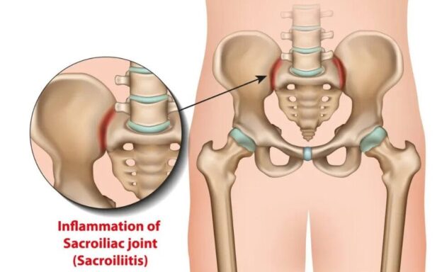
Definition of ankylosing spondylitis:

Ankylosis means joints fuse and become unmovable, and spondylitis is inflammation involving the spine’s vertebrae and facet joints.

The spine becomes stiff and immobile when vertebrae or other bones/joints fuse. AS commonly involves sacroiliac joints but may affect other joints like the spine and cause kyphosis.

    1. Ankylosing spondylitis is a form of arthritis that can affect the spine.

    2. The pain associated with ankylosing spondylitis can be severe.

    3. Ankylosing spondylitis is thought to be caused by a combination of genetic and environmental factors.

B. Demographics of ankylosing spondylitis

    1. Ankylosing spondylitis is most common in men.

    2. Ankylosing spondylitis is also more common in people between the ages of 30 and 60.

C. Causes of ankylosing spondylitis

1. It is believed that a combination of genetic (HLA B27) and environmental factors is what causes ankylosing spondylitis.

    2. Environmental factors that may increase your risk of developing ankylosing spondylitis include your family history and your overall health.

D. Risk factors for ankylosing spondylitis

    1. Age is one of the main risk factors for ankylosing spondylitis.

    2. Other risk factors for ankylosing spondylitis include smoking, obesity, and a sedentary lifestyle.

E. Symptoms of ankylosing spondylitis

  1. The classic symptoms of ankylosing spondylitis include severe pain in your spine, stiffness, and limited movement.
  2. Other symptoms of ankylosing spondylitis can include fatigue, mood swings, and trouble sleeping.
  3. Inflammation in key areas. AS could be to blame if you experience discomfort in different parts of your body. The places most often affected by AS include:
  4. SI joints
  5. Lower back vertebrae
  6. Hip and shoulder joints
  7. The entheses, or areas where tendons and ligaments attach to bones, are mainly in your spine but sometimes at the back of your heels
  8. The cartilage around your ribs and breastbone

F. Diagnosis of ankylosing spondylitis

    1. Ankylosing spondylitis can often be diagnosed based on your medical history, symptoms, and blood work with HLA B27.

    2. Your doctor may order tests to confirm the diagnosis, such as an x-ray or an MRI.

G. Treatment of ankylosing spondylitis

    1. The goal of treatment for ankylosing spondylitis is to reduce your pain and improve your mobility.

    2. Treatment for ankylosing spondylitis may include physical therapy, medications, and surgery.

  1. DMARDs prescribed to treat AS include methotrexate and sulfasalazine (Azulfidine)
  2. Biologics such as adalimumab (Humira), certolizumab (Cimzia), secukinumab (Cosentyx), and ixekizumab (Taltz).
  3. Laminectomy
  4. Osteotomy
  5. Spinal instrumentation and fusion
  6. Joint replacement

H. Prognosis of ankylosing spondylitis

    1. The prognosis for ankylosing spondylitis varies based on age, symptoms, and treatment response.

    2. In some people with ankylosing spondylitis, the condition worsens over time.

I. Precautions for ankylosing spondylitis: FIRST AND FOREMOST QUIT SMOKING

    1. take precautions to prevent falls in people with ankylosing spondylitis.

    2. Avoid heavy lifting, which can aggravate ankylosing spondylitis.

    3. Be sure to wear supportive shoes around people with ankylosing spondylitis.

III. Conclusion

Ankylosing spondylitis is a common form of arthritis that can cause severe pain in the spine. A combination of genetic and environmental factors may be to blame for the chronic condition ankylosing spondylitis. If you or a loved one is diagnosed with ankylosing spondylitis, be sure to discuss treatment options with your doctor, Dr Rao, the best neurosurgeon and spine surgeon in Andhra Pradesh, Guntur, and India. Dr. Raos Hospital is the best neurosurgery hospital in India; Guntur and Andhra Pradesh provide the best spine surgery, spinal surgery, or spine specialist care for ankylosing spondylitis. Call us at 9010056444 or 9010057444 for an appointment.
the-best-sciatica-treatment-in-Guntur at Dr Rao's Hospital by Dr Rao

The best sciatica treatment in Guntur

Sciatica


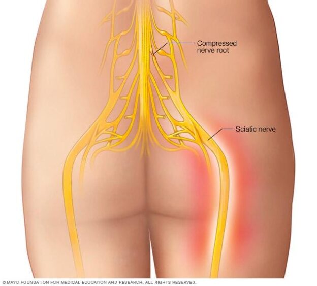
Sciatica is a pain caused in the back of the leg due to irritation and inflammation secondary to nerve compression. The two most common causes of sciatica are herniated discs or lumbar canal stenosis. 8 out of 10 people’s pain decreases without surgery. Sciatica is a common condition that can cause pain in the lower back and legs. This article will discuss the causes, symptoms, and treatment options for sciatica. Dr. Rao’s hospital is the place to go for spine surgery or spinal surgery treatment in Guntur, Andhra Pradesh, or India. Dr. Mohana Rao Patibandla is the best spine surgeon in Guntur and Andhra Pradesh.

What is sciatica?


The sciatic nerve is the thickest in the back, originating from the lower two lumbar or upper three sacral nerves. The irritation or inflammation leads to nerve pain and is called sciatica. The pain generally starts in the back or gluteals and then goes back to the bottom of the leg. Actual injury to the Sciatic nerve is rare, but the compression leads to pain. Sciatic compression may cause weakness of the leg and foot and tingling or unpleasant sensation in the leg and foot.

What does sciatica pain feel like?


Pain is burning or stabbing or electric or sharp shooting or jolts of pain. Pain may be intermittent or constant, more in the leg than the back, increasing with sitting or standing for a prolonged time, twisting the body, coughing, or sneezing.

Can sciatica occur down both legs?


Generally, bilateral nerve compression is rare but can occur sometimes and may present bilaterally.

Does it take time to develop?


Sciatica may be sudden or gradual.

How common is sciatica?


Back pain is one of the most common complaints to visit the hospital, and there is a 40% chance that every person will feel sciatica in their lifetime.

What are the risk factors for sciatica?

Accident or injury or trauma to the back
Aging
Overweight or obese
Weak core muscles, that is, the abdomen and back
Weight lifting
Prolonged sitting
Poor posture in Gym or workplace
Osteoarthritis
Diabetes
Sedentary lifestyle
Smoking

Is the weight of pregnancy the reason why so many pregnant women get sciatica?


Ligaments loosening due to hormones is the main reason for developing sciatica.

SYMPTOMS AND CAUSES


What causes sciatica?

A herniated or slipped disk – the most common cause
Spinal stenosis
Spondylolisthesis
Osteoarthritis of aging with bone spurs
Trauma
Tumors
Piriformis syndrome
Cauda equina syndrome

What are the symptoms of sciatica?


Pain radiating down the leg
tingling numbness, pins and needles sensation in the legs radiating downwards
pain worsens with movement
Bowel and bladder incontinence

DIAGNOSIS AND TESTS


Medical History
Straight leg raise test
FABER Test
Spinal X-rays – Flexion and extension
Magnetic resonance imaging (MRI) – will note detailed soft tissue problems, disc desiccation, disc prolapse, and ligamentum flavum hypertrophy.
Computed tomography (CT) scans fractures, infection, to know more about bony pathology.
Nerve conduction velocity studies (NCV) or electromyography (EMG)
Myelogram if MRI is contraindicated

MANAGEMENT AND TREATMENT

How is sciatica treated?


The goal of treatment is to increase mobility and decrease pain; 8 out of 10 cases don’t need surgery.

Conservative management or self-care treatments:
Appling ice and hot packs: 20 min of hot or cold packs, switching between the hot and cold several times a day.
Taking over-the-counter medicines: non-steroidal anti-inflammatory drugs (NSAIDs)
Performing gentle stretches: Core muscle strengthening

How long can a patient wait to go to a Neurosurgeon?


Every person with sciatic pain is different. Typically, six weeks of conservative management.
Muscle relaxants: cyclobenzaprine, tricyclic antidepressants, and anti-seizure medications.
Physical therapy: Stretching and aerobic exercises
Spinal injections: Corticosteroid and lidocaine mixture


When is surgery considered?


Indication of surgery is alarming symptoms like increasing pain severity even after six weeks, the development of weakness, or bowel or bladder incontinence. A neurosurgeon can do surgery immediately if a patient develops bowel or bladder symptoms or cauda equina syndrome.
The goal of the surgery is to decompress the nerve and stabilize the spine.

Surgical options to relieve sciatica include:
Microdiscectomy
Laminectomy
Transforaminal lumbar interbody fusion

How long does the surgery take, and what’s the typical recovery time?


Laminectomy and Discectomy (endoscopic or microscopic) – generally take one to two hours to perform. Recovery time is around six weeks.

What are the complications of spinal surgery?


Blood clots.
Bleeding.
Infection.
Spinal fluid leak
Nerve damage.
Bladder or bowel control incontinence.

What complications are associated with sciatica?


Developing chronic pain
Foot drop
Loss of feeling in the legs
bowel or bladder disturbance
call the Neurosurgeon immediately if anything develops

PREVENTION

Can sciatica be prevented?


Maintain good posture
Don’t smoke:
Maintain a healthy weight
Exercise regularly: strengthen your core
Activities that don’t hurt your back
Keep yourself safe from falls

OUTLOOK / PROGNOSIS

What can I expect if I have been diagnosed with sciatica?


The good news is that it goes on its own. Eight out of 10 people don’t need surgery, improve in the first six weeks, and rarely need surgery.

LIVING WITH

When should I contact my healthcare provider?

Severe leg pain increases even after six weeks, or excruciating pain lasting more than a few hours.
Muscle weakness or Numbness in the same leg.
Bowel or bladder incontinence
Cauda equina syndrome
Trauma
infection

Is the sciatic nerve the only source of “sciatica” pain?


No, upper or lower roots can also cause symptoms and are considered sciatica.

How can I tell if the pain in my hip is a hip issue or sciatica?


FABER test

Is radiculopathy the same as sciatica?


Radiculopathy and sciatica are interchangeable words.

Should I rest if I have sciatica?


2 to 3 days of activity restriction is advised, but not bed rest

Can sciatica cause my leg and ankle to swell?


Yes, it might cause swelling.

Sciatica is a pain in the back of the leg caused by irritation and inflammation secondary to nerve compression. The two most common causes of sciatica are herniated discs and lumbar canal stenosis. Six weeks of conservative treatment will reduce pain in most people. Eight out of 10 people’s pain decreases without surgery. Dr. Rao’s hospital is for spine surgery and spinal surgery treatment in Guntur, Andhra Pradesh, India. Dr. Mohana Rao Patibandla is the best spine surgeon in Guntur and Andhra Pradesh.

The Science of Brain-Computer Interfaces: How to Control Computers with Your Mind

Cauda Equina Syndrome (CES) – Dr Rao at Dr Rao’s Hospital

Cauda Equina Syndrome (CES) – The best treatment at Dr Rao’s Hospital

Get the best neurosurgeon in Guntur. Dr. Mohana Rao Patibandla is doing minimally invasive spine surgery with a fixation for a patient with cauda equine syndrome secondary to spine trauma.

Compression of the spinal cord results in Cauda Equina Syndrome, a severe neurological condition. Early diagnosis and treatment are essential to improving the chances of a good outcome. The cauda equina is a bundle of nerves at the base of the spinal cord that controls movement and sensation in the legs and lower body. When these nerves are compressed, it can cause pain, numbness, and weakness in the legs. Cauda Equina Syndrome is a medical emergency and requires immediate treatment. Guntur’s best spine surgery hospital offers round-the-clock emergency surgical services and care.

Causes:

Various factors, including a herniated disc, spinal tumors, or trauma to the spine, can cause this compression.

One of the most common causes of CES is a herniated disc. A herniated disc occurs when the outer layer of the disc ruptures, allowing the inner layer to bulge out and compress the nerves. This can happen due to various factors, including age-related wear and tear, injury, or lifting heavy objects.

Another common cause of CES is spinal tumors. Spinal tumors are growths that develop on or near the spine. They can be benign (non-cancerous) or malignant (cancerous). While benign tumors are not usually life-threatening, they can still compress the nerves and cause CES.

Symptoms

Symptoms of a herniated disc include pain, numbness, and weakness in the legs and lower body. These symptoms can vary depending on the location of the herniated disc. In some cases, a herniated disc can also cause urinary incontinence. Malignant tumors are more likely to cause CES because they grow quickly and can spread to other body parts. Spinal tumor symptoms include back pain, numbness, and weakness in the legs and lower body. In some cases, a spinal tumor can also cause paralysis.

Diagnosis and Treatment

Once the diagnosis of CES is made and the etiology is established through clinical history, physical examination, diagnostic tests like an MRI of the spine, CT myelogram, and emergent surgery are the treatments of choice. The goal of CES treatment is to free up the compressed nerve roots. if not treated in time, it will result in paralysis and incontinence. The duration of surgery symptoms is less than 48 hours, leading to a better prognosis and looking for the best neurosurgery or spine surgery hospital in Guntur. Look no further than Dr. Rao’s hospital! We’re the best in the business, and our neurosurgeon is the best in India!

Conclusion:

Cauda Equina Syndrome is a severe condition that can cause permanent nerve damage. The syndrome occurs when the cauda equina, a bundle of nerves at the base of the spine, is compressed. A herniated disc, spinal tumors, or spine trauma are just a few causes of this compression.

CES is a medical emergency and requires prompt treatment to prevent permanent nerve damage. Suppose you experience any of the symptoms of CES. In that case, it is essential to seek medical attention immediately and call Dr. Rao’s hospital at 9010056444 or 9010057444 for the best spine surgeon in India, Dr. Mohana Rao Patibandla.

First Biplane cath lab in Andhra Pradesh, India at Dr Rao's Hospital for the advanced interventional neurology procedures

The Best Neurosurgery Hospital in Guntur With The Latest State of the Art Technology

The Best Neurosurgery Hospital in Guntur With The Latest State of the Art Technology

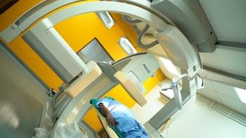
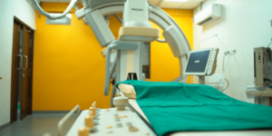
A hybrid operating theater is a type of operating room that combines the features of a traditional operating room and a catheterization lab. It is typically used for procedures that require both surgical and interventional radiology (IR) techniques. 

First biplane cathlab in Andhra Pradesh and Telangana diagnostic-tests-in-neurosurgery-the-best-at-dr-raos-guntur

first biplane cath lab in Andhra Pradesh and Telangana diagnostic-tests-in-neurosurgery-the-best-at-dr-Rao’s hospital-Guntur


Hybrid operation theater @ Dr. Rao’s hospital.

Some of the benefits of using a hybrid operating theater include: 

1. Increased efficiency: procedures typically requiring two separate rooms can be completed in one hybrid room, saving time and money. 

2. Better patient outcomes—Having all of the necessary equipment and personnel in one room can help ensure that procedures are performed safely and effectively. 

3. Enhanced safety: having a dedicated hybrid operating room can help reduce the risk of infection and other complications. 

4. Improved patient comfort—Patients may feel more comfortable knowing that they will not have to be moved between two different rooms during their procedure. 

Our hospital’s Biplane Cath Lab Hybrid operation theater is equipped with the latest 4K endoscopy and neuronavigation technology. This state-of-the-art equipment lets us provide our patients with the best possible care. 

Our hospital’s Biplane Cath Lab Hybrid operation theater is equipped with the latest 4K endoscopy and neuronavigation technology. This state-of-the-art equipment lets us provide our patients with the best possible care. 4K endoscopy provides superior image quality, which helps our doctors diagnose and treat conditions more accurately. 

This advanced equipment helps us to safely and effectively perform procedures such as brain surgery. Neuronavigation technology allows us to precisely target areas in the brain, while neuromonitoring helps us to monitor the patient’s neurological status during surgery. This information is essential to ensure that the procedure is being performed safely and effectively. 

Conclusion: Our hospital’s Biplane Cath Lab Hybrid operation theater is equipped with the latest 4K endoscopy and neuronavigation technology. This state-of-the-art equipment lets us provide our patients with the best possible care. Neuronavigation and neuromonitoring technology help us to safely and effectively perform procedures such as brain surgery.


Your brain and spine health with keyhole surgery

The best Keyhole Neurosurgery-All You Need to Know

Open surgery comes with various complications.

But these complications can now be avoided!

How?

By performing keyhole neurosurgery!

The concept of keyhole surgery is the safe removal of brain and skull base cancers through tiny holes that cause less collateral harm to the scalp, brain, and nerves. The best neurologist in Guntur performs keyhole neurosurgery, which is effective and painless. Keyhole neurosurgery allows patients to avoid the risks of open surgery. This procedure prevents needless surgical site investigation and brain retraction by minimizing the anatomic window and adequately exposing the lesions.

The process, kinds, and benefits of keyhole neurosurgery are discussed in this article.

What is keyhole neurosurgery,, and how does it work?

In keyhole surgery, a neurosurgeon at Rao Hospital Guntur will make a small incision in the location where the surgery will be performed. A light stick called an endoscope with a high-resolution camera is put into this opening. The device has an eyepiece that the neurosurgeon may look through. The camera sends photographs to your neurosurgeon in Guntur, and the light illuminates the inside of the channel. Delicate surgical equipment is inserted via another similar incision, and your neurosurgeon uses it to cut or amputate the tumor.

You will be unconscious for the length of the surgery because a general anesthetic will be used.

What are the different types of keyhole neurosurgery procedures?

The best neurology hospital in Guntur does the following keyhole neurosurgeries:.

  1. Keyhole brain surgery:

In this procedure, a hole is formed above the brows. Your neurosurgeon in Guntur can also insert an endoscope through the mouth. The following people are candidates for keyhole brain surgery:

  • Those with pituitary tumors:

A pituitary gland tumor is a collection of abnormal cells that have grown out of control in your pituitary gland.

  • Those diagnosed with Glioma

Glioma is a cancer that affects the brain and spinal cord.

  • Those diagnosed with chordoma or meningioma

Chordoma is an uncommon kind of bone cancer that most commonly affects the spine or skull bones. It usually appears where the skull sits atop the spine (skull base) or at the bottom of the spine (spine base) (sacrum). Conversely, a meningioma is a tumor that develops from the meninges, the membranes surrounding your brain and spinal cord.

2• Keyhole spinal surgery:

 Typically, open operations are used to treat spinal disorders. However, this procedure causes tears in the tissues of the spine. This condition can be solved with keyhole spinal surgery. A tubular retractor is used in keyhole spinal surgery to drill a hole in the spine while holding the muscles apart without causing a rupture. The following people are the best candidates for keyhole spinal surgery: –

  • Those with disc issues:

Disk issues cause discs to break down, causing excruciating discomfort. Herniated discs and degenerative disc disease are two common disc issues.

  • Those who have bone spurs on their spine:

Bone spurs indicate spinal degeneration and signify an expansion of the bony structure.

  • Those who suffer from spinal instability:

“Spinal instability” was coined to describe abnormal movement between vertebrae. When a disc degenerates, it loses tension or turgor, which allows the disc to bulge and allow more movement between the vertebrae.

What are the benefits of keyhole surgery in neurosurgery?

The following are some of the benefits of keyhole neurosurgery:

  1. • They lessen the chances of an infection developing at the operation site.
  2. • Healing time is reduced.
  3. • Keyhole neurosurgery avoids damaging the surrounding tissues and blood vessels.
  4. • Improved cosmetic outcomes

Conclusion

I hope this article has helped you understand keyhole neurosurgery, its types, procedures, and benefits. Keyhole surgery is a boon that overcomes the various complications of open surgery. Keyhole neurosurgery comes with multiple benefits. It decreases the risk of infections in the area of surgery and prevents the tissues from getting damaged. The healing time is also faster in keyhole neurosurgery, leading to better cosmetic results.

The top neurosurgeon in Guntur performs keyhole neurosurgery with great care and caution and has successfully performed many keyhole neurosurgeries.

So, visit the top neurosurgeon in Guntur right now to benefit from keyhole neurosurgery!

Dr. Rao’s Contact Information:

Craniosynostosis

Craniosynostosis- All you Need to Know

Have you seen a baby with an abnormally shaped skull in Guntur?

If so, they are likely to have craniosynostosis. Craniosynostosis is when a baby develops or is born with an abnormally shaped skull.

This happens when the natural spaces known as cranial sutures in the baby’s skull join earlier than they should.

The sutures should usually be joined only after the child reaches 2. This allows the brain to develop without any pressure from the skull.

Craniosynostosis affects the shape of the skull and the self-esteem of children. An odd-shaped head makes them feel odd and uncomfortable. They feel excluded from the rest of the crowd. It makes them feel lonely and isolated. They start feeling ashamed of their heads and ashamed of themselves.

But there’s a solution for this. Children with unusually shaped skulls no longer need to feel ashamed of their heads or themselves. They no longer need to feel excluded from the crowd. They no longer need to feel sad and isolated. Because there are treatment options available for craniosynostosis, by getting treatment for Craniosynostosis from the best neurology hospital in Guntur, one will not only get rid of craniosynostosis but also replace their isolation with happiness and shame with pride and confidence. They will no longer feel apart but will feel a part of society.

This article discusses craniosynostosis’s types, symptoms, causes, and treatment options.

First, let us understand the types of craniosynostosis.

What are the different types of craniosynostosis?

The different types of Craniosynostosis are as follows:

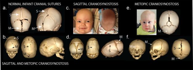
Sagittal synostosis

  • The sagittal suture, which runs along the top of the skull, unites relatively early in this condition. The head grows taller but narrower than it should in this kind of Craniosynostosis. Boys are more likely to experience this.

Coronal craniosynostosis

• The sutures that connect the top of the head to the ears fuse sooner than they should in this case. The kid will have a flat forehead if he or she has this form of Craniosynostosis. Girls are more likely to experience this.

Synostosis metopic

• The sutures that connect the sagittal suture to the nose fuse quite quickly. As a result, the scalp develops a triangle shape.

Synostosis of the lambdoid

This type of craniosynostosis affects the lambdoid suture at the back of the skull, forming a flat-topped head.

The best neurologist in Guntur can treat the types as mentioned above of craniosynostosis.

What are the symptoms of Craniosynostosis?

The common symptoms of Craniosynostosis are as follows:

  • An unusually shaped skull
  • The head grows slower than the body
  • Formation of a hard ridge along the suture.
  • The fontanel disappears soon.

If you experience any of the above symptoms of cranial synostosis, it is advisable to seek help from Rao Hospital in Guntur.

What are the causes and risk factors of Craniosynostosis?

Craniosynostosis can be either syndromic or nonsyndromic. Syndromic Cranial Synostosis occurs with other birth defects. With Cranial Synostosis, changes may be in genes that further affect different parts of the body, like fingers, heart, etc. Cell defects in the sutures may be the cause of Nonsyndromic Cranial Synostosis, which causes the sutures to fuse earlier than usual.

The following are the risk factors of craniosynostosis:

  • Taking valproic acid-like Depakote for epilepsy at the time of pregnancy may increase the risk of giving birth to a child having Craniosynostosis.
  • Babies whose mothers have thyroid disease or undergo thyroid treatment during pregnancy are more likely to be born with Craniosynostosis.
  • Babies whose mothers took fertility medicines like clomiphene citrate during pregnancy are more likely to be born with Craniosynostosis.

What are the treatment options for treating Craniosynostosis?

Surgery is an effective treatment option for cranial Synostosis. The best neurologist in Guntur can perform the surgery.

There are usually two types of surgeries that are preferred to treat Craniosynostosis. They are as follows-

• Conventional surgery:

In Guntur, a neurologist and a craniofacial surgeon perform the procedure. The neurosurgeon makes an incision across the top of the baby’s scalp and removes any sections of the skull that are out of form. The craniofacial surgeon then restores and replaces these pieces of the skull in the head. After that, stitches are applied. The doctor will later monitor the development of the skull.

• Endoscopic Surgery

In this procedure, your doctor in Guntur makes two small incisions in the scalp and uses an endoscope to cut the sutures. This procedure should be performed before the baby reaches the age of three months. After the treatment, wearing a molding helmet is recommended to allow your head to grow into its correct form.

It is essential to get timely treatment for Cranial Synostosis from the best neurosurgeon in Guntur to avoid further complications. If Cranial Synostosis is left untreated, the skull will continue to develop in an unusual manner, which may lead to additional complications and problems such as vision problems, etc.

Conclusion

This article explains craniosynostosis, its symptoms, causes, and treatment options. It is advisable to get proper and timely treatment for craniosynostosis from the best neurology hospital in Guntur to avoid further complications, to feel a part of the crowd and not feel apart from the crowd, and to develop a feeling of oneness and not feel different and odd! It is essential to get timely treatment for Craniosynostosis to replace discomfort with self-worth and self-esteem.

Dr. Rao’s Contact Information:


stroke-and-mechanical-thrombectomy at Dr Rao's Hosptial by Dr Rao

The best STROKE Treatment is MECHANICAL THROMBECTOMY

The most effective STROKE treatment is MECHANICAL THROMBECTOMY.

Stroke is dangerous and very damaging. It is caused by a blood clot that disrupts the flow of blood to the brain.

However, it is now possible to overcome the dangerous stroke with a procedure called mechanical thrombectomy. Yes, mechanical thrombectomy treatment by the best neurologist in Guntur can help one successfully defeat a stroke.


But what is a mechanical thrombectomy?

Mechanical Thrombectomy is an effective stroke treatment. It helps remove blood clots that disturb blood flow in the body. Blood clots slow down blood flow in the body, causing different complications. Mechanical Thrombectomy removes these blood clots. It helps treat stroke and other complications caused by the formation of blood clots. Mechanical Thrombectomy help one deal with strokes and confront them effectively. Now, one can courageously face and defeat problems like a stroke with the help of mechanical thrombectomy treatment from the best neurology hospital in Guntur.

What Causes Stroke?

The common risk factors that lead to stroke are as follows:

· Improper Sleep

· Fear and anxiety

· Irregular eating habits

· Air Pollution

· Smoking and drinking alcohol

In case you get a stroke, it is advisable to seek immediate help from neurologists at Rao Hospital, Guntur. With treatments like mechanical thrombectomy, you are sure to recover from a stroke.

What kinds of devices are used in mechanical thrombectomy procedures?

Neurosurgeons in Guntur carry out mechanical thrombectomies to remove blood clots using the following tools:

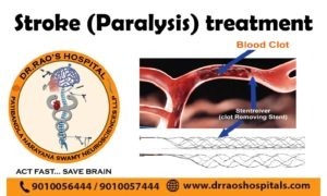
· Stentriever mechanical thrombectomy

The latest mechanical device is a stentriever. The device is a self-expanding mesh tube attached to a wire-guided through a catheter (thin tube). The surgeon inserts the catheter in an artery in the groin and guides the catheter through various blood vessels up to the blood clot in the brain.

· Aspiration thrombectomy –

Aspiration thrombectomy entails either manual cluster yearning or the use of catheter equipment to suck the coagulation out of the vessel.

How is the mechanical thrombectomy procedure performed?

In mechanical thrombectomy, neurosurgeons in Guntur use a vacuum or mechanical gear to remove blood clots. Thus, the blood flow is restored, and stroke gets treated.

To achieve effective results, it is advisable to get mechanical thrombectomy treatment from the best neurology hospital in Guntur.

Why go for a mechanical thrombectomy?

In mechanical thrombectomy treatment, one has a window of 24 hours from the onset of a stroke to have a chance to recover from a stroke. This will help more patients recover from stroke and save them from the harmful impact of stroke. Mechanical Thrombectomy is a ray of hope for people affected by stroke. Mechanical Thrombectomy give them hope and courage to fight strokes. It gives life to their dead hopes and courage to fight stroke. It is a powerful weapon that helps people to win against difficult stroke. Mechanical Thrombectomy also leads to a speedy recovery from stroke. So, going for mechanical thrombectomy treatment at the best neurology hospital in Guntur is advisable.

What is the problem with mechanical thrombectomy?

Though mechanical thrombectomy is an effective procedure to treat stroke, there aren’t sufficient neurology hospitals in Guntur that provide this stroke treatment option.

Conclusion

In this article, we learned about stroke and the risk factors that cause a stroke. We also learned about mechanical thrombectomy treatment, which helps treat stroke. We learned about the devices used in mechanical thrombectomy treatment and its procedure. We also learned about the advantages and problems of mechanical thrombectomy in Guntur. A stroke is a severe problem. Before it defeats you, you defeat it with the help of mechanical thrombectomy treatment from the best doctor in Guntur. Mechanical Thrombectomy not only gives you life but also gives life to your dead hopes and courage. Mechanical Thrombectomy is a powerful and effective weapon that helps you battle stroke. It enables you to defeat death and makes your life win this battle. So, now it’s time to make your life a winner, not the time to lose it and allow it to perish! Now it’s time to defeat and not get defeated! Now is the time to win! Now is the time to live!

Dr. Rao’s Contact Information:

the best migraine treatment in Andhra Pradesh at Dr Rao's Hosptial by Dr Rao

Migraine – Symptoms, Signs, Causes, Types, and Treatment

Migraine – Symptoms, Signs, Causes, Types, and Treatment

Migraine is a neurological disease that is common in people of Guntur. In migraines, people experience severe headaches. Vomiting and nausea are common side effects of headaches. In migraines, one may also experience sensitivity to sound and light, problems in speech, etc. Migraine can occur in any age group. If you have migraine problems running in your family, it is more likely you will also experience migraines. In case you experience migraine headaches, it is advisable to visit Rao hospital Guntur, which is one of the best neurology hospitals in Guntur. This article discusses migraine symptoms, causes, types, and treatment.

What is a migraine?

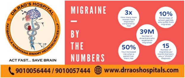
Migraine is a neurological condition in which one experiences severe headaches, nausea, and vomiting. One may also experience sensitivity to light and sound. Migraine is more common in women than men. The most common migraines are migraines with aura and without aura. We will study each of them in detail later in the article.

What are the common signs and symptoms of migraine?

Migraine occurs in stages. The symptoms of migraine may differ depending on the stage.

Prodrome stage:

In this stage of migraine, symptoms are experienced a few days before the headache starts. The common symptoms experienced in this stage include the following-

Increased hunger

Tiredness

Stiffness in the neck

Feeling depressed

Aura:

This occurs after the prodrome stage. The common problems experienced in this stage include the following-

Problems in speaking

Vision problems

Sensation problems

Attack:

In this phase, a severe headache is experienced. The common symptoms in this stage include the following-

Nausea

Vomiting

Very severe pain in your head

Sensitivity to light and sound

Postdrome Phase:

The following symptoms characterize this phase-

Extreme mood swings

Headaches

If you experience any of the above symptoms, you should visit the best neurologist in Guntur immediately for proper treatment. When left untreated, migraines may become severe and last longer.

What are the possible causes of migraine?

The actual cause of migraine is not yet known. However, certain risk factors may lead to migraine. Some of them are as follows-

  • Changes in weather
  • Less water intake in the body
  • Hormone changes
  • Increased stress
  • Irregular eating schedule and skipping meals
  • Improper sleep
  • Specific medications
  • Smoking and alcohol

Your neurologist may ask you in Guntur to keep a record of the things that you ate or the medicines you took before the onset of your migraine. This may help identify the triggers that lead to migraines and thus avoid them to reduce and treat them.

How can migraines be diagnosed?

Doctors in Guntur can diagnose migraines by examining symptoms, medical conditions, and family history. Various tests are used to diagnose migraines.

What are the different types of migraines?

The following are the different types of migraine seen in people of Guntur

Migraine without Aura:

This is one of the most common types of migraine. People with this type of migraine have had at least five attacks that have these characteristics:

Headaches lasting for 4-72 hours if timely treatment is not provided.

Moderate or severe headache on one side of the head

Headaches accompanied by sensitivity to light and sound and nausea.

Migraine with Aura:

This is also one of the common types of migraine. People with this type of migraine have had at least 2 attacks that have these characteristics:

Vision problems

Speech problems

Vertigo

Sensory problems

Chronic migraines:

People with chronic migraines experience severe headaches. They may also experience the following-

Depression

Other medical problems

Other chronic pain

Acute migraines:

This type of migraine is not chronic. People experience less severe headaches than chronic migraines in this type of migraine.

There are other types of migraines, such as an optical migraine(affects one of the eyes), Acephalgic migraine(migraine with no headaches), hormonal migraines(caused by changes in hormones like estrogen), abdominal migraine(experiencing stomach-ache), among others.

How to treat migraines?

Migraine is a very fatal condition. So, there are treatment options available to treat migraines. Migraines can’t be completely cured, but these treatment options can help control and reduce migraines to a great extent.

  • Lifestyle changes include proper sleep, regular exercise, stress management through yoga and meditation, avoiding triggers that lead to migraine attacks, and drinking plenty of water.
  • Medications like NSAIDs, Triptans, and CGRP analogs Are superior.
  • Surgical interventions
  • Acupressure and acupuncture also help treat migraines
  • Home remedies like lying in a quiet, dark room and placing a cold cloth over your head may also help.

It is advisable to visit a specialist in Guntur in case you experience severe migraine headaches to get proper treatment and to manage your migraines.

Conclusion

This article helped us understand migraines’ symptoms, causes, types, treatments, etc. Regular exercise, drinking plenty of water, and managing stress through yoga and meditation can help greatly in controlling and managing migraines. If you experience severe migraine headaches, kindly visit a neurologist in Guntur immediately to get proper treatment and relief from the severe headache that hinders you from performing your day-to-day activities.

Dr. Rao’s Contact Information:

Your health and well-being are our top priorities. For the best migraine care, trust in the expertise of Dr. Rao and the other leading neurosurgeons in India.

the best Hydrocephalus-treatment at Dr Rao's Hospital by Dr Rao blog

Hydrocephalus- All you need to know

Hydrocephalus: All you need to know

Various illnesses and diseases have an impact on the brain. This article discusses a type of brain condition known as ‘Hydrocephalus.’

What is Hydrocephalus, and how does it affect you?

We may deduce from the name that it relates to water/fluid because it contains the word hydro.

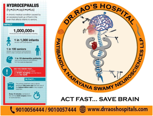
Yes, Hydrocephalus is a condition in which the brain has excess water. It is a disorder in which the brain accumulates too much cerebrospinal fluid, causing increased pressure in the skull. The tissues that line the brain’s ventricles produce cerebrospinal fluid. The fluid travels around the brain and spinal column, filling gaps. It’s absorbed chiefly via blood vessels in tissues around the brain’s base.

Cerebrospinal fluid is necessary because it eliminates waste products from the brain’s metabolism and allows the brain to float within the skull. However, too much of this fluid is damaging to the brain, resulting in disorders like Hydrocephalus. But don’t worry, Hydrocephalus can be treated with therapy from the best neurology hospital in Guntur.

Who is at risk for Hydrocephalus?

Hydrocephalus is more common in infants and those over the age of 60.

What are the signs and symptoms of Hydrocephalus?

The signs and symptoms of Hydrocephalus differ depending on the age at which it develops.

The following are some of the most prevalent symptoms of Hydrocephalus in infants:

  • Head size changes, such as a sudden increase in the size of the head
  • Nausea and vomiting
  • Feeling sleepy
  • Downward-looking eyes
  • Lack of sensitivity to touch
  • Inadequate development

The following are some of the most prevalent symptoms of Hydrocephalus  in older children:

  • You have a headache.
  • Visual impairment
  • Feeling sleepy
  • Nausea and vomiting
  • An abnormal increase in the head’s size
  • Appetite loss.
  • Changes in personality
  • Issues with speaking, walking, and other activities
  • A poor academic record

The following are some of the most prevalent Hydrocephalus symptoms in young and middle-aged adults:

  • You have a headache.
  • Tiredness and sluggishness
  • Visual impairment
  • Issues with balance
  • Issues with memory

The following are some of the most prevalent symptoms of Hydrocephalus  in elderly adults:

  • Urge to urinate regularly
  • Issues with memory
  • Issues with balance
  • Walking difficulties

Please see a neurologist in Guntur if you have any of the above-mentioned symptoms. Numerous therapies and facilities are available in Guntur to treat such illnesses.

We now have a better understanding of the symptoms that people with hydrocephalus encounter. Let’s now look at some of the most common causes of hydrocephalus.

What causes Hydrocephalus, and how may it be treated?

An increase in CSF fluid in the ventricles causes Hydrocephalus. The following factors can induce an excess of cerebrospinal fluid in the ventricles:

A blockage in the flow of cerebrospinal fluid from one ventricle to other areas of the brain causes hydrocephalus.

Problems with Absorption: One of the most common causes of Hydrocephalus is poor cerebrospinal fluid absorption.

Hydrocephalus is caused by excessive cerebrospinal fluid production, which is greater than the absorption rate.

Some factors enhance the risk of developing Hydrocephalus. The following are a few of them:
  • Tumors of the brain
  • A head injury causes bleeding in the brain.
  • Other types of traumatic brain damage
The following are some of the reasons that may cause Hydrocephalus in newborns:
  • The abnormal development of the central nervous system causes a blockage in cerebrospinal fluid flow.
  • Bleeding in the ventricles

Hydrocephalus Treatment

Hydrocephalus can be treated effectively with the help of experts in Guntur. In the case of severe Hydrocephalus, however, there may be severe consequences. The severity of the initial symptoms, how long it takes to diagnose and treat the condition, and other factors all affect the severity of complications.

Final Thoughts

This article helped us understand hydrocephalus and who is at risk of developing it. It also taught us about the signs and symptoms of hydrocephalus, which vary depending on whether the patient is an infant, a kid, or an adult. We also discovered that cerebrospinal fluid production and absorption imbalances cause hydrocephalus. We also learned about the most prevalent cause of hydrocephalus and the difficulties that come with it.

On the other hand, Hydrocephalus can be effectively treated with no severe problems if treated promptly and adequately by a neurologist in Guntur. So, if you’re experiencing any of the hydrocephalus symptoms listed above, see a neurologist in Guntur immediately. “The sooner, the better,” as the saying goes. The sooner an issue is identified and treated, the better, as it decreases the severity of treatment-related consequences.

In Conclusion

Choosing the right neurosurgeon for brain tumor treatment is crucial for ensuring the best possible outcomes. Dr. Rao at Dr. Rao’s Hospitals in Guntur and the other top specialists listed represent the pinnacle of expertise and compassionate care in neurosurgery. If you or a loved one are seeking treatment for a brain tumor, consider reaching out to these esteemed professionals.

Contact Information:

Dyskinesia - the best treatment at Dr Rao's hospital, Guntur

Dyskinesia – All you need to know

Dyskinesia: All You Need To Know

Dyskinesia is an uncontrollable, spontaneous reaction. It can involve one portion of the body, such as the head or arm, or the whole body.

Dyskinesia can vary from mild to extreme and can be very painful. It can also make performing your everyday work challenging. It may also vary in terms of frequency and time of day.

If you are from Guntur and searching for a proper treatment for dyskinesia, you should visit Dr. Mohana Rao’s hospital, the best neurosurgeon in Guntur. 

Dyskinesia is a common complication of long-term levodopa therapy in people with Parkinson’s disease. Dyskinesia may also occur in conditions other than Parkinson’s disease, such as coordination disorders.

Now let us learn about the different signs and symptoms of dyskinesia:

The signs and symptoms of dyskinesia differ from person to person. They may be mild, with a slight head, arm, or leg twitch. They can also be severe, causing various body parts to move involuntarily. Dyskinesia can happen in a variety of ways, including:

  • fidgeting
  • boobing in head
  • restlessness
  • writhing
  • twitching 
  • the swaying of the body 

Dyskinesia is not the same as tremor but is common in Parkinson’s disease. 

Now let us see the reasons behind this:

The most frequent cause of dyskinesia is long-term use of the drug levodopa. Because of its efficacy, levodopa is the recommended drug for the treatment of Parkinson’s disease.

Dopamine levels in the brain rise due to the use of levodopa. A lack of dopamine-producing brain cells causes Parkinson’s disease. For those with Parkinson’s disease and other disorders involving reduced dopamine levels, levodopa increases the dopamine level. 

When you take levodopa, as per your neurologist in Guntur, the dopamine levels increase and then decrease as the drug wears off. Variations in dopamine levels are thought to be the source of dyskinesia’s spontaneous gestures.

Some antipsychotic drugs cause tardive dyskinesia, a form of dyskinesia.

Conditions that are related to dyskinesia:

Dystonia:

Dystonia and dyskinesia are often confused. Dystonia induces excessive muscle tightening rather than the involuntary movements of dyskinesia.

Dystonia is a drug complication. Low levels of dopamine, which are seen in patients with Parkinson’s disease, induce dystonia. Dystonia often affects the feet, vocal cords, hands, and eyelids. It often impacts only one side of the body.

Since levodopa causes dopamine levels to fluctuate, it can cause dystonia. Dystonia can occur as dopamine levels fall and the levodopa wears off. 

Tardive dyskinesia:

People with severe psychiatric disorders that need long-term antipsychotic therapy suffer from tardive dyskinesia. Since it induces repetitive gestures, tardive dyskinesia is similar to dyskinesia.

On the other hand, Tender dyskinesia typically affects the tongue, lips, jaw, or eyelids. The following repetitive gestures can be a sign of tardive dyskinesia:

  • licking your mouth repeatedly 
  • grimacing continuously 
  • blinking rapidly, 
  • puckering your mouth  
  • poking out your tongue

Now, let us see what the treatment for dyskinesia is.

Each person’s dyskinesia treatment needs to be unique. Any of the following considerations influence treatment:

  • The seriousness of the symptoms
  • When do the effects worsen (for example, when the levodopa wears off)?
  • Age
  • Amount of time on levodopa, and 
  • Time after getting a Parkinson’s diagnosis

Below are some possible recovery options:

  • Adjusting your levodopa dosage to prevent the significant fluctuation of dopamine levels in your bloodstream
  • You can use a continuous infusion or a sustained-release formulation of levodopa
  • Take amantadine extended-release (Gocovri), a well-approved treatment for tardive dyskinesia
  • valbenazine, a newly approved drug for tardive dyskinesia
  • Taking levodopa in smaller doses 
  • Take the levodopa 30 minutes before your meal 
  • According to your neurosurgeon in Guntur, exercising, such as walking and swimming, would be best.
  • It will help eliminate extra stress, which can harm you. 

Before starting the proper medication for you, discuss every detail about your treatment with the doctor.

When dyskinesia progresses, speaking with the doctor about the different treatments is essential. When you are first diagnosed with Parkinson’s, talk about the benefits and drawbacks of taking levodopa. It would be better for you if you could delay starting levodopa, as it increases the chances of dyskinesia.