Category Archives: Minimally Invasive Spine Surgery

minimally-invasive-spine-surgery-working-treatment-and-spinal-fusion

The best Minimally Invasive Spine Surgery in Guntur

Minimally invasive spine surgery is a relatively new treatment that allows surgeons to shun the traditional, open approach. By performing this procedure with a tiny incision, the surgeon can avoid disrupting muscles, nerves and blood vessels. We, humans are walking because of the muscles and the minimally invasive spine surgery will not damage the muscles unlike the traditional open surgery.

The results are dramatic: shorter operation time, less pain for the patient, and fewer complications for your surgeon. What’s even more amazing is that the best spine doctor at the best spine surgery hospital in Guntur agrees that Minimally Invasive Spine Surgery provides more benefits than traditional Surgery like less blood loss, less recovery time, faster resumption of activities, less hospital stay, e.t.c.

The first aspect of minimally invasive surgery is reducing surgical exposure to a minimum while maintaining adequate surgical exposure. A spine surgeon in Guntur has learned that a patient’s neurological function and minimizing the collateral damage during surgery are more important than anything else.

It is no longer acceptable to put patients at risk by not making every effort to preserve sensitive structures around their spinal cord. This concern has led to fewer extensive surgeries and smaller incisions which use neuronavigation, neuromontioring with precision and accuracy.

In addition, spine specialists in Guntur are now able to initiate procedures in patients who used to be considered untreatable because of their degree of disability.

Diseases Cured by Minimally Invasive Spine Surgery

  • Lumbar spinal stenosis
  • Spinal infections
  • Spinal deformities such as scoliosis, Kyphosis
  • Degenerative disc disease
  • Spondyloarthritis of spine
  • Cervical discectomy
  • Cervical foraminotomy
  • Minimally invasive spine reonstruction procedures
  • Conditions that require full spinal fusion

How does Minimally Invasive Spine Surgery Works?

Minimally invasive spine surgery uses a scope to guide the surgeon during spinal operations. To perform minimally invasive spine surgery, an incision is made in the skin on either side of the spine.

The doctor inserts a scope into this incision and then uses it to manipulate and guide the surgical instruments. The scope is also used by surgeons to view internal injuries, open blood vessels and perform special procedures. Because there is less incision and only a small piece of bone removed, spinal surgeons can operate on patients who would otherwise be considered difficult cases for open procedures.

Using small incisions in combination with instrumentation allows the best spine surgeon in Andhra Pradesh to perform MIS procedures on patients. Whose vertebral bodies would have otherwise been beyond reach with traditional approaches.

These are areas such as the thoracic spine involved in lung cancer surgery. Several methods can be used to minimize trauma during MIS surgery. Some of the more common techniques are outlined here.

Use of Tubular Retractor

The tubular retractors are made of a variety of plastics or metals which are used during spine surgeries. These devices are threaded rods which the surgeon inserts through small incisions in the patient’s back to hold the vertebrae apart from each other. The retractors also allow for easy access to delicate nerves and blood vessels.

These devices ensure that certain spinal segments do not move during surgery by holding them in place. Although these devices can be used with traditional open procedures. They have been found to have great success with minimally invasive navigation solutions.

  1. Percutaneous Placement of Screws and Rods: The surgeon uses the above devices to create an open space through which they can operate. The surgeon inserts a small cage and then inserts pieces of titanium into it which are used to exercise the vertebrae and allow for an in-depth vision.
    Percutaneous means that the surgeon is using a needle to insert rods, screws and instruments. The advantage of using the percutaneous placement of screws and rods is that they can be placed without the need for open invasive surgery.
  2. Direct Lateral Access Routes: This is one of the most common methods used to perform MIS procedures and is similar to the lateral access approach. The patient, who is lying on their side, undergoes an X-ray or using navigation  under neuromonitoring.
    This procedure ensures that there are enough pathways in place to allow them to operate. After making sure that there are no significant nerves on the way to the patient’s spine, they proceed with the surgery.
  3. Thoracoscopic Access Route: Thoracoscopic access is similar to lateral access. In this case, the surgeon uses a camera and a small needle to create an open space on the front side of the thoracic spine.
    The main advantage of this method is that it averts exposure to the patient’s lungs and surrounding muscles during the procedure.

Using these minimally invasive procedures, surgeons can operate on patients who would otherwise be considered “untreatable”.
This minimally invasive technique is more effective than traditional methods in achieving results for many diseases that were previously beyond reach.

Treatment

  1. Endoscopic Discectomy: We are removing herniated discs with endoscopic techniques (with a small scope). This requires that the patient have an X-ray in the spinal suite which shows the disc space well enough for the surgeon to see what he needs to do. Herniated discs are removed through a small incision from above.
  2. Spinal decompression: We are performing spinal decompressions with a minimally invasive approach. These types of surgeries are used to treat herniated discs (which bulge or burst out of their normal location).
    Another condition that can be treated with this method is spinal stenosis, which is the narrowing of the space surrounding the spinal cord or nerves.
  3. Transforaminal Lumbar Interbody Fusion (TLIF): This is a minimally invasive procedure that is done to treat the disc disease called lumbar spinal stenosis or LSS. It is done through a small incision in the patient’s back 2 to 5 cm away from the midline.
    After making sure there are no other issues in the spine, a surgeon inserts a thin Cage into the spine. And then fuses (fixation with rods and screws along with bone) several vertebrae where they have degenerated (to stabilize their structure and improve mobility).

Instruments Used

Some of the more commonly used instruments in minimally invasive spine surgery are:

Screws: These are used to help stabilize and fuse vertebrae. They are usually made of titanium.

Endoscope: These instruments have small heads with a camera attached to them. An endoscope is used to see the inside of the body while performing minimally invasive spine surgeries.

Minimally invasive tubular retractor (MITR): Minimally invasive tubular retractors are used to stabilize vertebrae, mobilize the muscles out of the surgical way and allow for surgical procedure of discectomy, spinal stenosis and others.

Discectomy retractors: Discectomy retractors are used to stabilize vertebrae, and visualize the surgical field. And protect the patient’s back during discectomy surgery.

Bladed Spinal Instrumentation: Many different types of blades can be used during a minimally invasive surgery including spinal bars, wedges, discs and instruments that can remove bone or discs.

Portals: Ports are small incisions that are used to improve visualization and allow for easy placement of instruments. These can be placed through the back, front or side of a patient’s body. It separates tissue during a minimally invasive surgery so that the surgeon can get a clear view of the procedure. And perform it with minimal damage to the surrounding tissue.

Spinal Fusion

Spinal Fusion is a minimally invasive spine surgery that is used to treat herniated discs or spinal stenosis. This procedure involves the removal of the disc. And then fusing it with one or more vertebral bodies to both stabilize them and increase fusion.

The main goal of this procedure is to bring the back body centre closer to the head so that it can be more flexible and easier for patients to move. The procedure requires a patient to be in good physical health, have no infection in their spine and be able to withstand stress from spinal fusion surgeries.

Final Words

Minimally invasive spine surgery is a way for patients to gain back their quality of life. It becomes a total solution to many problems or disadvantages that come with the traditional surgery and recovery process.

Minimally invasive spine surgery should be advised in patients who are willing to undergo spine surgery with less pain, less scarring, less hospital stay, immediate mobilization, less time to work and less blood loss. We, at Guntur in Andhra Pradesh providing all the minimally invasive spine surgeries with utmost care, high technology and efficient technique s at Dr. Rao’s hospital, The best neurosurgery hospital for the minimally invasive spine surgeries.

Dyskinesia - the best treatment at Dr Rao's hospital, Guntur

Dyskinesia: Signs, Symptoms, Causes, Diagnosis and the best treatment

Dyskinesia: Signs, Symptoms, Causes, Diagnosis and the best treatment

Dyskinesia is a chronic neurological disorder that causes involuntary body movements. It is characterized by uncontrollable movements of the joints and muscles, often with a simultaneous movement disorder.

Dyskinesia can affect individuals at all levels of society. Although it frequently manifests as a behavioral symptom, overexposure to toxic substances or diseases like Huntington’s disease and encephalitis can also be the cause.

This post aims to provide information about dyskinesia, how it’s diagnosed, how it’s treated, and why you should take this condition seriously.

Dr Rao Hospital is the best spine surgery hospital in India. It provides the best treatment and surgery for all types of spine diseases.

Diagnosis of Dyskinesia

The first impression of a patient with Dyskinesia is that of a behavioral problem. However, repeated episodes may contradict this impression. In most cases, patients with this disorder present an intractable condition to medical professionals.

A diagnostic test is used to detect the presence of Dyskinesia. This may include muscle tone and reflex tests, neurological motor tests, and coordination assessments.

An MRI is the best way to identify if there are any other types of disorders.

Parkinson’s or Huntington’s disease, as well as any brain lesions that could affect the neuromuscular system. Moreover, Dr Rao Hospital is the best spine surgery hospital in India, and it provides the best treatment and test facilities for MRI, CT scans, and more.

Causes of Dyskinesia

Although many medical experts think that damage to the basal ganglia causes dyskinesia, the etiology and cause are still under investigation.

The best Neurosurgeon in Guntur, Andhra Pradesh, suggests that the cause is neurological in nature and damage to the basal ganglia may be linked to other neurological disorders, such as Alzheimer’s disease or Parkinson’s disease.

Symptoms that tell about Dyskinesia

Depending on the underlying cause and severity of the condition, dyskinesia can have mild or severe symptoms. Some symptoms include sudden muscular movements, uncontrolled muscle jerking or spasms, and sudden movements repeated over a short period.

Best neurologist in Guntur say that the symptoms may appear purposeless, but in most cases, the movement is functional (referred to as functional dystonia).

Dyskinesia’s involuntary movements may range from mild tics (twitches) to severe spasticity. People with dyskinesia often experience pain and other symptoms related to their condition.

People with this disorder must receive a thorough evaluation by a medical specialist, such as a spine surgeon, who can determine what other causes of movement are present and take appropriate treatment steps.

Some signs of Dyskinesia

a. Rapid, jerky movements of part or all of the body
b. Muscle spasms
c. Abnormal movements that are repeated and appear purposeless
d. Spontaneous, uncontrolled muscle activity in one or both arms, legs, or face
e. Fluid-filled cavities within muscles
f. Uncontrollable tremor or shaking.

Conditions Linked to Dyskinesia

Treatment is tailored to the underlying cause and severity of the condition, which the evaluation of a medical specialist such as a spine surgeon in Guntur may determine.

Best spine surgeon in Guntur suggest that Dyskinesia can be linked to the following conditions:

  1. Dystonia: Dystonia is a term for movement disorders that affect voluntary muscle groups (usually the muscles of facial expression). It can cause a variety of symptoms, including muscle cramps, spasms, and unusual postures.
  2. Parkinson’s disease: Parkinson’s is a disorder that often affects the basal ganglia system, which is responsible for controlling coordinated movement. In individuals with Parkinson’s disease, movement coordination is lost, and involuntary movements occur (see dyskinesia).
  3. Tardive dyskinesia: Tardive dyskinesia is a rare, irreversible movement disorder that usually appears after the long-term use of antipsychotic medications. Symptoms include involuntary jerking and repetitive movements.
  4. Huntington’s disease: Huntington’s disease is a hereditary neurodegenerative disorder that causes involuntary movements (see dystonia).

Treatment Options for Dyskinesia

The goal of dyskinesia treatment is to eliminate or reduce the symptoms. Different treatments are used for different types of dyskinesias.

Most treatment options have limitations and should be discussed with a medical professional before implementation. The likelihood of success varies depending on the person with dyskinesia, the type of dyskinesia, and how well it is treated.

There are no effective treatments for all types of dyskinesias. However, if you experience dystonic movements, you may be able to take medication to prevent them.

From occurring without causing serious side effects, such as low blood pressure or sudden death (central nervous system toxicity).

Some steps to lower the chances of Dyskinesia and increase Recovery

  • Don’t take too much alcohol, especially if you are prone to having a seizure
  • Use a high-quality sleeping pill or herbal sleeping pill.
  • Good sleep hygiene is essential. If you drink too much coffee, don’t drink any at night for at least 24 hours.
  • Take a supplement that promotes the enzymatic breakdown of lipid compounds in human cells.
  • Do not take it rectally, if possible.
  • If you take it rectally, you will see that the supplement’s concentration is high and that you can absorb the amount needed.
  • Use a lot of fiber, antifungal fiber, etc., in combination with glutamine, especially for those with H. pylori or any other kind of gastric bacterial overgrowth in the stomach.

It is important to realize that dyskinesia does not have to be permanent and cannot be cured with surgery alone. Physical therapy and other behavioural therapies can sometimes avoid or reduce dyskinesia.

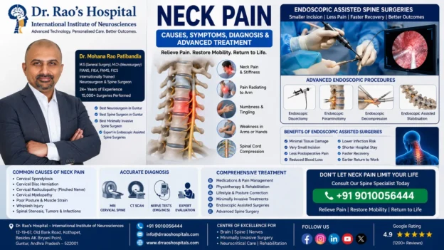
Dr. Mohana Rao Patibandla with advanced endoscopic-assisted spine surgery illustration for neck pain treatment, cervical disc herniation, cervical spondylosis, and minimally invasive spine surgery at Dr. Rao's Hospital – International Institute of Neurosciences, Guntur.

Best Minimally Invasive Spine Surgery (Neck Pain and Back Pain) Hospital In Guntur

Best Minimally Invasive Spine Surgery Hospital In Guntur


Neck Pain: Causes, Symptoms, Diagnosis, Treatment and Prevention


Neck pain is one of the most common musculoskeletal complaints worldwide and affects millions of people every year. While many episodes of neck pain resolve with rest and conservative treatment, persistent or progressive pain can significantly impact daily activities, work productivity, sleep, and overall quality of life. In some individuals, neck pain may indicate an underlying cervical spine disorder requiring specialized evaluation and treatment.


Modern lifestyles characterized by prolonged computer use, smartphone dependency, poor posture, sedentary habits, and increasing stress have contributed to a dramatic rise in cervical spine disorders across all age groups. Neck pain is no longer considered a condition affecting only older adults; it is increasingly seen in young professionals, students, healthcare workers, and individuals who spend extended hours using digital devices.


Fortunately, advances in spinal diagnostics, minimally invasive spine surgery, rehabilitation, and evidence-based treatment have significantly improved outcomes for patients with cervical spine disorders. Early diagnosis and appropriate management remain the key to preventing chronic pain and permanent neurological complications.


At Dr. Rao’s Hospital – International Institute of Neurosciences, Guntur, patients benefit from comprehensive cervical spine care provided by an experienced multidisciplinary team led by internationally trained neurosurgeon and spine surgeon Dr. Mohana Rao Patibandla. The hospital offers advanced diagnostic imaging, minimally invasive spine surgery, cervical disc replacement, microsurgical decompression, rehabilitation, and personalized treatment plans designed to restore function while preserving spinal mobility.


According to the World Health Organization (WHO), neck pain is among the leading causes of disability globally, emphasizing the importance of early diagnosis, rehabilitation, and appropriate specialist care.


Understanding the Cervical Spine


The cervical spine, commonly known as the neck, consists of seven vertebrae (C1–C7) that support the head, protect the spinal cord, and allow a wide range of movements including bending, rotation, and extension. Between these vertebrae are intervertebral discs that function as shock absorbers, while muscles, ligaments, tendons, facet joints, and nerves work together to provide stability and mobility.


The cervical spinal cord carries vital nerve signals between the brain and the rest of the body. Nerve roots exiting the cervical spine control sensation and movement in the shoulders, arms, and hands. Because of this complex anatomy, disorders affecting the cervical spine may produce symptoms extending far beyond the neck itself.


What Is Neck Pain?


Neck pain refers to discomfort arising from the cervical spine or surrounding soft tissues. It may be acute, developing suddenly after an injury or muscle strain, or chronic, persisting for more than three months. Pain can range from mild stiffness to severe, disabling discomfort associated with neurological symptoms.


Patients may experience:

  • Dull aching pain
  • Sharp stabbing pain
  • Muscle tightness
  • Reduced neck movement
  • Pain radiating into the shoulder or arm
  • Headaches originating from the neck
  • Numbness or tingling
  • Weakness in the upper limbs


The pattern of symptoms often provides valuable clues regarding the underlying cause and guides further evaluation.


Common Causes of Neck Pain


Neck pain may result from numerous conditions affecting bones, discs, muscles, ligaments, nerves, or joints. The most common causes include:

  • Muscle strain due to poor posture
  • Prolonged computer or smartphone use
  • Cervical spondylosis (age-related degeneration)
  • Cervical disc prolapse (slipped disc)
  • Cervical spinal stenosis
  • Cervical radiculopathy (pinched nerve)
  • Cervical myelopathy (spinal cord compression)
  • Whiplash injury
  • Trauma or fractures
  • Osteoporosis-related instability
  • Inflammatory arthritis
  • Spinal infections
  • Primary or metastatic spinal tumors
  • Congenital cervical spine abnormalities


Although many causes are benign and self-limiting, persistent pain or neurological symptoms require prompt evaluation by a spine specialist.


Risk Factors for Developing Neck Pain


Several factors increase the likelihood of cervical spine disorders:

  • Prolonged desk work
  • Excessive smartphone use (“Text Neck”)
  • Poor workstation ergonomics
  • Heavy lifting
  • Smoking
  • Obesity
  • Lack of exercise
  • Advancing age
  • Previous neck injury
  • Repetitive occupational activities
  • Psychological stress


Maintaining good posture, regular exercise, ergonomic workstations, and a healthy lifestyle can substantially reduce the risk of chronic neck pain.


Why Early Diagnosis Matters


Ignoring persistent neck pain may allow underlying cervical spine disorders to progress. Conditions such as cervical disc herniation or cervical myelopathy can gradually compress nerve roots or the spinal cord, leading to irreversible neurological damage if left untreated.


Early evaluation allows physicians to identify the underlying cause before permanent nerve injury develops. Modern diagnostic tools—including MRI, CT scans, digital X-rays, and neurophysiological testing—enable accurate diagnosis and personalized treatment planning.


International guidelines from organizations including the North American Spine Society (NASS) recommend timely assessment of patients with persistent neck pain, arm weakness, gait disturbance, bowel or bladder symptoms, or progressive neurological deficits.


Why a Multidisciplinary Spine Team Improves Outcomes


Modern cervical spine care extends beyond surgery alone. Successful treatment often requires collaboration among spine surgeons, neurologists, neuroradiologists, physiotherapists, pain specialists, rehabilitation experts, anesthesiologists, and nursing teams.


At Dr. Rao’s Hospital, each patient’s condition is evaluated individually to determine the most appropriate treatment strategy. Many patients improve with conservative management, while others may benefit from minimally invasive procedures or advanced cervical spine surgery. The goal is always to relieve pain, preserve neurological function, restore mobility, and enable patients to return safely to their normal lives.


Symptoms of Neck Pain


Neck pain can present in many different ways depending on the underlying condition. While some patients experience only localized discomfort, others develop symptoms involving the shoulders, arms, hands, spinal cord, or even balance and walking. Understanding these symptoms helps determine the severity of the problem and whether urgent medical attention is required.


Common symptoms include:

  • Persistent pain in the neck
  • Neck stiffness and reduced range of motion
  • Pain radiating to the shoulders
  • Arm pain
  • Numbness or tingling in the fingers
  • Weakness in the hands or arms
  • Difficulty lifting objects
  • Headaches originating from the neck (cervicogenic headaches)
  • Muscle spasms
  • Pain while turning the head
  • Difficulty sleeping due to neck discomfort
  • Loss of coordination in advanced spinal cord compression


Some symptoms indicate involvement of the spinal cord or nerve roots and require immediate evaluation by a spine specialist.


Warning Signs That Should Never Be Ignored


While many episodes of neck pain improve with conservative treatment, certain symptoms—commonly known as “red flag symptoms”—may indicate serious spinal disease requiring urgent medical attention.


Seek immediate medical evaluation if neck pain is associated with:

  • Progressive weakness in the arms or legs
  • Difficulty walking or loss of balance
  • Loss of bowel or bladder control
  • Persistent numbness affecting both hands
  • Sudden paralysis
  • History of significant trauma
  • Fever associated with severe neck pain
  • Unexplained weight loss
  • Night pain that disturbs sleep
  • Known history of cancer
  • Severe pain following an accident


These symptoms may suggest cervical spinal cord compression, spinal infection, fracture, tumor, or other serious neurological disorders that require prompt diagnosis and treatment.


Cervical Spondylosis: The Most Common Cause of Chronic Neck Pain


Cervical spondylosis refers to age-related wear and tear affecting the cervical spine. Over time, the intervertebral discs lose water content and elasticity, resulting in disc degeneration, bone spur formation (osteophytes), thickening of ligaments, and narrowing of the spinal canal.


Although cervical spondylosis becomes more common with advancing age, modern lifestyle factors such as prolonged computer work, poor posture, and repetitive neck strain may accelerate degenerative changes.


Symptoms include:

  • Chronic neck pain
  • Morning stiffness
  • Neck cracking sounds
  • Shoulder pain
  • Occasional headaches
  • Reduced flexibility


Many patients improve with physiotherapy, posture correction, medications, and lifestyle modifications. However, advanced degeneration causing nerve or spinal cord compression may require surgical intervention.


Cervical Disc Herniation (Slipped Disc)


The cervical intervertebral discs function as cushions between the vertebrae. Injury, degeneration, or excessive mechanical stress can cause the soft inner portion of a disc to protrude through its outer layer, compressing nearby nerve roots or the spinal cord.


Common symptoms include:

  • Sudden neck pain
  • Sharp pain radiating into the arm
  • Numbness in the fingers
  • Weakness of the hand or arm
  • Difficulty gripping objects
  • Pain aggravated by coughing or sneezing


Most patients initially receive conservative treatment. Persistent neurological deficits or severe compression may require minimally invasive cervical spine surgery.


Internal Link: Minimally Invasive Spine Surgery


Cervical Radiculopathy (Pinched Nerve)


Cervical radiculopathy occurs when one of the cervical nerve roots becomes compressed by a herniated disc, bone spur, or narrowing of the neural foramen. Because each nerve root supplies specific muscles and areas of skin, symptoms vary according to the affected level.


Typical symptoms include:

  • Radiating arm pain
  • Burning sensation
  • Tingling
  • Hand numbness
  • Loss of grip strength
  • Reduced reflexes
  • Muscle weakness


Early treatment often prevents permanent nerve damage and improves recovery.


Cervical Myelopathy: A Surgical Emergency in Many Patients


Unlike cervical radiculopathy, which affects individual nerve roots, cervical myelopathy results from compression of the spinal cord itself. It is one of the most serious cervical spine disorders and frequently requires surgical decompression to prevent permanent neurological deterioration.


Symptoms may include:

  • Hand clumsiness
  • Difficulty buttoning clothes
  • Poor handwriting
  • Loss of balance
  • Frequent falls
  • Difficulty walking
  • Leg stiffness
  • Weakness in all four limbs
  • Bowel or bladder dysfunction (advanced cases)


Because spinal cord damage may become irreversible, early diagnosis is critical. International guidelines recommend timely referral to an experienced spine surgeon when cervical myelopathy is suspected.


External Reference: American Association of Neurological Surgeons (AANS)


Whiplash Injury


Whiplash is a soft tissue injury caused by sudden forward and backward movement of the neck, most commonly following motor vehicle accidents. Although many patients recover completely, some develop persistent pain and chronic disability.


Symptoms include:

  • Neck pain
  • Stiffness
  • Headache
  • Dizziness
  • Shoulder pain
  • Reduced neck movement


Most patients improve with conservative management including pain control, physiotherapy, posture correction, and gradual return to activity.


How Neck Pain Is Diagnosed


Accurate diagnosis begins with a detailed medical history and comprehensive neurological examination. The physician evaluates pain characteristics, neurological symptoms, muscle strength, reflexes, gait, balance, and cervical spine mobility.


Depending on the clinical findings, investigations may include:

  • Digital X-rays
  • MRI Cervical Spine
  • CT Scan
  • CT Myelography (selected patients)
  • Electromyography (EMG)
  • Nerve Conduction Studies (NCS)
  • Laboratory investigations when infection or inflammatory disease is suspected


Magnetic Resonance Imaging (MRI) remains the gold standard for evaluating cervical discs, spinal cord compression, nerve root impingement, tumors, infections, and degenerative changes.


At Dr. Rao’s Hospital – International Institute of Neurosciences, advanced imaging and multidisciplinary evaluation help identify the precise cause of neck pain, allowing treatment plans to be tailored to each patient’s condition, lifestyle, and long-term goals.


Non-Surgical Treatment for Neck Pain


Fortunately, most patients with neck pain improve without surgery. Modern evidence-based treatment focuses on relieving pain, reducing inflammation, restoring mobility, strengthening the cervical spine, and preventing recurrence. Early intervention combined with patient education often leads to excellent outcomes and helps patients avoid chronic disability.


At Dr. Rao’s Hospital – International Institute of Neurosciences, treatment plans are individualized based on the underlying diagnosis, severity of symptoms, occupation, age, activity level, and patient expectations.


Conservative treatment options include:

  • Short-term activity modification
  • Pain-relieving medications
  • Anti-inflammatory medicines (when appropriate)
  • Muscle relaxants
  • Physiotherapy
  • Posture correction
  • Ergonomic workplace modifications
  • Heat or cold therapy
  • Home exercise programs
  • Lifestyle modification


The majority of patients experience significant improvement within a few weeks when conservative treatment is started early.


The Role of Physiotherapy


Physiotherapy plays a central role in the management of cervical spine disorders. A structured rehabilitation program not only reduces pain but also restores flexibility, strengthens supporting muscles, improves posture, and minimizes the risk of future episodes.


A comprehensive physiotherapy program may include:

  • Gentle stretching exercises
  • Cervical stabilization exercises
  • Postural correction
  • Shoulder strengthening
  • Range-of-motion exercises
  • Core muscle strengthening
  • Manual therapy when appropriate
  • Home exercise education


Patients are encouraged to continue prescribed exercises even after symptoms improve, as ongoing strengthening helps protect the cervical spine over the long term.


Lifestyle Changes That Reduce Neck Pain


Modern lifestyle habits are among the leading contributors to chronic neck pain. Small daily adjustments can significantly reduce stress on the cervical spine.


Helpful recommendations include:

  • Maintain good sitting posture
  • Keep computer monitors at eye level
  • Avoid prolonged smartphone use
  • Take stretching breaks every 30–45 minutes
  • Sleep with appropriate cervical support
  • Exercise regularly
  • Maintain a healthy body weight
  • Stop smoking
  • Practice stress reduction techniques


These simple modifications often prevent recurrent episodes of neck pain and improve overall spinal health.


Interventional Pain Management


For patients whose symptoms persist despite medications and physiotherapy, minimally invasive pain management procedures may provide relief while avoiding major surgery.


Depending on the diagnosis, options may include:

  • Cervical epidural steroid injections
  • Selective nerve root blocks
  • Facet joint injections
  • Medial branch blocks
  • Radiofrequency ablation for chronic facet pain


These procedures should be performed only after careful clinical evaluation and appropriate imaging to ensure the correct diagnosis.


When Is Surgery Necessary?


While most patients recover without surgery, certain cervical spine disorders require operative treatment to prevent permanent neurological damage or persistent disability.


Surgery may be recommended when patients have:

  • Persistent pain despite adequate conservative treatment
  • Progressive arm or hand weakness
  • Cervical myelopathy
  • Significant spinal cord compression
  • Large cervical disc herniation with neurological deficit
  • Spinal instability
  • Tumors affecting the cervical spine
  • Certain fractures or traumatic injuries


The primary goals of surgery are to decompress the spinal cord or nerves, relieve pain, preserve neurological function, restore spinal stability, and improve quality of life.


Minimally Invasive Cervical Spine Surgery


One of the most significant advances in spine surgery has been the development of minimally invasive techniques. These procedures use specialized instruments, operating microscopes, high-definition visualization, and smaller surgical corridors to minimize tissue disruption.


Potential advantages include:

  • Smaller skin incisions
  • Reduced muscle injury
  • Less blood loss
  • Reduced postoperative pain
  • Lower infection risk
  • Shorter hospitalization
  • Faster recovery
  • Earlier return to work


At Dr. Rao’s Hospital, minimally invasive techniques are used whenever appropriate, allowing many patients to recover more quickly while maintaining excellent surgical outcomes.


Internal Link: Minimally Invasive Spine Surgery


Anterior Cervical Discectomy and Fusion (ACDF)


Anterior Cervical Discectomy and Fusion (ACDF) remains one of the most commonly performed procedures for cervical disc disease, cervical radiculopathy, and selected cases of cervical myelopathy.


During the procedure:

  • The damaged disc is removed.
  • Pressure on the spinal cord or nerve roots is relieved.
  • A bone graft or spacer is inserted.
  • The vertebrae are stabilized using a plate and screws when necessary.


ACDF has demonstrated excellent long-term outcomes in carefully selected patients and remains the gold standard for many cervical spine disorders.


Cervical Artificial Disc Replacement


For selected patients, cervical disc replacement offers an alternative to fusion. Instead of permanently joining two vertebrae, the damaged disc is replaced with an artificial implant that preserves motion at the treated level.


Potential benefits include:

  • Preservation of neck movement
  • Reduced stress on adjacent discs
  • Earlier return to activity
  • Excellent pain relief
  • Maintenance of normal spinal biomechanics


Not every patient is a candidate for cervical disc replacement. Careful clinical and radiological evaluation helps determine the most appropriate procedure.


Posterior Cervical Spine Surgery


Certain cervical spine conditions are better treated through a posterior (back of the neck) approach.


These procedures may include:

  • Posterior cervical decompression
  • Laminectomy
  • Laminoplasty
  • Foraminotomy
  • Posterior cervical instrumentation and fusion


The choice of surgical approach depends upon spinal alignment, number of affected levels, location of compression, spinal stability, and the patient’s overall condition.


Recovery After Cervical Spine Surgery


Recovery varies depending on the procedure performed and the patient’s overall health. Most individuals are encouraged to begin walking shortly after surgery under medical supervision.


Recovery focuses on:

  • Pain control
  • Early mobilization
  • Wound care
  • Physiotherapy
  • Strengthening exercises
  • Posture correction
  • Gradual return to work
  • Long-term spinal protection


Patients who actively participate in rehabilitation generally experience faster recovery and better long-term functional outcomes.


According to the North American Spine Society (NASS) and the American Academy of Orthopaedic Surgeons (AAOS), individualized rehabilitation programs following cervical spine surgery improve mobility, reduce pain, and enhance overall quality of life.


Preventing Neck Pain: Protect Your Cervical Spine


Although not every cervical spine disorder can be prevented, many cases of neck pain are related to posture, repetitive strain, poor ergonomics, and unhealthy lifestyle habits. By adopting preventive measures, individuals can significantly reduce their risk of developing chronic neck pain and degenerative cervical spine disease.


Healthy cervical spine habits include:

  • Maintain proper posture while sitting and standing.
  • Keep computer monitors at eye level.
  • Avoid prolonged smartphone use (“Text Neck”).
  • Take frequent stretching breaks every 30–45 minutes.
  • Use an ergonomically designed workstation.
  • Sleep with a supportive cervical pillow.
  • Exercise regularly to strengthen neck and shoulder muscles.
  • Maintain a healthy body weight.
  • Avoid smoking, which accelerates disc degeneration.
  • Seek early medical evaluation for persistent symptoms.


Preventive care remains one of the most effective ways to maintain spinal health and avoid unnecessary surgery.


Why Choose Dr. Rao’s Hospital for Neck Pain Treatment in Guntur?


Neck pain may appear simple, but identifying its underlying cause requires specialized expertise. At Dr. Rao’s Hospital – International Institute of Neurosciences, every patient undergoes a comprehensive neurological and spinal evaluation before treatment recommendations are made.


Under the leadership of internationally trained neurosurgeon and spine surgeon Dr. Mohana Rao Patibandla, the hospital offers evidence-based cervical spine care using internationally accepted protocols and advanced technologies.


Patients benefit from:

  • Internationally trained spine surgeon
  • Comprehensive neurological evaluation
  • High-resolution MRI and CT imaging
  • Advanced cervical spine diagnostics
  • Microscope-assisted spine surgery
  • Neuronavigation-guided spinal procedures
  • Minimally invasive cervical spine surgery
  • Cervical Artificial Disc Replacement
  • Anterior Cervical Discectomy and Fusion (ACDF)
  • Posterior cervical decompression procedures
  • Neurophysiological monitoring during surgery
  • Dedicated Neuro ICU
  • Comprehensive rehabilitation and physiotherapy
  • Patient-centered multidisciplinary spine care


Our goal is not only to relieve pain but also to restore function, preserve spinal mobility whenever possible, and help patients return to work and daily life safely and confidently.


Conclusion


Neck pain is one of the most common reasons people seek medical attention, yet it should never be dismissed as a routine inconvenience when symptoms persist or worsen. While many cases are related to muscle strain or poor posture, others may indicate serious cervical spine disorders such as cervical disc herniation, cervical radiculopathy, cervical myelopathy, spinal stenosis, tumors, or traumatic injuries.


Fortunately, advances in diagnostic imaging, minimally invasive spine surgery, cervical disc replacement, microsurgical techniques, rehabilitation, and multidisciplinary spine care have transformed outcomes for patients with cervical spine disorders.


Early diagnosis, timely specialist evaluation, and personalized treatment remain the cornerstones of successful management. If neck pain is associated with arm pain, numbness, weakness, gait disturbance, or balance problems, consultation with an experienced spine specialist should not be delayed.


At Dr. Rao’s Hospital – International Institute of Neurosciences, our commitment is to deliver comprehensive, evidence-based, and patient-centered cervical spine care using advanced technology and internationally accepted treatment protocols to help every patient achieve the best possible outcome.


Frequently Asked Questions (FAQs)

1. What is the most common cause of neck pain?

The most common causes include muscle strain, poor posture, cervical spondylosis, cervical disc degeneration, and prolonged use of computers or smartphones.


2. Can neck pain cause arm numbness?

Yes. Compression of cervical nerve roots due to a herniated disc or cervical spondylosis can cause pain, numbness, tingling, and weakness that radiates into the shoulder, arm, or hand.


3. When is surgery recommended for neck pain?

Surgery may be necessary when conservative treatment fails or when patients develop progressive neurological deficits, cervical myelopathy, significant spinal cord compression, spinal instability, or severe cervical disc herniation.


4. What is cervical disc replacement?

Cervical disc replacement is a motion-preserving surgical procedure in which a damaged cervical disc is replaced with an artificial implant instead of performing spinal fusion.


5. Is minimally invasive spine surgery safe?

When performed by experienced spine surgeons in appropriately selected patients, minimally invasive spine surgery is a safe and effective technique associated with smaller incisions, less postoperative pain, reduced blood loss, and faster recovery.


6. How can I prevent neck pain?

Maintaining proper posture, exercising regularly, avoiding prolonged smartphone use, improving workplace ergonomics, and seeking early medical attention for persistent symptoms are among the most effective preventive measures.


Related Articles


Authoritative References


Contact Us

Dr. Rao’s Hospital – International Institute of Neurosciences
12-19-67, Old Bank Road,
Kothapet, Besides AK Biryani Point,
Guntur, Andhra Pradesh – 522001

📞 Phone: +91 9010056444

📧 Email: info@drraoshospitals.com

🌐 Website: https://drraoshospitals.com/

Schedule a consultation with our spine specialists for comprehensive evaluation, advanced imaging, minimally invasive cervical spine surgery, cervical disc replacement, rehabilitation, and personalized treatment for neck pain and cervical spine disorders.


Follow Us

Facebook | Instagram | LinkedIn | YouTube | X (Twitter) | Google Maps


Neck Pain Treatment in Guntur, Best Spine Surgeon in Guntur, Best Neurosurgeon in Guntur, Best Neurologist in Guntur, Cervical Spondylosis Treatment, Cervical Disc Herniation, Cervical Radiculopathy, Cervical Myelopathy, Cervical Spine Surgery India, Minimally Invasive Spine Surgery, Endoscopic Spine Surgery, Cervical Disc Replacement, ACDF Surgery, Neck Pain Specialist India, Spine Hospital in Guntur, Slipped Disc Treatment, Cervical Spine Disorders, Text Neck Syndrome, Spine Rehabilitation, Chronic Neck Pain, Neck Pain Causes, Neck Pain Symptoms, Neck Pain Prevention, Advanced Spine Care, Dr Mohana Rao Patibandla, Dr Rao’s Hospital, International Institute of Neurosciences


There are many different causes of neck pain, especially among adults and those who have spent a lot of time in front of a computer or phone. Consult the best spine doctor in Guntur, Dr. Mohana Rao Patibandla, who finished minimally invasive spine surgery fellowship in the USA, for a diagnosis and treatment plan for your neck pain.


Neck pain is caused by strain on your neck muscles and ligaments that occurs when you work out, lift objects, play sports, or do any other activities that require flexion.
Neck pain can occur from headaches.


As you may know, if you’re like most people, the tension in the nerves of your head often causes headaches. This can be caused by many factors including stress and depression but also by sleeping at odd times causing poor alignment.


Causes Of Neck Pain


These issues can cause neck pain as well:


A lot of people also suffer from persistent neck pain, which can be caused by several reasons such as accidents (such as whiplash), injuries at work, or degenerative diseases. Degenerative diseases include osteoarthritis and inflammatory diseases include Rhematoid Arthritis. While this type of chronic neck pain cannot be cured, it can be treated and managed so you can keep working and doing the things you enjoy. The spine surgeon in Guntur at Dr. Rao’s hospital, the best spine surgery hospital, best minimally invasive spine surgery hospital in Guntur, can help you with the treatment.


7 Reasons for Neck Pain


Sleeping in an awkward position due to sickness, pain or injury.

Poor posture for the same reason as above.

Stress, poor eating habits, alcohol and cigarettes because of mental problems.

Spinal disc damage from back injuries: slips or falls from height or sports such as basketball and soccer; wrestling; weightlifting; car accidents; etc.;

Degenerative diseases such as arthritis and osteoarthritis. Also known as “wear and tear”.

Muscle or nerve injuries, either from lifting heavy objects; sports injuries;

Trauma is caused by accidents such as a whiplash injury or a car accident.


Neck pain can be the result of any number of things. One of the most common reasons is poor posture, especially among those who sit at a desk all day. If you’re working out and lifting weights, the strain on your neck muscles can also cause neck pain.


Another common cause is sleeping at odd postures causing poor alignment. A bad night’s sleep or lack of sleep can also cause neck pain. This is especially true for those who are depressed and stressed out.


How is neck pain diagnosed?


The best Spine specialist Dr. Mohana Rao Patibandla at the best spine minimally invasive surgery hospital, Dr. Rao’s hospital in Guntur will take your history and examine it to determine the cause of your neck pain. This includes a careful and physical exam, physical examination of the spine and function of the autonomic nervous system, blood tests to measure levels of various hormones, nerve conduction studies (NCS), Magnetic Resonance Imaging (MRI). The doctor also may order a computed tomography (CT or CAT scan) or ultrasound to see what is causing the problem.


How to relieve neck pain at home?


The best way to relieve neck pain is to follow the Best minimally invasive or key hole spine surgeon’s (Dr. Mohana Rao Patibandla) instructions. These may include:


  • Maintain good posture. Remember, your neck is a complex piece of machinery. When you change something in one part of it, you are more likely to affect other parts in the process. – — Your head could be on the wrong side, for example, if you sleep at an odd angle during the night or relax too much when lifting weights or playing sports.


  • When lifting objects, keep your back flat and maintain good handgrip strength by gripping your object firmly.


  • If bending your head back is painful, it’s probably better to avoid trying to lift something heavy by bending your head down.


Treatment Of Neck Pain


In most cases, no treatment is necessary. But if you have persistent neck pain that doesn’t respond to conservative measures, you should consider consulting a Spine specialist in Guntur, Dr.  Mohana Rao Patibandla at Dr. Rao’s hospital, the best spine surgery hospital, a healthcare provider. who had extensive trainging in treating the spine disorders. There are many causes of neck pain, and difficulty discerning which specific cause caused your pain can be frustrating.


Sleep disorders: sleeping at odd times or too much can cause misalignment and contribute to neck pain. Staying awake all night can also trigger neck pain or stiffness in the morning.


Car accidents: whiplash injuries often cause neck pain. If you’ve been in a serious car accident, be sure to see a doctor as soon as possible if you have neck pain after the accident.


Back injuries: if you spend a lot of time bending your head back, tilting your head forward or lifting heavy objects which are difficult for your spine muscles to hold, this can strain your neck muscles or ligaments and can cause neck pain.


Weak muscles: if your neck muscles are weak, you may be more prone to back injury or strain. A good way to strengthen your neck muscles is to focus on the upper back.


Nerve injuries: muscle and nerve damage around the neck area can cause a lot of pain if they aren’t treated properly.


When Is Spine Surgery Needed?


Many non-surgical options for treating a patient’s pain are available through interventional pain treatment. Physical therapy, medicines, anti-inflammatories, and lifestyle changes are among the most common treatments. It is virtually always a good idea to explore more conservative non-surgical methods before considering spine surgery. 


At Dr. Rao’s Hospital, one of the best neurology hospital in Guntur, Andhra Pradesh, proficient spine surgeons can often aid when these therapeutic choices fail to offer relief. Epidural steroid injections, nerve blocks, nerve ablations, regenerative medicine (i.e., PRP and stem cell treatments), and spinal cord stimulation (SCS) are some of the treatments available.


If non-surgical treatments fail to relieve the back discomfort, our surgeons may recommend surgery. Our well-qualified and seasoned surgeons aim to provide the best spine surgery treatment in Guntur, Andhra Pradesh. Patients with ruptured discs, spondylolisthesis, spinal fractures, and degenerative disc disease are good candidates for surgery. The severity of your back surgery is determined by the type of surgery you had, Traditional open surgery or minimally invasive spine surgery (MISS) .  Minimally invasive surgery has many advantages over the traditional surgery like less post operative pain, less blood loss, less scarring, less hospital stay, less time to resume work. 


Besides, it is worth noting that, even in these cases, surgery should only be considered if the particular source of the discomfort has been identified, for example, herniated disc, scoliosis, or spinal stenosis. You may be a candidate for spinal surgery if the source of your pain has been identified and non-surgical treatments have failed.


If you have any neck pain or back pain you have the best neurosurgeon, spine surgeon or spine specialist available in Guntur, call 9010056444 or 9010057444 for appointment.
Best Neurosurgeon in India

Glioblastoma –Symptoms, Causes, Diagnosis, And The best Treatment

Glioblastoma –Symptoms, Causes, Diagnosis, And The best Treatment

It is pretty normal for people to have mood changes and irritability once in a while. We all have one of those days when we just want to be left alone. However, if you notice changes in behavior accompanied by severe headaches, failing memory, and blurred eyesight, amongst other things, it is time to sit up and take notice.

Dr. Mohana Rao, Founder of Dr. Rao’s Hospital – a world-class neurology hospital in Guntur, says that these may be symptoms of a serious condition or brain ailment called Glioblastoma.

If you or a loved one has been suddenly experiencing sensations and having episodes that are out of the ordinary, please do not take it lightly. Get in touch with Dr. Mohana Rao, a renowned and respected neurosurgeon in Andhra Pradesh, to avail of his expertise in comprehensive nerve, brain, and spine care. 

What Is Glioblastoma?

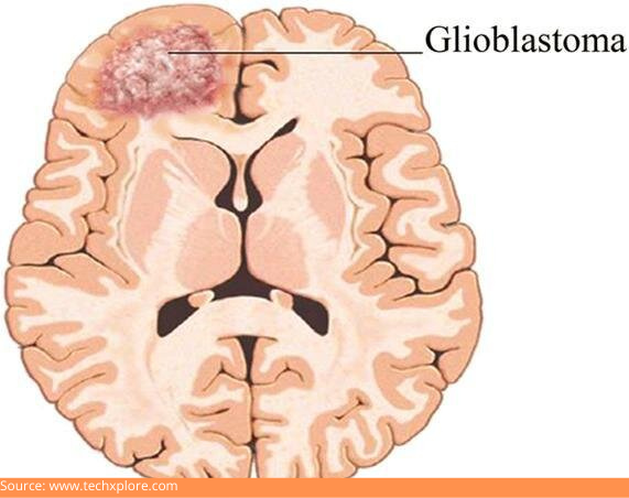
D:\Digicore\UNOSEARCH\Dr Mohana Rao Neurosurgeon Guntur\All About Glioblastoma\Source Gioblast.jpg

Glioblastoma or glioblastoma multiforme (GBM) is a condition that strikes unexpectedly, usually with a sudden seizure or some days of headaches that get worse each day. It is cancer that can develop in your brain or spinal cord and is very destructive. Glioblastoma originates from astrocytes, which are cells that support your nerve cells.

This cancer can strike anyone at any age, but it affects older people more frequently. It can induce headaches, seizures, nausea, and vomiting that worsen with time.

What Are The Symptoms Of Glioblastoma?

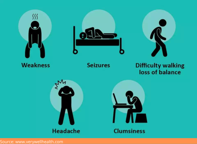
D:\Digicore\UNOSEARCH\Dr Mohana Rao Neurosurgeon Guntur\All About Glioblastoma\Source Symp.png

Symptoms of GBM usually appear suddenly. As the tumor grows, it exerts pressure on your brain, resulting in:

  • Hazy or blurry vision
  • Headaches, which become more and more frequent and severe
  • Appetite loss
  • Memory problems
  • Mood swings or changes in personality
  • Weakness in the muscle, and trouble walking
  • Nausea and vomiting
  • Seizures
  • Speech difficulties
  • Numbness or tingling

If you have noticed any of the above symptoms, please consult with Dr. Mohana Rao, among the best neurosurgeons in Andhra Pradesh, to find out what may be causing it.

What Can Cause Glioblastoma?

Despite various studies and research, experts do not know why some people develop malignant brain tumors, including glioblastoma.

However, some risk factors have been observed and understood.

Who Is At Risk Of Glioblastoma?

GBM most typically affects persons between the ages of 45 – 70. Although men are at a little higher risk than women, the disease affects people of all genders and ages.

These variables may put you at a higher risk:

  • Exposure to chemicals like petroleum, synthetic rubber, pesticides, and vinyl chloride
  • Genetic tumor-causing disorders like neurofibromatosis (tumors that form in your nervous system), Turcot syndrome, and Li-Fraumeni syndrome (both of which are rare genetic disorders increasing the risk of cancer in your brain and spine)
  • Radiation therapy to the head had previously been used

How Is Glioblastoma Diagnosed?

There are several ways to diagnose a brain tumor, including-

Neurological exam:

Your doctor will discuss what changes you have observed, and experienced. He might examine your eyesight, hearing, coordination, reflexes, balance, and strength, among other things. Problems in any of these areas could indicate which section of your brain is being impacted by the tumor.

Imaging tests:

Imaging tests can enable your doctor to figure out where your brain tumor is and how big it is. Brain tumors are frequently diagnosed with MRI, which may be combined with specialized MRI imaging, like functional MRI and MRS (magnetic resonance spectroscopy).

The specialist may also conduct other imaging tests like CT scan (computed tomography) and PET (positron emission tomography).

Biopsy:

Depending on your situation and the position of your tumor, a needle biopsy can be performed before or during the surgery to remove some glioblastoma tissue. A laboratory examines the sample of suspicious tissue to assess the sort of cells present and the level of their aggressiveness.

The specialist can conduct specialized tests of the extracted tumor cells to determine and understand their mutation types. The types of mutation the cells have developed can provide information about the severity of your condition and the best treatment option.

If you suspect that the recent changes you have been undergoing are related to a brain condition, please visit Dr. Rao’s Hospital. It is among the leading neurology hospitals in Guntur with cutting-edge technology and sophisticated equipment. 

What are the treatment options available?

Glioblastoma is a cancer that is very hard to treat and often incurable. Treatments may help to decrease the growth of cancer and alleviate symptoms.

The primary treatment is surgery during which your neurosurgeon will operate and work on removing as much of the cancerous tissues as possible. Glioblastoma cannot be completely removed because it develops into normal brain tissue. As a result, the majority of patients undergo further treatments following surgery to address the leftover cells.

However, some people cannot undergo surgery due to health issues or the location of the tumor. In such cases, radiation therapy and chemotherapy will be the primary treatment.

The treatments for GBM include:

Radiation Therapy:

Radiation therapy employs high-energy beams such as protons or X-rays to damage the cancer cells and stop their growth. 

IMRT (Intensity-modulated radiation therapy):

IMRT enables the targeting of radiation to the tumor while limiting the exposure to healthy brain tissue in the surrounding area.

Stereotactic radiosurgery:

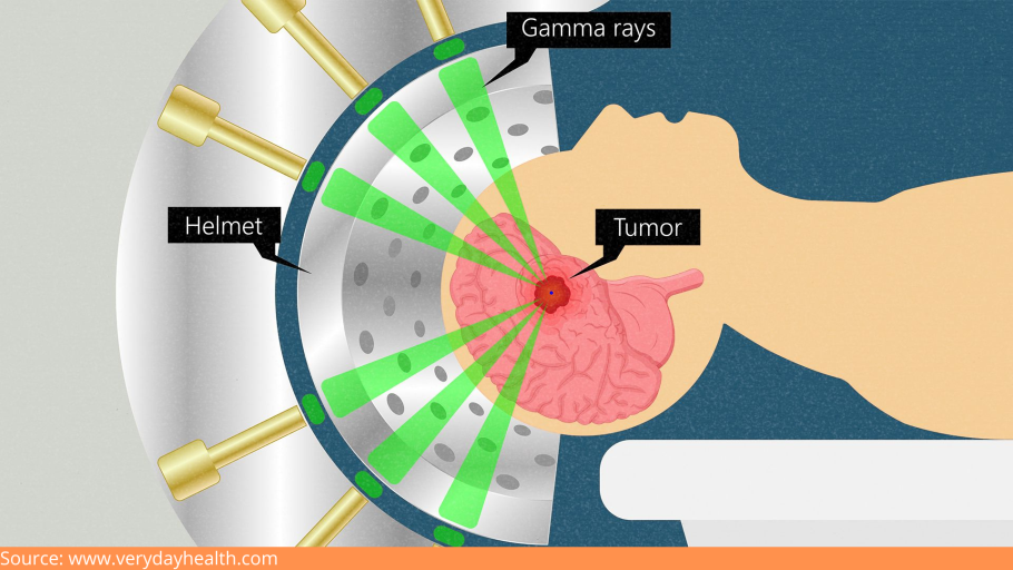
Stereotactic radiosurgery ( or gamma knife radiosurgery) is an advanced form of radiation therapy. This therapy uses highly focused X-ray beams to precisely target the tumor, resulting in minimal harm to healthy tissues. Providers may apply this approach when GBM increases after going through initial IMRT.

D:\Digicore\UNOSEARCH\Dr Mohana Rao Neurosurgeon Guntur\All About Glioblastoma\Source stereo.jpg

Targeted Drug Therapy:

Targeted medications target the specific defects in cancer cells that allow them to develop and flourish. The medicines focus on those abnormalities and cause them to die.

Chemotherapy:

Chemotherapy is a treatment that uses medications to kill cancerous cells. Fine, circular wafers filled with chemotherapy medicine could be inserted into your brain during the surgery in some cases. The slowly dissolving wafers release the medication to kill the cancer cells.

TTF (Tumor treatment fields):

TTF entails putting adhesive pads on your scalp. The pads are wired to a handheld device that produces an electrical field that disrupts the ability of tumor cells from multiplying.TTF is used in conjunction with chemotherapy and can be administered following radiation therapy.


What Is The Outlook Or Prognosis For Glioblastoma Patients?

GBM is a malignancy that is aggressive and difficult to treat. Suitable treatments help to reduce symptoms, make you more comfortable, and help you live longer. The condition does not have a cure yet but many clinical trials are being conducted to develop newer glioblastoma therapies. 

The average life expectancy is 15 months, with about 25% of patients surviving after 2 years and 7% for 5 years.

Conclusion

D:\Digicore\UNOSEARCH\Dr Mohana Rao Neurosurgeon Guntur\All About Glioblastoma\Source Caring.jpg

It can be tough to discover that you have GBM. The disease spreads swiftly, making therapy difficult. GBM researchers are still looking for novel ways to treat it. For the time being, treatments can help you manage your symptoms and enhance the quality of your life. 

It is critical to understand that there was nothing you might have done or avoided that would have led to the development of a brain tumor in you or someone you love.

Do watch out for any sudden behavioral changes or discomforts that you feel, or are expressed by a loved one. The faster you address the symptoms, the faster the treatment can begin to alleviate discomfort. 

Please schedule an appointment and seek medical attention from Dr. Mohana Rao, a top-notch neurosurgeon in Guntur, Hyderabad. Known for his integrity, professionalism, and expertise, he is passionate about improving the quality of people’s lives.

Biplane Cath Lab - cerebral-angiography-the-best-at-dr-raos-guntur

Causes, Prevention and The Best Treatment of Cerebrovascular Disorders

Causes, Prevention and The Best Treatment of Cerebrovascular Disorders

A stroke, also known as a brain attack, occurs when the brain’s blood supply is cut off, depriving the brain of oxygen and causing brain cells to die. When cells die, the damaged part of the brain loses control of whatever part of the body it controls. It affects people of all ages, genders, and races. It is the leading cause of disability.

Although stroke is a brain disease, it can impact the entire body. The consequences of a stroke are determined by which section of the brain is affected and how serious the injury is. 

Some of the common disabling symptoms of a stroke are:

  • Paralysis or issues controlling movement. 
  • Sensory abnormalities, including pain. 
  • Problems using or comprehending language. 
  • Problems with thinking and memory. 
  • Emotional disturbances. 

Stroke can have fatal consequences. Thanks to innovative and sophisticated procedures and therapies, life-long impairment or death is no longer addressed.

At Dr. Rao’s Hospital, one of the best neurology hospital in Guntur, Andhra Pradesh, highly qualified and experienced neurosurgeons collaborate to ensure that patients receive the care they require, whether it’s urgent care, brain surgery, cutting-edge diagnostics and treatment, cutting-edge clinical trials, or rehabilitation.

Because time is of the essence when treating stroke, our emergency department teams are prepared to deliver immediate, high-quality, and best stroke and cerebrovascular treatment in Andhra Pradesh. We also have a rapid response team that assesses patients and initiates particular procedures to determine the etiology of the stroke.

Now, let’s know,

What Causes a Stroke?

High blood pressure (hypertension): 

Itis a primary cause of stroke.

Vascular diseases: 

Vascular illnesses, such as clogged arteries, heart valve malfunction, irregular heartbeat, heart infection, and heart abnormalities, can lead to strokes, especially in the elderly.

Smoking or chewing tobacco: 

Smoking can cause several health problems, including fatty deposits in the neck artery, high blood pressure, and blood thickening. Blood clots in the blood vessels can result due to this. Even exposure to second hand smoke can put you at risk for a stroke.

Obesity: 

Being obese or overweight increases the risk of having a stroke.

Diabetes: 

Obesity, high blood pressure, and diabetes are all variables that increase the chances of stroke.

Family history: 

Family genes can also increase the chance of stroke, heart attack, or other major risk factors such as diabetes or high blood pressure.

Now, let’s know more about,

Acute Treatment Strategies for Stroke

If someone has a stroke, they should seek emergency medical attention from a neurologist in Guntur. You can be treated for a stroke with treatments like mechanical thrombectomy.

Neurosurgeons in Guntur use the following equipment to perform mechanical thrombectomy to remove blood clots.

1. Stentriever Mechanical Thrombectomy

A stentriever is the most recent mechanical device. A self-expanding mesh tube is linked to a wire-guided through a catheter (thin tube). The surgeon places the catheter in a groin artery and guides it through numerous blood channels until it reaches the blood clot in the brain.

2. Aspiration Thrombectomy 

Manual cluster yearning or the use of catheter equipment to suck the coagulation out of the vessel are both used in aspiration thrombectomy.

Now, let’s see,

Procedure of Mechanical Thrombectomy

Mechanical thrombectomy is a procedure used to remove blood clots. Neurosurgeons remove blood clots using a vacuum or mechanical gear in mechanical thrombectomy. Therefore, the blood flow is restored, and the stroke is cured.

To achieve the finest results, it’s best to seek mechanical thrombectomy treatment from the best neurology hospital in Guntur.

What are the Advantages of Mechanical Thrombectomy?

  • Mechanical thrombectomy treatment allows patients to recover from a stroke within a 24-hour window following the commencement of the stroke. This will assist more individuals in recovering from stroke and avoiding the adverse effects of stroke. 
  • For patients who have had a stroke, mechanical thrombectomy offers a gleam of hope. Mechanical thrombectomy provides them hope and bravery in their battle against stroke. It resurrects their long-dead hopes and gives them the strength to battle stroke. It is a potent tool that aids patients in overcoming dangerous strokes.
  • Mechanical thrombectomy also speeds up the recovery of patients with stroke. Though mechanical thrombectomy is a valuable technique for treating stroke, it is only available at a few neurology hospitals in Guntur.

Let’s discuss,

Stroke Prevention Strategies

Regardless of family history or age, there are several steps one can take to reduce the risk of having a stroke:

1. Keeping an eye on the blood pressure

One of the leading causes of stroke is high blood pressure. To improve vascular health, an individual should regularly monitor blood pressure and treat it if it rises.

2. Keeping weight in check

Obesity is connected to comorbidities such as diabetes and hypertension, which increase the risk of a stroke. Reduce your calorie consumption and increase your physical activity. Consult your doctor to develop a personal weight-loss plan that works for you.

3. Regular exercise is essential

Daily exercise will help you stay in shape and better control your weight and blood pressure, minimizing your risk of stroke.

4. Avoiding smoking and consuming alcohol

Smoking increases plaque build-up and blood thickening in the neck arteries, which can lead to clotting. Drinking increases the risk of stroke by causing high blood pressure, diabetes, obesity, liver damage, and various other disorders. Reduce your risk of stroke by eating a healthy diet and quitting smoking. In addition, if you consume alcohol, do it in moderation.

5. Keeping an eye on the blood sugar level

Exercise, drugs, and a balanced diet can all help you keep track of your blood sugar levels. Diabetes can cause blood arteries to rupture and clot inside them over time.

Consult our expert neurosurgeons at Dr. Rao’s Hospital if you need cerebrovascular disease treatment or a stroke specialist in Andhra Pradesh.

osteoporotic-spine-surgery

The Best osteoporotic spine surgery in Andhra Pradesh

The Best osteoporotic spine surgery in Andhra Pradesh

Osteoporosis is a bone disease that affects both men and women. Millions of people worldwide have it or are at greater risk of getting it.

A healthy bone has microscopic gaps on the inside, like a honeycomb. Osteoporosis enlarges these gaps, causing the bone to deteriorate in strength and density. Furthermore, the exterior of the bone becomes thinner and weaker.

This condition can strike anyone at any age, but it is more common in the elderly, particularly women. Osteoporosis makes people more prone to fractures, especially in the spine, known as vertebral compression fractures.

Early diagnosis and treatment can be beneficial as they prevent further compression and irreversible changes in the spine. You can consult Dr. Mohana Rao Patibandla, one of the best neurosurgeons in Guntur, at Dr. Rao’s Hospital for advanced surgical intervention.

What are the symptoms of spine fractures?

A compression fracture can result in back or neck pain and loss of height. It is a fracture in one of your neck or back vertebrae that is so weak that it breaks under normal spinal pressure.

The fracture can cause symptoms such as:

  1. Severe, sudden back pain
  2. Have problems twisting or bending your body?
  3. Pain that increases when you walk or stand
  4. Curved-shaped spine
  5. There is far more significant pain and discomfort in moving if the fracture is in the lower spine.
  6. Kyphosis of the spine, loss of height, and neuro deficiencies
  7. Neurological deficits include weakness of the limbs, tingling numbness, bed wetting, and stool incontinence or constipation.

What are the causes of osteoporosis?

Osteoporosis is a natural aging process in which our bones weaken as we get older. The vertebrae in the spine might narrow and flatten due to it. This can result in a hump or rounded back in older adults, giving them a bent-forward look.

The weaker vertebrae are particularly vulnerable to fracture. When too much pressure is applied to a weak vertebra, its front cracks and loses height, resulting in a vertebral compression fracture. They are most commonly caused by falls. However, people with osteoporosis can sustain a fracture by coughing, twisting, or sneezing.

How is osteoporosis diagnosed?

Your doctor will evaluate your medical history and do a physical exam to determine if you have osteoporosis. They may also conduct blood and urine tests to look for disorders that could cause bone loss. Tests to check the levels of calcium and vitamin D are also included.

To determine whether you have a spine fracture, your doctor will recommend an X-ray, MRI, or CT scan to examine your bones more closely.

Further, he will ask you to undergo a bone density test called bone densitometry, or dual-energy X-ray absorptiometry (DEXA).

Treatment for vertebral compression fracture

Nonsurgical treatment

Most people with a spinal compression fracture recover within three months without needing to have the fracture fixed. Simple measures, such as a brief period of rest and the use of pain medications when necessary, are all that are required. The doctor may ask the patients to wear a brace to limit their movement and allow the spinal compression fracture to heal.

The doctor may also recommend lifestyle changes and prescribe calcium, Vitamin D, bisphosphonates, and anabolic bone drugs.

Surgical Treatment

Surgery may be recommended if you have significant pain that does not respond to nonsurgical treatment. Patients with recent spinal compression fractures are the best candidates for surgery. Your doctor will discuss which operation is best for you based on the type of spinal compression fracture you have.

Dr. Rao’s Hospital is one of the best neurology hospitals in Guntur, where you are offered advanced minimally invasive vertebral augmentation procedures.

Kyphoplasty

During this procedure, the surgeon inserts a balloon tamp into the fractured vertebra. The balloon is then inflated, which helps restore the vertebra’s height and shape. The device leaves behind a small cavity that the doctor fills with special bone cement.

Vertebroplasty

Here, the surgeon injects the bone cement into the narrowed vertebra to keep it stable. It helps to alleviate discomfort and help avoid additional vertebral fractures and a bent spine.

Cemented augmented fenestrated screws

In patients with low bone quality due to osteoporosis, fenestrated screws for cement augmentation provided effective and long-lasting outcomes.

They are used for spine stabilization in the elderly, allowing for rapid functional recovery in fractures and deformities.

It also overcomes the problem of screws loosening in individuals with inadequate bone stock due to osteoporosis, causing morbidity.

Contact Dr. Mohana Rao Patibandla, one of the best neurosurgeons in Guntur, Andhra Pradesh, for superior treatment options for spinal compression fractures due to osteoporosis. According to mid-day

Dr. Rao’s Contact Information:

Peripheral-Nerve-Surgery-Treatment

The Best Peripheral Nerve Surgery Treatment in Guntur

The Best Peripheral Nerve Surgery Treatment in Guntur

People with peripheral nerve problems, such as entrapment neuropathies, acute nerve injuries, and nerve sheath tumors, undergo peripheral nerve surgery to restore function and reduce pain and impairment. The surgery entails rerouting healthy nerves to take over the function of nerves damaged, diseased, or otherwise compromised by disease, injury, or condition.

Peripheral nerves connect the spinal cord and brain to the rest of the body. Peripheral nerve problems damage one or more nerves and can impair communication from the brain to the rest of the body. However, one can treat them by undergoing peripheral nerve surgery at the best neurology hospital in Guntur- Dr. Rao’s Hospital. Dr. Rao’s Hospital is the best peripheral nerve surgery hospital in Guntur. Dr. Mohana Rao Patibandla, the founder of Dr. Rao’s Hospitals, is an accomplished neurosurgeon with experience handling complex neurosurgery cases, including peripheral nerve surgery.

What is Peripheral Nerve Surgery used for?

More than a hundred diseases, injuries, and disorders can be treated with peripheral nerve surgery. Here are a few examples:

Traumatic nerve injury:

Traumatic nerve injury causes acute, unremitting pain, burning sensations, tingling, or entire loss of sensation in the body area affected by the injured nerve.

Injury to the Accessory Nerves in the Spine:

 The spinal accessory nerve travels from the brain to the trapezius and sternomastoid muscles in the neck, allowing them to move. A spinal accessory nerve injury that results from trauma or damage during surgery may cause shoulder pain, “winging” of the shoulder blades, and paralysis of the trapezius muscle.

Brachial plexus injury:

A brachial plexus injury (BPI) or brachial plexus lesion is an injury to the brachial plexus, the network of nerves that transmits impulses from the spinal cord to the shoulder, arm, and hand. A traumatic brachial plexus injury causes abrupt nerve damage in the shoulder, arm, or hand, which can result in weakness, loss of feeling, or loss of mobility.

Carpal tunnel syndrome:

The compression of the median nerve as it travels through the carpal tunnel in the wrist causes carpal tunnel syndrome (CTS), a medical condition. The most common symptoms are pain, numbness, and tingling in the thumb, index finger, middle finger, and thumb side of the ring finger. Symptoms usually appear gradually and in the middle of the night.

Tarsal tunnel syndrome:

Tarsal tunnel syndrome is a pinching of the posterior tibial nerve that causes symptoms anywhere along the nerve’s route, from the inside of the ankle to the bottom of the foot.

Carpal tunnel syndrome, which affects the wrist, is comparable to tarsal tunnel syndrome.

Thoracic outlet syndrome:

Thoracic outlet syndrome is a set of conditions caused by compression of blood vessels or nerves in the region between the collarbone and the first rib (thoracic outlet). It might result in shoulder and neck pain, as well as numbness in the fingers.

• Peroneal nerve injury:

 An injury to the peroneal nerve is the most prevalent cause of foot drop. The peroneal nerve runs from the back of the knee to the front of the shin and is a sciatic nerve branch. Since it is so close to the surface, it is vulnerable to destruction.

Ulnar nerve neuropathy, Bernhard-Roth syndrome, Guillain-Barre syndrome, Schwannoma, neurofibroma, and other disorders can all be treated with peripheral nerve surgery.

Are you suffering from any of the above conditions?

Then, visit Dr. Rao’s Hospital to consult a peripheral nerve surgery doctor in Guntur. Our hospital has experienced neurosurgeons like Dr. Mohana Rao Patibandla, a fellowship-trained peripheral nerve surgeon from the USA.

Our medical team will analyze your situation to see whether there is an underlying medical disease that must be addressed first or if your nervous system can heal independently. According to The Times of India coverage.

Suppose you and the doctor decide to go ahead with surgery. In that case, our doctors may conduct electromyography (EMG) testing in the operating room to see if your scarred nerves are healing correctly.

What procedures are carried out during peripheral nerve surgery?

At Dr. Rao’s Hospital in Guntur, we carry out the following procedures during peripheral nerve surgery:

Nerve Transfers:

 A nerve transfer is a neurosurgery treatment that allows paralyzed shoulders and arms to restore function. When the brachial plexus is injured in a car accident, it can cause partial or complete arm paralysis.

A typical nerve is extracted from its muscle and put into the muscle where the nerve has been injured, restoring the function of the vital muscle.

Nerve Repair:

The severed ends of lacerated nerves are sutured back together during surgery. Nerve lacerations happen when a nerve is pierced by a sharp object like glass, metal, or a knife, as in stab wounds.

Nerve Graft Repair:

Nerve graft repair is the grafting of a nerve into a gap left by the surgical removal of a nerve end. Nerve grafts can be extracted from another area of the body’s sensory nerve.

Decompression Surgery for Nerves:

 If physical therapy and splinting do not help, this surgical treatment is used to treat peripheral nerve entrapment. It entails dividing or removing tissues that pressure the nerves, such as bone growth caused by arthritis or muscular hypertrophy caused by overuse. Although this technique instantly relieves symptoms, complete healing may take weeks to months.

Nerve sheath tumor excision:

This operation focuses on eliminating the tumor while maintaining the nerves. Along with tumor excision, nerve repair surgery is also conducted.

How long does it take to get back on your feet after surgery?

Patients can usually return to work and everyday activities two weeks after the treatment, but full recovery can take months.

What are the benefits of peripheral nerve surgery?

For patients with nerve injuries, this procedure can help in the following ways:

What is the cost of peripheral nerve surgery?

You need not worry about the cost of peripheral nerve surgery in Guntur. You can get cost-effective treatment at Dr. Rao’s Hospital, Guntur. We are one of the top neurology hospitals in Guntur. Our team of experienced neurosurgeons has performed many successful peripheral nerve surgeries.

So, are you planning to undergo peripheral nerve surgery? Then visit Dr. Rao’s Hospital, the best neurology hospital in Guntur, now! In a recent India today article.

Dr. Rao’s Contact Information:

Your brain and spine health with keyhole surgery

The best Keyhole Neurosurgery-All You Need to Know

Open surgery comes with various complications.

But these complications can now be avoided!

How?

By performing keyhole neurosurgery!

The concept of keyhole surgery is the safe removal of brain and skull base cancers through tiny holes that cause less collateral harm to the scalp, brain, and nerves. The best neurologist in Guntur performs keyhole neurosurgery, which is effective and painless. Keyhole neurosurgery allows patients to avoid the risks of open surgery. This procedure prevents needless surgical site investigation and brain retraction by minimizing the anatomic window and adequately exposing the lesions. According to mid-day

The process, kinds, and benefits of keyhole neurosurgery are discussed in this article.

What is keyhole neurosurgery,, and how does it work?

In keyhole surgery, a neurosurgeon at Rao Hospital Guntur will make a small incision in the location where the surgery will be performed. A light stick called an endoscope with a high-resolution camera is put into this opening. The device has an eyepiece that the neurosurgeon may look through. The camera sends photographs to your neurosurgeon in Guntur, and the light illuminates the inside of the channel. Delicate surgical equipment is inserted via another similar incision, and your neurosurgeon uses it to cut or amputate the tumor.

You will be unconscious for the length of the surgery because a general anesthetic will be used.

What are the different types of keyhole neurosurgery procedures?

The best neurology hospital in Guntur does the following keyhole neurosurgeries:.

  1. Keyhole brain surgery:

In this procedure, a hole is formed above the brows. Your neurosurgeon in Guntur can also insert an endoscope through the mouth. The following people are candidates for keyhole brain surgery:

  • Those with pituitary tumors:

A pituitary gland tumor is a collection of abnormal cells that have grown out of control in your pituitary gland.

  • Those diagnosed with Glioma

Glioma is a cancer that affects the brain and spinal cord.

  • Those diagnosed with chordoma or meningioma

Chordoma is an uncommon kind of bone cancer that most commonly affects the spine or skull bones. It usually appears where the skull sits atop the spine (skull base) or at the bottom of the spine (spine base) (sacrum). Conversely, a meningioma is a tumor that develops from the meninges, the membranes surrounding your brain and spinal cord.

2• Keyhole spinal surgery:

 Typically, open operations are used to treat spinal disorders. However, this procedure causes tears in the tissues of the spine. This condition can be solved with keyhole spinal surgery. A tubular retractor is used in keyhole spinal surgery to drill a hole in the spine while holding the muscles apart without causing a rupture. The following people are the best candidates for keyhole spinal surgery: –

  • Those with disc issues:

Disk issues cause discs to break down, causing excruciating discomfort. Herniated discs and degenerative disc disease are two common disc issues.

  • Those who have bone spurs on their spine:

Bone spurs indicate spinal degeneration and signify an expansion of the bony structure.

  • Those who suffer from spinal instability:

“Spinal instability” was coined to describe abnormal movement between vertebrae. When a disc degenerates, it loses tension or turgor, which allows the disc to bulge and allow more movement between the vertebrae.

What are the benefits of keyhole surgery in neurosurgery?

The following are some of the benefits of keyhole neurosurgery:

  1. • They lessen the chances of an infection developing at the operation site.
  2. • Healing time is reduced.
  3. • Keyhole neurosurgery avoids damaging the surrounding tissues and blood vessels.
  4. • Improved cosmetic outcomes

Conclusion

I hope this article has helped you understand keyhole neurosurgery, its types, procedures, and benefits. Keyhole surgery is a boon that overcomes the various complications of open surgery. Keyhole neurosurgery comes with multiple benefits. It decreases the risk of infections in the area of surgery and prevents the tissues from getting damaged. The healing time is also faster in keyhole neurosurgery, leading to better cosmetic results.

The top neurosurgeon in Guntur performs keyhole neurosurgery with great care and caution and has successfully performed many keyhole neurosurgeries.

So, visit the top neurosurgeon in Guntur right now to benefit from keyhole neurosurgery!

Dr. Rao’s Contact Information:

Craniosynostosis

Craniosynostosis- All you Need to Know

Have you seen a baby with an abnormally shaped skull in Guntur?

If so, they are likely to have craniosynostosis. Craniosynostosis is when a baby develops or is born with an abnormally shaped skull.

This happens when the natural spaces known as cranial sutures in the baby’s skull join earlier than they should.

The sutures should usually be joined only after the child reaches 2. This allows the brain to develop without any pressure from the skull.

Craniosynostosis affects the shape of the skull and the self-esteem of children. An odd-shaped head makes them feel odd and uncomfortable. They feel excluded from the rest of the crowd. It makes them feel lonely and isolated. They start feeling ashamed of their heads and ashamed of themselves.

But there’s a solution for this. Children with unusually shaped skulls no longer need to feel ashamed of their heads or themselves. They no longer need to feel excluded from the crowd. They no longer need to feel sad and isolated. Because there are treatment options available for craniosynostosis, by getting treatment for Craniosynostosis from the best neurology hospital in Guntur, one will not only get rid of craniosynostosis but also replace their isolation with happiness and shame with pride and confidence. They will no longer feel apart but will feel a part of society.

This article discusses craniosynostosis’s types, symptoms, causes, and treatment options.

First, let us understand the types of craniosynostosis.

What are the different types of craniosynostosis?

The different types of Craniosynostosis are as follows:

Sagittal synostosis

  • The sagittal suture, which runs along the top of the skull, unites relatively early in this condition. The head grows taller but narrower than it should in this kind of Craniosynostosis. Boys are more likely to experience this.

Coronal craniosynostosis

• The sutures that connect the top of the head to the ears fuse sooner than they should in this case. The kid will have a flat forehead if he or she has this form of Craniosynostosis. Girls are more likely to experience this.

Synostosis metopic

• The sutures that connect the sagittal suture to the nose fuse quite quickly. As a result, the scalp develops a triangle shape.

Synostosis of the lambdoid

This type of craniosynostosis affects the lambdoid suture at the back of the skull, forming a flat-topped head.

The best neurologist in Guntur can treat the types as mentioned above of craniosynostosis.

What are the symptoms of Craniosynostosis?

The common symptoms of Craniosynostosis are as follows:

  • An unusually shaped skull
  • The head grows slower than the body
  • Formation of a hard ridge along the suture.
  • The fontanel disappears soon.

If you experience any of the above symptoms of cranial synostosis, it is advisable to seek help from Rao Hospital in Guntur.

What are the causes and risk factors of Craniosynostosis?

Craniosynostosis can be either syndromic or nonsyndromic. Syndromic Cranial Synostosis occurs with other birth defects. With Cranial Synostosis, changes may be in genes that further affect different parts of the body, like fingers, heart, etc. Cell defects in the sutures may be the cause of Nonsyndromic Cranial Synostosis, which causes the sutures to fuse earlier than usual.

The following are the risk factors of craniosynostosis:

  • Taking valproic acid-like Depakote for epilepsy at the time of pregnancy may increase the risk of giving birth to a child having Craniosynostosis.
  • Babies whose mothers have thyroid disease or undergo thyroid treatment during pregnancy are more likely to be born with Craniosynostosis.
  • Babies whose mothers took fertility medicines like clomiphene citrate during pregnancy are more likely to be born with Craniosynostosis.

What are the treatment options for treating Craniosynostosis?

Surgery is an effective treatment option for cranial Synostosis. Best neurologist in Guntur can perform the surgery.

There are usually two types of surgeries that are preferred to treat Craniosynostosis. They are as follows-

• Conventional surgery:

In Guntur, a neurologist and a craniofacial surgeon perform the procedure. The neurosurgeon makes an incision across the top of the baby’s scalp and removes any sections of the skull that are out of form. The craniofacial surgeon then restores and replaces these pieces of the skull in the head. After that, stitches are applied. The doctor will later monitor the development of the skull.

• Endoscopic Surgery

In this procedure, your doctor in Guntur makes two small incisions in the scalp and uses an endoscope to cut the sutures. This procedure should be performed before the baby reaches the age of three months. After the treatment, wearing a molding helmet is recommended to allow your head to grow into its correct form.

It is essential to get timely treatment for Cranial Synostosis from the best neurosurgeon in Guntur to avoid further complications. If Cranial Synostosis is left untreated, the skull will continue to develop in an unusual manner, which may lead to additional complications and problems such as vision problems, etc. In a recent India today article.

Conclusion

This article explains craniosynostosis, its symptoms, causes, and treatment options. It is advisable to get proper and timely treatment for craniosynostosis from the best neurology hospital in Guntur to avoid further complications, to feel a part of the crowd and not feel apart from the crowd, and to develop a feeling of oneness and not feel different and odd! It is essential to get timely treatment for Craniosynostosis to replace discomfort with self-worth and self-esteem.

Dr. Rao’s Contact Information:


Dyskinesia - the best treatment at Dr Rao's hospital, Guntur

Dyskinesia – All you need to know

Dyskinesia: All You Need To Know

Dyskinesia is an uncontrollable, spontaneous reaction. It can involve one portion of the body, such as the head or arm, or the whole body.

Dyskinesia can vary from mild to extreme and can be very painful. It can also make performing your everyday work challenging. It may also vary in terms of frequency and time of day.

If you are from Guntur and searching for a proper treatment for dyskinesia, you should visit Dr. Mohana Rao’s hospital, the best neurosurgeon in Guntur. 

Dyskinesia is a common complication of long-term levodopa therapy in people with Parkinson’s disease. Dyskinesia may also occur in conditions other than Parkinson’s disease, such as coordination disorders.

Now let us learn about the different signs and symptoms of dyskinesia:

The signs and symptoms of dyskinesia differ from person to person. They may be mild, with a slight head, arm, or leg twitch. They can also be severe, causing various body parts to move involuntarily. Dyskinesia can happen in a variety of ways, including:

  • fidgeting
  • boobing in head
  • restlessness
  • writhing
  • twitching 
  • the swaying of the body 

Dyskinesia is not the same as tremor but is common in Parkinson’s disease. 

Now let us see the reasons behind this:

The most frequent cause of dyskinesia is long-term use of the drug levodopa. Because of its efficacy, levodopa is the recommended drug for the treatment of Parkinson’s disease.

Dopamine levels in the brain rise due to the use of levodopa. A lack of dopamine-producing brain cells causes Parkinson’s disease. For those with Parkinson’s disease and other disorders involving reduced dopamine levels, levodopa increases the dopamine level. 

When you take levodopa, as per your neurologist in Guntur, the dopamine levels increase and then decrease as the drug wears off. Variations in dopamine levels are thought to be the source of dyskinesia’s spontaneous gestures.

Some antipsychotic drugs cause tardive dyskinesia, a form of dyskinesia.

Conditions that are related to dyskinesia:

Dystonia:

Dystonia and dyskinesia are often confused. Dystonia induces excessive muscle tightening rather than the involuntary movements of dyskinesia.

Dystonia is a drug complication. Low levels of dopamine, which are seen in patients with Parkinson’s disease, induce dystonia. Dystonia often affects the feet, vocal cords, hands, and eyelids. It often impacts only one side of the body.

Since levodopa causes dopamine levels to fluctuate, it can cause dystonia. Dystonia can occur as dopamine levels fall and the levodopa wears off. 

Tardive dyskinesia:

People with severe psychiatric disorders that need long-term antipsychotic therapy suffer from tardive dyskinesia. Since it induces repetitive gestures, tardive dyskinesia is similar to dyskinesia.

On the other hand, Tender dyskinesia typically affects the tongue, lips, jaw, or eyelids. The following repetitive gestures can be a sign of tardive dyskinesia:

  • licking your mouth repeatedly 
  • grimacing continuously 
  • blinking rapidly, 
  • puckering your mouth  
  • poking out your tongue

Now, let us see what the treatment for dyskinesia is.

Each person’s dyskinesia treatment needs to be unique. Any of the following considerations influence treatment:

  • The seriousness of the symptoms
  • When do the effects worsen (for example, when the levodopa wears off)?
  • Age
  • Amount of time on levodopa, and 
  • Time after getting a Parkinson’s diagnosis

Below are some possible recovery options:

  • Adjusting your levodopa dosage to prevent the significant fluctuation of dopamine levels in your bloodstream
  • You can use a continuous infusion or a sustained-release formulation of levodopa
  • Take amantadine extended-release (Gocovri), a well-approved treatment for tardive dyskinesia
  • valbenazine, a newly approved drug for tardive dyskinesia
  • Taking levodopa in smaller doses 
  • Take the levodopa 30 minutes before your meal 
  • According to your neurosurgeon in Guntur, exercising, such as walking and swimming, would be best.
  • It will help eliminate extra stress, which can harm you. 

Before starting the proper medication for you, discuss every detail about your treatment with the doctor.

When dyskinesia progresses, speaking with the doctor about the different treatments is essential. When you are first diagnosed with Parkinson’s, talk about the benefits and drawbacks of taking levodopa. It would be better for you if you could delay starting levodopa, as it increases the chances of dyskinesia.