Tag Archives: Skull Base Surgery

Dr. Mohana Rao Patibandla demonstrating White Matter-Sparing Brain Surgery using minimally invasive cylindrical corridor surgery and neuronavigation for deep brain lesions.

White Matter-Sparing Brain Surgery: Minimally Invasive Brain Surgery for Deep-Seated Brain Lesions | Dr. Mohana Rao Patibandla

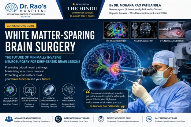
White Matter-Sparing Brain Surgery: The Future of Minimally Invasive Neurosurgery for Deep-Seated Brain Lesions

Featured in The Hindu (Vijayawada Edition, 30 August 2026) following Dr. Mohana Rao Patibandla’s keynote presentation at the World Neurosciences Summit (WNS) 2026, New Delhi.

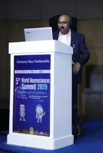
Dr. Mohana Rao Patibandla delivering the keynote lecture on White Matter-Sparing Brain Surgery at the 5th World Neurosciences Summit 2026 in New Delhi, India.

Dr. Mohana Rao Patibandla presenting his keynote lecture, “Minimally Invasive Cylindrical Corridor Surgery – Modern Microsurgery Strategy for Deep-Seated Brain Lesions,” at the 5th World Neurosciences Summit 2026 in New Delhi, highlighting advanced White Matter-Sparing Brain Surgery techniques for preserving critical neural pathways.
Cornerstone Article

White Matter-Sparing Brain Surgery: The Future of Minimally Invasive Neurosurgery for Deep-Seated Brain Lesions

Author: Dr. Mohana Rao Patibandla, Neurosurgeon, Founder & Chief Neurosurgeon, Dr. Rao’s Hospital – International Institute of Neurosciences, Guntur, Andhra Pradesh, India

Published: September 2026

Reading Time: 18–22 minutes

Target Keywords: White matter sparing surgery, minimally invasive brain surgery, cylindrical corridor surgery, deep-seated brain tumor surgery, brain lesion surgery, neurosurgeon India, Dr. Mohana Rao Patibandla.

Executive Summary

The human brain contains billions of neurons connected through highly organized white matter tracts—the communication highways that allow movement, speech, memory, vision, sensation, and cognition. Traditional surgery for deep-seated brain tumors or lesions often requires passing through healthy brain tissue, increasing the risk of neurological deficits.

White Matter-Sparing Surgery, also called Minimally Invasive Cylindrical Corridor Surgery, represents a transformative advancement in neurosurgery. Instead of cutting through healthy brain tissue, surgeons navigate between natural white matter pathways, preserving critical neural connections while reaching tumors or lesions deep inside the brain.

This technique was the focus of Dr. Mohana Rao Patibandla’s keynote lecture at the World Neurosciences Summit 2026, where he presented modern microsurgical strategies for safely treating deep-seated brain lesions. The approach was subsequently highlighted by The Hindu in a feature article discussing the future of minimally invasive brain surgery.

This comprehensive guide explains everything patients, families, neurologists, and neurosurgeons need to know about white matter-sparing surgery.

Table of Contents

  • What is White Matter-Sparing Brain Surgery?
  • Understanding White Matter and Brain Networks.
  • Why Traditional Brain Surgery Can Damage White Matter.
  • Evolution of Minimally Invasive Cylindrical Corridor Surgery.
  • How White Matter-Sparing Surgery Works.
  • Neuronavigation and Brain Mapping.
  • Who Needs White Matter-Sparing Surgery?
  • Conditions Treated.
  • Benefits Compared to Conventional Brain Surgery.
  • Surgical Planning and Technology.
  • Risks and Limitations.
  • Recovery After White Matter-Sparing Surgery.
  • Frequently Asked Questions.

Understanding White Matter: The Brain’s Information Highway

White matter tracts in the brain [IMAGE] | EurekAlert! Science News Releases

Diffusion Tensor Imaging 101 | Diffusion Imaging; Introduction, tutorials and background on diffusion tensor imaging and techniques

European Journal of Translational and Clinical Medicine

Most people think of the brain as a gray organ filled with neurons. However, white matter is equally important.

What is White Matter?

White matter consists of millions of insulated nerve fibers called axons. These fibers connect different brain regions and enable rapid communication.

White Matter Controls

Brain Function

Major White Matter Pathway

MovementCorticospinal tract
Speech productionArcuate fasciculus
Language comprehensionSuperior longitudinal fasciculus
VisionOptic radiations
MemoryFornix and cingulum
EmotionUncinate fasciculus

Damage to these pathways may result in:

  • Paralysis

  • Speech difficulty

  • Vision loss

  • Memory impairment

  • Cognitive dysfunction

  • Personality changes

Protecting these pathways has become one of the central goals of modern neurosurgery.

Why Traditional Brain Surgery Isn’t Always Enough

Neuroinfo Japan�F�J����p

Brain Pterional approach | UpSurgeOn

Minimally Invasive Brain Surgery | Keyhole Surgery | Pacific Brain Tumor Center

Historically, surgeons approached deep tumors by creating a path through the brain.

The Challenge

Deep lesions may lie:

  • Near motor pathways.

  • Adjacent to speech centers.

  • Beneath healthy cortex.

  • Close to visual pathways.

  • Near memory circuits.

Traditional surgery may require:

  • Brain retraction.

  • Cortical incision.

  • Traversing healthy tissue.

Even with excellent technique, these maneuvers may increase neurological risk.

Potential Consequences

Potential Injury

Possible Deficit

Motor tract injuryWeakness or paralysis
Language tract injurySpeech impairment
Visual tract injuryVisual field loss
Frontal network injuryBehavioral or cognitive changes

Modern neurosurgery seeks to avoid these injuries rather than simply treat them afterward.

What is White Matter-Sparing Surgery?

Our Integrated System - NICO Corporation

Surgical Neurology International

Frontiers | Minimally invasive treatment for glioblastoma through endoscopic surgery including tumor embolization when necessary: a technical note

Definition

White Matter-Sparing Surgery is a minimally invasive microsurgical technique that uses:

  • Natural anatomical corridors.

  • Cylindrical tubular retractors.

  • Advanced neuronavigation.

  • White matter tractography.

  • High-definition operating microscopy.

Instead of retracting large portions of the brain, surgeons gently separate white matter fibers along their natural orientation.

The Philosophy

Preserve normal brain. Remove abnormal tissue. Protect neural connectivity.

This is fundamentally different from older approaches focused primarily on lesion access.

The Science Behind Cylindrical Corridor Surgery

A New Way into the Brain | Johns Hopkins Medicine

Sobre Dr. Alexis Palpán

courses:456:2021:projects:456-2021-01:project-01 – CIIS Wiki

Why a Cylinder?

A cylindrical corridor distributes pressure evenly.

Traditional Retractor

Tumor retractor: a simple and novel instrument for brain tumor surgery | World Journal of Surgical Oncology | Springer Nature Link

Brain Pterional approach | UpSurgeOn

Principles of Intraventricular Surgery | Cohen Collection | Volumes | The Neurosurgical Atlas

  • Flat blades.

  • Uneven pressure.

  • Greater cortical retraction.

Cylindrical Corridor

  • Gentle circumferential dilation.

  • Less tissue distortion.

  • Smaller surgical footprint.

  • Better preservation of white matter fibers.

This concept has become increasingly adopted in minimally invasive neurosurgery worldwide.

How White Matter-Sparing Surgery Works

Step-by-Step Surgical Strategy

Stereotactic neurosurgery | MedLink Neurology

How Can New Imaging Modalities Help in the Practice of Radiology? - PMC

O nás | FNOL

Step

Purpose

1Advanced MRI with tractography identifies functional white matter pathways.
2Neuronavigation selects the safest trajectory.
3Small craniotomy is created.
4Tubular corridor gently separates white matter fibers.
5Microsurgical instruments remove lesion through the corridor.
6Hemostasis is achieved while preserving surrounding tissue.

The result is maximal safe lesion removal with minimal disruption.

White Matter Tractography: GPS for the Brain

Visualização Multimodal de Imagens de Tensores de Difusão | Visualização de Imagens de DTI

#mri #dti #corticospinal | Vahid Shahmaei

Correlation between language function and the left arcuate fasciculus detected by diffusion tensor imaging tractography after brain tumor surgery – Neurosurgery Blog

What is DTI Tractography?

Diffusion Tensor Imaging (DTI) visualizes white matter pathways before surgery.

It Helps Identify

  • Motor tracts.

  • Speech tracts.

  • Visual pathways.

  • Sensory pathways.

  • Memory networks.

Why It Matters

Surgeons can:

  • Avoid eloquent pathways.

  • Choose safer entry points.

  • Predict postoperative risks.

  • Tailor surgery to each patient.

This represents precision neurosurgery rather than generalized anatomy.

Neuronavigation: GPS Inside the Brain

Home - Neurochirurgie | Uniklinikum Erlangen

Удаление опухолей головного мозга с применением нейронавигации

Vall d'Hebron: un navegador para mejorar el diagnóstico con ecógrafo

Neuronavigation provides real-time anatomical guidance.

It Allows Surgeons To

  • Locate lesions accurately.

  • Avoid blood vessels.

  • Maintain surgical trajectory.

  • Navigate deep structures safely.

Combined with tractography, neuronavigation creates a personalized surgical roadmap.

Brain Mapping: Protecting Function During Surgery

Neurosurgeons are really good at removing brain tumors—they're about to get even better

Low Grade Glioma and Glioblastoma Outcomes Research | Brain Tumor Center

神经电生理监测在胶质瘤手术中的意义|运动|功能|神经|监测|手术|-健康界

Modern white matter-sparing surgery often incorporates:

Intraoperative Monitoring

  • Motor evoked potentials.

  • Somatosensory evoked potentials.

  • Cranial nerve monitoring.

  • Speech mapping when required.

This provides continuous feedback about neurological function.

Which Brain Conditions Can Be Treated?

Deep-Seated Brain Tumors

PPT - Clinical MRI Insights by Dr. Ravichandra: Brain and Vascular Imaging PowerPoint Presentation - ID:9635346

a, b Preoperative axial and sagittal T1-weighted MRI of posterior type... | Download Scientific Diagram

Examples include:

  • Gliomas

  • Metastatic tumors

  • Low-grade glioma

  • High-grade glioblastoma

  • Thalamic tumors

  • Basal ganglia tumors

Goal

Maximal safe resection while preserving neurological function.

Cavernomas

cerebral cavernous venous malformation | pacs

Brainstem Cavernoma - Neuropedia

海綿状血管腫 | 社会医療法人寿会 富永病院(大阪)

White matter-sparing corridors are valuable for:

  • Thalamic cavernoma.

  • Brainstem cavernoma.

  • Deep cerebral cavernomas.

Colloid Cysts

Checking your browser - reCAPTCHA

Surgical management of colloid cysts of the third ventricle: a single-institution comparison of endoscopic and microsurgical resection – Neurosurgery Blog

Checking your browser - reCAPTCHA

Small minimally invasive approaches reduce brain manipulation around the ventricles.

Intraventricular Tumors

intraventricular neoplasms and lesions | pacs

Central neurocytoma | Radiology Reference Article | Radiopaedia.org

Examples include:

  • Central neurocytoma.

  • Ependymoma.

  • Choroid plexus tumors.

  • Subependymoma.

Brain Abscesses

New Page 1

Infections of Brain Parenchyma I Flashcards | Quizlet

Intracranial Infections: Bacterial, Viral, Fungal, Parasitic -

Targeted tubular access allows drainage with limited cortical injury in selected patients.

Epilepsy Lesions

Displasia cortical focal en 7 Tesla - Mayo Clinic

Minimally Invasive Surgery for Cerebral Cavernous Malformations

Let the Lesion Do the Talking :: Behance

Certain deep epileptogenic lesions can be approached through white matter-preserving techniques.

Advantages Over Conventional Brain Surgery

Why White Matter-Sparing Surgery Matters

Traditional Surgery

White Matter-Sparing Surgery

Larger craniotomySmaller craniotomy
More brain retractionMinimal brain retraction
Cortical disruptionNatural white matter corridor
Greater tissue manipulationTargeted microsurgical access
Higher postoperative swellingReduced tissue trauma
Longer recoveryFaster recovery in selected patients

Key Patient Benefits

East Carolina Heart Institute at ECU Health Medical Center - Greenville, NC 27834

Dr. Alejandro Ceja Espinosa Neurocirujano, Morelia - Agenda cita | Doctoralia.com.mx

I've been diagnosed with a brain tumor. Now what? | Michigan Medicine

Potential benefits include:

  • Smaller incision.

  • Less pain.

  • Reduced blood loss.

  • Less brain swelling.

  • Better cosmetic outcome.

  • Earlier mobilization.

  • Shorter ICU stay in selected cases.

  • Preservation of neurological function.

These benefits depend on careful patient selection and surgical expertise.

Who is a Candidate?

Ideal Candidates

Suitable Patients

Why

Deep brain tumorsAccessible through safe anatomical corridors.
Small-to-medium lesionsMinimal disruption possible.
Functional area lesionsWhite matter preservation is especially valuable.
Selected hemorrhagesTubular evacuation may be appropriate in selected cases.
Deep cavernomasNatural corridor approach.

Patients Who May Need Conventional Surgery

  • Very large tumors.

  • Diffuse infiltrative lesions.

  • Extensive vascular malformations.

  • Multiple compartment lesions.

Every patient requires individualized evaluation.

Surgical Planning at Dr. Rao’s Hospital

Equipment and medical devices in modern operating room

3T MRI | Book Your Scan Today — Rezolut

Grupo Policlínica incorpora el microscopio más avanzado del mercado en su unidad de neurocirugía | Onda Cero Radio

A white matter-sparing operation begins long before entering the operating room.

Comprehensive Planning Includes

Advanced Imaging

3 Tesla MR Fiyatları 2026 | Fulya Açık Emar

브런치

How Can New Imaging Modalities Help in the Practice of Radiology? - PMC

  • MRI Brain

  • Contrast MRI

  • DTI tractography

  • Functional MRI when indicated.

  • MR angiography/venography.

Digital Planning

Frontiers | Neuronavigation in glioma resection: current applications, challenges, and clinical outcomes

Stryker Launches Q Guidance System with Cranial Guidance Software | OR Today

Brain Tumor Program | Neurosurgery

  • Surgical trajectory simulation.

  • White matter tract preservation.

  • Vascular avoidance.

  • Cortical entry planning.

Microsurgical Technology Used

High-End Neurosurgical Equipment

Doctors Hospital installs the first Kinevo 900 microscope in Mexico. - Doctors Hospital

M720 OH5 Operationsmikroskop der Spitzenklasse - Medien | Produkte | Leica Microsystems

Neurokirurgi: sätter standarden inom modern neurokirurg

Technology may include:

  • Operating microscope.

  • Neuronavigation.

  • Ultrasonic aspirator.

  • Endoscope-assisted visualization.

  • Neurophysiological monitoring.

  • High-definition imaging.

Each tool contributes to safer microsurgery.

White Matter-Sparing Surgery in Brain Tumor Management

Low-Grade Gliomas

The silent phase of diffuse low-grade gliomas. Is it when we missed the action? – Neurosurgery Blog

Subject-Specific Automatic Reconstruction of White Matter Tracts | Journal of Imaging Informatics in Medicine | Springer Nature Link

MRI Tractography - Brain Tumor

Goal:

  • Preserve language.

  • Preserve cognition.

  • Maximize tumor removal.

High-Grade Glioblastoma

Glioblastoma, IDH-wildtype | Radiology Reference Article | Radiopaedia.org

Checking your browser - reCAPTCHA

Brain Tumour Resection Surgery Johannesburg

Goal:

  • Maximal safe resection.

  • Protect functional pathways.

  • Improve quality of life.

White Matter Preservation in Pediatric Neurosurgery

Tumores cerebrales en niños | Pediatría integral

Zabieg usunięcia olbrzymiego guza mózgu u miesięcznego dziecka w IPCZD - Aktualności - Centrum Zdrowia Dziecka

Pediatric Hemato-Oncology Department – TAMC

Children have developing neural pathways.

Advantages include:

  • Better developmental preservation.

  • Reduced tissue injury.

  • Smaller surgical footprint.

  • Functional protection during growth.

White Matter-Sparing Surgery for Brainstem Lesions

Cavernomas do Sistema Nervoso Central :: Clinica Maddalozzo

511-2018-09-05-anatomy-II

【北总神外论道 】经安全区切除脑干海绵状血管瘤系列2 - 脑医汇

Brainstem surgery is among the most challenging areas in neurosurgery.

The technique focuses on:

  • Safe entry zones.

  • Natural fiber orientation.

  • Cranial nerve preservation.

  • Microsurgical precision.

Recovery After White Matter-Sparing Surgery

What Patients Can Expect

Patient’s ER visit turns into brain surgery, cancer diagnosis - UNC Lineberger

Post-Craniotomy Recovery & Care in Pune

Consultation after MRI examination in medical clinic. MRI Doctor is showing x-ray and of brain tumors in the patient head.

Hospital Stay

Stage

Typical Course

ICU1 day in many selected cases.
Ward2–5 days depending on condition.
WalkingOften within 24 hours if neurologically appropriate.
Return HomeDepends on recovery and pathology.

Recovery varies by diagnosis, lesion location, age, and overall health.

Rehabilitation

Speech Therapy After Stroke - Sheltering Arms Institute

Fisioterapia Neuro Funcional: Restaure a Qualidade de Vida com Corpo Ativo

Neurologie — Ergothérapie de la Prévôté

Some patients benefit from:

  • Physiotherapy.

  • Speech therapy.

  • Occupational therapy.

  • Cognitive rehabilitation.

Risks and Limitations

No brain surgery is risk-free.

Potential Risks

EPOS™

Continuous EEG Monitoring | Nihon Kohden

Checking your browser - reCAPTCHA

  • Bleeding.

  • Infection.

  • Seizures.

  • Neurological deficits.

  • Brain swelling.

  • CSF leak.

The goal of white matter-sparing surgery is to reduce unnecessary injury—not eliminate all surgical risk.

Evidence Supporting White Matter-Sparing Approaches

Why This Technique is Growing Worldwide

Modern literature supports:

  • Less cortical injury.

  • Improved preservation of white matter tracts.

  • Better functional outcomes in selected lesions.

  • Reduced surgical morbidity in carefully chosen patients.

International adoption continues to increase alongside advances in imaging and neuronavigation.

Dr. Mohana Rao Patibandla’s Keynote at World Neurosciences Summit 2026

NeuroScience Frontiers Symposium 2025 | Neurogen Therapeutic

Groundbreaking Research on Minimally Invasive Brain Tumor Surgery Presented by Dr. Rao at Andhra Pradesh Neuroscientists Association Conference

Society for Neuroscience - Neuroscience 2026

At the World Neurosciences Summit 2026, Dr. Mohana Rao Patibandla presented a keynote lecture titled:

“Minimally Invasive Cylindrical Corridor Surgery – Modern Microsurgery Strategy for Deep-Seated Brain Lesions.”

The presentation emphasized:

  • White matter preservation.

  • Modern microsurgical planning.

  • Neuronavigation-guided access.

  • Safe treatment of deep brain lesions.

  • Functional outcome optimization.

The lecture highlighted the growing importance of minimally invasive microsurgical corridors in contemporary neurosurgery.


Featured in The Hindu: National Recognition of White Matter-Sparing Surgery

The Hindu reported Dr. Mohana Rao Patibandla’s press conference explaining how preserving normal brain tissue and neural pathways has become a key objective of modern minimally invasive microsurgery. The article also described cylindrical corridor surgery as a white matter-sparing strategy for deep-seated brain lesions.


This coverage reflects growing public awareness of advanced brain-preserving surgical techniques in India.

Why This Matters for Patients in India

Advanced Brain & Spine Neurosurgery Treatments in Chennai

Competent medical expert explaining diagnosis

Neurologist in Pimpri & Brain Specialist in Pimpri-Chinchwad

Patients increasingly seek:

  • Safer surgery.

  • Smaller incisions.

  • Faster recovery.

  • Preservation of neurological function.

  • International-standard neurosurgical care within India.

White matter-sparing surgery aligns with these goals when clinically appropriate.

Why Choose Dr. Rao’s Hospital for White Matter-Sparing Brain Surgery?

Best Neurosurgery & Spine Care at Dr. Rao’s Hospital, Guntur

Brain Surgery in Pune | Dr. Jaydev Panchawagh, Neurosurgeon

Home - Neurochirurgie | Uniklinikum Erlangen

Dr. Rao’s Hospital – International Institute of Neurosciences, Guntur, offers comprehensive brain and spine care with advanced neurosurgical technology and internationally trained expertise.

Areas of Expertise

  • Brain Tumor Surgery

  • Skull Base Surgery

  • Endoscopic Brain Surgery

  • Pediatric Neurosurgery

  • Cerebrovascular Surgery

  • Epilepsy Surgery

  • Functional Neurosurgery

  • Minimally Invasive Neurosurgery

Advanced Technologies

  • Operating Microscope

  • Neuronavigation

  • Neuroendoscopy

  • Neurophysiological Monitoring

  • Advanced MRI Planning

  • Minimally Invasive Brain Surgery

Frequently Asked Questions (FAQ)

1. What is White Matter-Sparing Brain Surgery?

White matter-sparing brain surgery is a minimally invasive microsurgical approach that reaches deep brain lesions through natural white matter corridors while minimizing injury to healthy neural pathways.

2. What is Cylindrical Corridor Surgery?

It is a minimally invasive technique using a tubular retractor to create a gentle corridor through the brain, allowing surgeons to access deep lesions with less tissue disruption.

3. Is White Matter-Sparing Surgery safer than traditional brain surgery?

It may reduce disruption to healthy brain tissue in carefully selected patients, but safety depends on lesion type, location, patient health, and surgical expertise.

4. Which brain tumors can be treated?

Selected gliomas, metastases, intraventricular tumors, cavernomas, colloid cysts, and certain deep-seated lesions may be suitable candidates.

5. What is tractography?

Tractography is an MRI technique (DTI) that maps white matter pathways before surgery to help surgeons avoid critical neural connections.

6. Does every patient need tractography?

Not always. It is especially useful when lesions are close to eloquent brain pathways.

7. Is recovery faster?

Many selected patients experience smaller incisions, less pain, and earlier mobilization, although recovery varies with the underlying disease and surgery performed.

8. Is this surgery available in India?

Yes. Advanced minimally invasive white matter-sparing brain surgery is available in specialized neuroscience centers including Dr. Rao’s Hospital in Guntur.

9. Can brain function be preserved completely?

The goal is maximal preservation of neurological function, but outcomes depend on lesion location, disease biology, and individual patient factors.

10. How do I know if I’m a candidate?

A neurosurgical consultation with MRI review, tractography when indicated, and individualized surgical planning is necessary.

Key Takeaways

White matter tracts in the brain [IMAGE] | EurekAlert! Science News Releases

A team of surgeons performing brain surgery to remove a tumor.

Elements Fibertracking | Brainlab

White Matter-Sparing Surgery represents one of the most significant advances in modern neurosurgery because it shifts the surgical philosophy from simply reaching a lesion to protecting the brain’s communication networks while treating disease.

For carefully selected patients with deep-seated brain tumors, cavernomas, intraventricular lesions, and other complex pathologies, minimally invasive cylindrical corridor surgery offers a highly precise approach centered on preserving neurological function whenever possible.

As highlighted during Dr. Mohana Rao Patibandla’s keynote lecture at the World Neurosciences Summit 2026 and subsequently featured in The Hindu, the future of neurosurgery lies in combining advanced imaging, neuronavigation, microsurgical precision, and white matter preservation to achieve better patient outcomes.


About the Author

Dr. Mohana Rao Patibandla is an internationally fellowship-trained neurosurgeon and the Founder & Chief Neurosurgeon of Dr. Rao’s Hospital – International Institute of Neurosciences, Guntur, Andhra Pradesh, India.

His advanced training includes skull base surgery, pediatric neurosurgery, minimally invasive brain surgery, neuro-oncology, cerebrovascular and endovascular neurosurgery, and functional neurosurgery in India and the United States. He regularly serves as invited faculty at national and international neuroscience conferences and delivered a keynote lecture at the World Neurosciences Summit 2026 on White Matter-Sparing Minimally Invasive Cylindrical Corridor Surgery.


Authoritative Neurosurgical Resources

Patients with deep-seated gliomas, meningiomas, metastatic tumors, and other complex lesions may benefit from

Brain Tumor Surgery 
performed using modern white matter-sparing microsurgical techniques. Many lesions located near the skull base require specialized Skull Base Surgery 
combined with advanced neuronavigation and minimally invasive approaches.

Our comprehensive Neuro-Oncology Program focuses on personalized treatment for benign and malignant brain tumors. Children with deep-seated brain tumors may benefit from advanced Pediatric Neurosurgery that prioritizes preservation of developing neural pathways.

Selected intraventricular tumors and colloid cysts can often be treated using Endoscopic Brain Surgery or minimally invasive cylindrical corridor techniques.

White matter-sparing surgery relies heavily on Neuronavigation-Guided Brain Surgery 
to identify the safest surgical pathway while protecting eloquent brain regions.

Patients with epilepsy caused by cavernomas or focal cortical lesions may also be candidates for Epilepsy Surgery using minimally invasive neurosurgical strategies.

Learn more about the international training, experience, research, and surgical philosophy of Dr. Mohana Rao Patibandla Founder of Dr. Rao’s Hospital – International Institute of Neurosciences.

YouTube

Facebook

Instagram

Google Business Page

LinkedIn

X / Twitter

Need Expert Care for a Brain Tumor or Deep-Seated Brain Lesion?

At Dr. Rao’s Hospital – International Institute of Neurosciences (IIN),
we provide advanced White Matter-Sparing Brain Surgery,
Minimally Invasive Brain Tumor Surgery,
Skull Base Surgery, Endoscopic Brain Surgery,
Neuro-Oncology, Pediatric Neurosurgery,
Stroke Care, and Spine Surgery using internationally
recognized technology and evidence-based treatment protocols.

Every patient receives a personalized surgical plan based on advanced MRI,
neuronavigation, white matter tractography, intraoperative neuromonitoring,
and minimally invasive microsurgical techniques designed to preserve neurological function whenever possible.


Why Choose Dr. Rao’s Hospital?

  • Internationally Fellowship-Trained Neurosurgeon – Dr. Mohana Rao Patibandla
  • Dedicated Super-Specialty Neurosciences Hospital in Guntur, Andhra Pradesh.
  • Advanced White Matter-Sparing & Minimally Invasive Brain Surgery.
  • Brain Tumor, Skull Base, Pediatric, Spine, Stroke & Endovascular Neurosurgery.
  • Neuronavigation, Neurophysiological Monitoring & High-End Microsurgical Technology.
  • Comprehensive Neuro-Oncology, Neurocritical Care and Rehabilitation.
  • 24×7 Emergency Brain, Stroke and Spine Care.

Contact Dr. Rao’s Hospital

Dr. Rao’s Hospital – International Institute of Neurosciences (IIN)
12-19-67, Old Bank Road, Kothapet,
Opposite Sravani Hospital, Besides AK Biryani point,
Guntur – 522001, Andhra Pradesh, India.

📞 +91 90100 56444
✉️ info@drraoshospitals.com
🌐 www.drraoshospitals.com

The best Minimally Invasive Keyhole Endoscopic Spine Surgery in Guntur

How Keyhole Neurosurgery Is Transforming Brain and Spine Care in India – Dr. Rao’s IIN


How Minimally Invasive Keyhole Neurosurgery Is Revolutionizing Brain and Spine Care in India


Modern neurosurgery has entered an era of remarkable transformation driven by advanced imaging, precision-guided technologies, minimally invasive surgical systems, and sophisticated neurosciences innovation.


Historically, many neurological and spine surgeries required large surgical openings and extensive tissue manipulation to reach delicate structures deep within the brain or spine.


While traditional surgical techniques saved countless lives, they were often associated with longer hospitalization, greater tissue disruption, postoperative pain, and extended rehabilitation periods.


Today, minimally invasive keyhole neurosurgery is changing this landscape by enabling surgeons to perform selected procedures through smaller openings while preserving surrounding neurological structures and improving recovery outcomes.


A recent Times of India feature highlighted this transformation through the recognition awarded to Dr. Mohana Rao Patibandla as the Best Minimally Invasive Keyhole Neurosurgeon and Spine Surgeon at the prestigious Radiocity FM 91.1 Icon Awards.


Primary Source: Times of India – Dr. Mohana Rao Patibandla Honoured as Best Minimally Invasive Keyhole Neurosurgeon and Spine Surgeon


The Complexity of Brain and Spine Surgery


Neurosurgery remains one of the most technically demanding specialties in medicine because it involves structures responsible for:


  • Movement
  • Speech
  • Memory
  • Vision
  • Cognition
  • Sensory function


The brain and spinal cord contain highly delicate neural pathways and vascular structures where even small surgical inaccuracies may potentially affect long-term neurological function.


Conditions commonly requiring neurosurgical or spine intervention include:


  • Brain tumors
  • Spinal cord compression
  • Disc prolapse
  • Stroke-related disorders
  • Traumatic injuries
  • Neurovascular diseases
  • Degenerative spine disease


Modern neurosurgery therefore depends heavily on precision, technology integration, multidisciplinary collaboration, and continuous innovation.


American Association of Neurological Surgeons


What Is Minimally Invasive Keyhole Neurosurgery?


Keyhole neurosurgery is an advanced minimally invasive surgical technique that allows surgeons to access neurological pathology through smaller surgical openings using specialized instruments, microscopes, endoscopes, and neuronavigation systems.


According to the Times of India article, Dr. Mohana Rao Patibandla has become widely recognized for advancing minimally invasive keyhole neurosurgery and spine surgery in India.


Unlike traditional open surgery, keyhole approaches focus on:


  • Smaller incisions
  • Microsurgical precision
  • Reduced tissue disruption
  • Enhanced visualization
  • Function preservation


These techniques often utilize:


  • Operating microscopes
  • Endoscopic systems
  • Advanced neuroimaging
  • Neuronavigation
  • Intraoperative monitoring


Modern keyhole surgery represents one of the most important advancements in contemporary neurosciences.


The Benefits of Minimally Invasive Neurosurgery


The evolution of minimally invasive surgery has significantly improved patient care in selected neurological and spinal conditions.


Potential advantages may include:


  • Reduced blood loss
  • Less postoperative pain
  • Shorter hospitalization
  • Smaller surgical scars
  • Faster recovery
  • Reduced tissue trauma


The Times of India article emphasizes Dr. Rao’s dedication toward advancing minimally invasive surgery to improve patient outcomes and accessibility.


Modern healthcare increasingly prioritizes preserving function while minimizing surgical disruption.


Minimally Invasive Neurosurgery at Dr. Rao’s Hospital


The Evolution of Minimally Invasive Spine Surgery


Spine surgery has also undergone major transformation through minimally invasive approaches.


Traditional spine procedures often involved:


  • Larger muscle dissection
  • Greater blood loss
  • Longer recovery time
  • Extended rehabilitation


Modern minimally invasive spine surgery now enables selected procedures through smaller corridors while preserving muscles and surrounding tissues.


Conditions commonly treated using minimally invasive spine techniques may include:


  • Lumbar disc prolapse
  • Cervical disc disease
  • Spinal stenosis
  • Degenerative spine disorders
  • Selected spinal tumors


These advancements have significantly improved postoperative rehabilitation and patient mobility.


North American Spine Society


International Training and Global Exposure


The Times of India feature highlights Dr. Rao’s international training experience in the United States, Europe, and Japan.


Global exposure often provides neurosurgeons experience with:


  • Advanced surgical technologies
  • Research-oriented healthcare systems
  • Complex multidisciplinary care
  • Modern operating room infrastructure
  • Evidence-based medicine


Such international training frequently influences long-term institutional vision and innovation-driven healthcare development.


The article emphasizes how these experiences shaped Dr. Rao’s patient-centered approach focused on precision and innovation.


The Importance of Regional Neurosciences Infrastructure


One of the most important themes reflected throughout the article is the expansion of advanced neurological care beyond metropolitan cities.


Historically, many patients from smaller towns traveled long distances seeking:


  • Complex brain surgery
  • Advanced spine surgery
  • Stroke intervention
  • Neuro-oncology care
  • Endovascular treatment


This often created:


  • Financial burden
  • Treatment delays
  • Interrupted rehabilitation
  • Accessibility challenges


According to the Times of India article, Dr. Rao’s dedication toward accessible and high-quality care has benefited patients from Andhra Pradesh, Telangana, and neighboring states.


Regional neuroscience systems improve:


  • Emergency accessibility
  • Continuity of treatment
  • Patient convenience
  • Long-term rehabilitation support


Technology Is Transforming Neurosciences


Modern neurological care increasingly depends on sophisticated technologies including:


  • Neuronavigation systems
  • Advanced MRI imaging
  • Microsurgical operating microscopes
  • Endoscopic systems
  • Intraoperative neuromonitoring
  • Precision-guided surgical platforms


These technologies improve:


  • Surgical planning
  • Precision
  • Safety
  • Neurological preservation
  • Patient recovery


The future of neurosurgery will increasingly depend on integrating technology with human expertise and ethical patient-centered care.


The Human Side of Neurosurgery


Despite technological advancements, neurological care remains deeply human.


Patients facing brain or spine disorders often experience:


  • Fear
  • Anxiety
  • Concerns regarding disability
  • Emotional uncertainty
  • Questions about long-term recovery


Modern healthcare therefore requires not only technical expertise but also:


  • Compassion
  • Communication
  • Empathy
  • Trust
  • Long-term rehabilitation support


The Times of India article repeatedly reflects themes of dedication toward innovation, patient care, and medical excellence.


Recognition and Healthcare Leadership


Healthcare awards frequently recognize broader contributions toward innovation, accessibility, and patient-centered care.


The article notes several recognitions associated with Dr. Rao’s contributions, including:


  • Brain Lab Neurosurgery Award
  • Leaders Award
  • Atal Achievement Award


Such recognitions highlight the growing importance of innovation-driven neurological care within India’s healthcare ecosystem.


The Future of Keyhole Neurosurgery in India


India’s neurosciences ecosystem continues evolving rapidly through:


  • Artificial intelligence-assisted diagnostics
  • Robotic surgery
  • Advanced neuroimaging
  • Precision-guided surgery
  • Minimally invasive systems
  • Endovascular intervention


At the same time, future healthcare transformation will depend heavily on:


  • Regional accessibility
  • Advanced training
  • Ethical healthcare systems
  • Continuous innovation
  • Patient-centered treatment


Institutions capable of combining advanced technology with compassionate care are likely to shape the future of neurological treatment across India.


Conclusion


The recognition awarded to Dr. Mohana Rao Patibandla as Best Minimally Invasive Keyhole Neurosurgeon and Spine Surgeon reflects a broader transformation occurring within India’s neurosciences ecosystem.


Modern neurological and spinal care increasingly depends on:


  • Minimally invasive surgery
  • Advanced imaging technologies
  • Precision-guided systems
  • Multidisciplinary expertise
  • Patient-centered care


Institutions such as Dr. Rao’s Hospital – International Institute of Neurosciences represent an important part of India’s expanding commitment toward accessible, innovation-driven, and ethically focused neurosciences care.


As minimally invasive surgery continues evolving globally, the integration of precision technology, surgical expertise, and compassionate healthcare will likely define the next era of brain and spine treatment in India.


Frequently Asked Questions (FAQ)


1. What is keyhole neurosurgery?

Keyhole neurosurgery is a minimally invasive surgical technique using smaller openings and advanced imaging systems to reduce tissue disruption.


2. What are the benefits of minimally invasive spine surgery?

Benefits may include smaller incisions, less pain, shorter hospitalization, and faster recovery.


3. Why is neuronavigation important in neurosurgery?

Neuronavigation helps surgeons identify safer surgical pathways and improve precision during brain and spine surgery.


4. Why are regional neuroscience centers important?

They improve accessibility to advanced neurological and spine care outside major metropolitan cities.


5. Who is Dr. Mohana Rao Patibandla?

Dr. Mohana Rao Patibandla is an internationally trained neurosurgeon and founder of Dr. Rao’s Hospital – International Institute of Neurosciences.


Internal Links


Call To Action

If you or your loved one is experiencing neurological symptoms such as persistent headaches, seizures, spinal pain, weakness, stroke warning signs, or neurological disorders, timely expert evaluation is essential.


Dr. Rao’s Hospital – International Institute of Neurosciences
12-19-67, Old Bank Road, Kothapet, Besides AK Biryani Point, Guntur, Andhra Pradesh

Phone: +91 9010056444

Email: info@drraoshospitals.com

Website: https://drraoshospitals.com/


Connect With Us

“`

Dr. Mohana Rao Patibandla, Top Neurosurgeon in India 2024, featured on The Enterprise World magazine cover for leadership in minimally invasive neurosurgery at Dr. Rao’s Hospital, Guntur.

Emerging Innovations in Neurosurgery: From Endoscopic to AI


Emerging Innovations in Neurosurgery: From Endoscopic Brain Surgery to AI-Guided Spine Procedures

The field of neurosurgery is undergoing a transformative evolution, driven by cutting-edge technologies that redefine what’s possible in brain and spine care. From endoscopic brain surgery to AI-guided spine procedures, these innovations promise more precise, less invasive, and highly personalized treatments.

At Dr. Rao’s Hospital, one of India’s most advanced centers for neurosurgical excellence, innovation meets compassion. Led by Dr. Mohana Rao Patibandla — one of the best neurosurgeons in Guntur — the hospital integrates artificial intelligence, robotics, and endoscopic techniques to deliver the highest standards of care.


1. Endoscopic Brain Surgery: A New Window into the Brain

What is Endoscopic Brain Surgery? It’s a minimally invasive approach where neurosurgeons use a small camera (endoscope) and micro-instruments to access deep-seated areas of the brain through tiny openings or natural pathways like the nasal cavity. This allows precise tumor removal with minimal disruption to healthy tissues.

Benefits of Endoscopic Brain Surgery

  • Smaller incisions and reduced scarring
  • Less blood loss and faster recovery
  • Shorter hospital stay and reduced infection risk

At the Neurosurgery Department of Dr. Rao’s Hospital, advanced endoscopic procedures are performed routinely for brain tumors, colloid cysts, hydrocephalus, and skull base lesions. These surgeries combine technology with surgical mastery for optimal patient outcomes.

[Source: WHO – Surgical Safety and Innovation]


2. AI-Guided Spine Surgery: Where Precision Meets Technology

How does AI improve spine surgery? Artificial Intelligence assists spine surgeons by analyzing preoperative imaging, planning precise screw placements, and guiding robotic systems during the procedure. AI tools reduce human error and optimize patient-specific surgical plans.

At the Spine Surgery Unit at Dr. Rao’s Hospital, AI-integrated navigation and robotic assistance are redefining complex spinal deformity corrections and fusion surgeries. Patients benefit from enhanced safety, accuracy, and quicker recovery times.

[Source: NIH – Artificial Intelligence in Neurosurgery]


3. Intraoperative Neuromonitoring (IONM): Real-Time Safety

Intraoperative Neuromonitoring (IONM) has become essential in modern neurosurgery. It allows continuous monitoring of nerve function during surgery, helping prevent permanent neurological damage.

  • Protects critical brain and spinal pathways
  • Detects early warning signals during operations
  • Reduces postoperative complications

Dr. Mohana Rao presented his clinical experience on the efficacy of IONM in over 1000 neurosurgical cases, showcasing its vital role in improving safety across brain and spine surgeries.


4. Minimally Invasive Skull Base Surgery: Access Without Trauma

Traditionally, skull base surgery involved large openings and long recoveries. Today, with endoscopic transnasal approaches, neurosurgeons can reach complex regions through the nasal passage, minimizing trauma and preserving facial structures.

These approaches are particularly effective for:

  • Pituitary tumors
  • Craniopharyngiomas
  • Meningiomas
  • Chordomas

Dr. Rao’s Hospital is among the few centers in India where such minimally invasive skull base surgeries are routinely performed — including advanced procedures like endoscopic transnasal odontoidectomy.

[Source: PubMed – Endoscopic Skull Base Surgery]


5. Neuro-navigation and 3D Brain Mapping: The GPS of Neurosurgery

Neuro-navigation systems use real-time imaging to create 3D brain maps, allowing surgeons to precisely locate and remove tumors while avoiding vital brain areas. This technology acts as a “GPS for the brain,” improving both precision and safety.

Combined with intraoperative MRI and 3D mapping, these systems allow Dr. Rao’s Hospital’s Neurosurgery Department to offer unmatched accuracy in delicate brain tumor resections and epilepsy surgeries.


6. Robotics in Neurosurgery: Precision Beyond the Human Hand

Robotics has made its mark in neurosurgery by enhancing accuracy and stability during intricate procedures. With robotic assistance, neurosurgeons can perform deep-brain and spinal stabilization surgeries with sub-millimeter precision.

At Dr. Rao’s Hospital’s Neurology and Neurosurgery Units, robotic-assisted procedures are revolutionizing patient safety and postoperative outcomes through automation and artificial intelligence.


7. AI in Predictive and Personalized Neurosurgery

Artificial Intelligence is not limited to the operating room — it now extends to predictive modeling, outcome forecasting, and personalized rehabilitation. AI helps doctors estimate patient recovery times, surgical risks, and long-term mobility outcomes with remarkable accuracy.

[Source: Mayo Clinic – AI in Personalized Medicine]


8. The Future of Neurosurgical Care at Dr. Rao’s Hospital

The integration of AI, robotics, and endoscopy marks a new era in neurosurgery — one that prioritizes precision, speed, and safety. Patients at Dr. Rao’s Hospital benefit from:

  • Real-time intraoperative neuromonitoring
  • Minimally invasive surgical techniques
  • Shorter hospital stays and faster recovery
  • Multidisciplinary neurological care under one roof

To book an appointment or consultation, visit the Contact Page or call 📞 090100 56444.


Conclusion: Pioneering the Future of Neurosurgery in India

Neurosurgery is evolving from open procedures to precision-guided, minimally invasive solutions powered by AI and robotics. At the forefront of this revolution is Dr. Mohana Rao Patibandla, one of the best neurosurgeons in Guntur.

Under his leadership, Dr. Rao’s Hospital stands as a national hub for advanced neurosurgical innovation, offering safer and smarter treatments for complex brain and spine disorders.

If you’re searching for the best neurosurgeon in Guntur or expert care for complex neurological conditions, visit Dr. Rao’s Hospital. Contact us at 📞 090100 56444 or 📧 info@drraoshospitals.com.


What is endoscopic brain surgery?

Endoscopic brain surgery is a minimally invasive technique using a small camera and instruments through tiny openings to treat brain tumors, cysts, and hydrocephalus with less recovery time.

How does AI assist in spine surgery?

AI analyzes scans and guides surgical tools to enhance precision and safety during spinal procedures. It reduces errors and improves postoperative outcomes.

Why choose Dr. Rao’s Hospital for advanced neurosurgery?

At Dr. Rao’s Hospital, led by Dr. Mohana Rao Patibandla, one of the best neurosurgeons in Guntur, patients receive world-class minimally invasive, AI-assisted, and compassionate neurosurgical care.


Follow Dr. Rao’s Hospital

Dr. Mohana Rao Patibandla standing at the podium at SIONCON 2025 in Bengaluru, with a large screen displaying his professional profile, qualifications, and fellowships from leading global neurosurgery centers.

How Intraoperative Neuromonitoring (IONM) Is Transforming Brain and Spine Surgery in India: Insights from Dr. Mohana Rao’s 1008-Case Study Presented at SIONCON 2025, NIMHANS Bengaluru

Advancing Surgery with Intraoperative Neuromonitoring (IONM) in India

By Dr. Mohana Rao Patibandla, Chief Neurosurgeon, Dr. Rao’s Hospital, Guntur


Introduction: The Future of Neurosurgery Is Here

Brain and spine surgeries are among the most delicate and high-risk procedures in all of medicine. Surgeons work within millimeters of the structures that control movement, speech, vision, memory, and breathing. Even a minor surgical disruption can lead to paralysis, speech loss, or permanent disability.

For decades, neurosurgeons relied solely on their anatomical knowledge, experience, and the microscope to navigate these high-risk regions. But in the last few years, a revolutionary tool has transformed neurosurgical safety worldwide:
Intraoperative Neuromonitoring (IONM).

In November 2025, at SIONCON 2025, held at the prestigious NIMHANS Convention Centre, Bengaluru, I had the honor of presenting one of India’s most comprehensive IONM datasets—a 1008-case analysis covering diverse brain and spine surgeries performed over 5 years at Dr. Rao’s Hospital, Guntur.

Find the news on the Business World:

Guntur Neurosurgeon Presents India’s Largest IONM Dataset, Sets New Benchmark In Brain & Spine Safety

https://www.businessworld.in/article/guntur-neurosurgeon-presents-india-s-largest-ionm-dataset-sets-new-benchmark-in-brain-spine-safety-583438

The presentation demonstrated how real-time neuromonitoring can significantly reduce neurological complications and improve outcomes, even in the most complex neurosurgical cases.

This blog explains the entire study in a clear, patient-friendly way, detailing how IONM protects patients, helps surgeons make safer decisions, and is shaping the future of neurosurgery in India.


Chapter 1: What Exactly Is Intraoperative Neuromonitoring (IONM)?

Imagine having a real-time safety system during surgery that continuously checks how your brain, spine, and nerves are functioning—even while under anesthesia.
That is the power of IONM.

IONM monitors:

  • Motor function

  • Sensory pathways

  • Cranial nerve function

  • Language and speech centers (in awake surgeries)

  • Brainstem responses

  • Electrical activity of nerves and muscles

If anything harmful begins to happen—such as nerve stretching or decreased blood supply—the system alerts the surgical team immediately.

It is like having a “nerve safety alarm” during surgery.


Why IONM Is Critical in Modern Neurosurgery

Without IONM, nerve injury might be detected only after surgery, when it is too late to reverse the damage.
With IONM, however, risks are identified immediately, allowing the surgeon to respond and prevent permanent injury.

This simple concept can save movement, speech, vision, swallowing ability, and quality of life.


Chapter 2: Why This 1008-Case Study Is a Landmark for India

IONM is still growing in India.
Most hospitals use it only for select cases.
Very few centers have standardized, protocol-driven multimodal IONM across multiple years of surgical work.

At Dr. Rao’s Hospital, we took a different approach:

We used IONM in over 1000 surgeries across:

  • Brain tumors (including eloquent cortex tumors)

  • Skull base and brainstem lesions

  • Spine surgeries

  • Pediatric neurosurgery

  • Epilepsy surgery

  • Awake craniotomy

  • Vascular and endovascular neurosurgery

We also implemented a strict, step-by-step protocol to ensure quality, accuracy, and meaningful intervention whenever signals changed.

This dataset is among the largest single-center IONM studies from India, and the presentation at SIONCON 2025 highlighted the clinical impact of every modality.


Chapter 3: Study Results Explained Simply

Below is a simplified explanation of the key results we presented at SIONCON:


1. Diagnostic Accuracy: IONM Predicts Danger Early

Sensitivity: 85.7%

This means IONM correctly detected 85.7% of cases where something was going wrong.

Specificity: 98.8%

This means IONM rarely gave false alarms.

These numbers show the system is both reliable and precise. Out of 1008 surgeries:

  • True Positives (TP): 186

  • True Negatives (TN): 781

  • False Positives (FP): 10

  • False Negatives (FN): 31

IONM accurately predicted neurological outcomes in 967 out of 1008 cases.


2. Neurological Recovery & Complication Reduction

Key highlights:

  • 78.5% prevention rate for neurological deficits

  • 94% of temporary deficits recovered within 3 months

  • Only 2.1% permanent deficits, mostly in complex eloquent tumors

  • Permanent signal loss: 0.7%

  • Functional independence (KPS ≥80): 91.6% at 3 months

These outcomes match or exceed global neurosurgical benchmarks—reinforcing that India can deliver world-class results.


Chapter 4: Pathology-Wise Outcomes

Eloquent Area Brain Tumors (Movement/Speech Regions)

  • Very high risk surgeries

  • Permanent deficits only 2.1%

  • Safe maximal tumor removal possible

Epilepsy Surgery

  • 88.4% deficit prevention

  • Excellent long-term functional outcomes

Spine Surgery

  • 83.1% deficit prevention rate

  • EMG and MEP monitoring prevented nerve-root injury

Brainstem and Posterior Fossa Surgeries

  • Extremely high-risk surgeries

  • 76.9% neurological protection

Vascular Lesions (AVMs, Aneurysms)

  • 74.2% prevention rate

This shows IONM’s benefit across all neurosurgical domains.


Chapter 5: Pediatric Neurosurgery—Why Children Benefit More

Children’s brains have an amazing ability to recover—known as neuroplasticity.

With IONM:

  • Surgeons can safely operate on epilepsy-causing lesions

  • Brain tumors near critical regions can be removed precisely

  • Early intervention improves long-term development

The study showed better recovery rates in children than adults.


Chapter 6: Awake Brain Surgery—Speaking or Moving During Surgery

Awake craniotomy is a specialized surgery where:

  • The patient is awake during part of the operation

  • They speak, count, or move their hands/legs

  • The surgeon maps critical functions using DES/DCS stimulation

  • Tumors near speech/motor regions can be removed more safely

Results from 227 Awake Surgeries:

  • 11% transient problems

  • 0% long-term complications

  • Best outcomes achieved with DES/DCS mapping

This is among the largest awake craniotomy series in the region.


Chapter 7: What Happens When the Signal Drops?

A small percentage of patients show irreversible signal loss during surgery.

At our hospital, we follow a 5-step Signal Loss Protocol:

  1. Recheck connections, anesthesia, temperature

  2. Increase blood pressure (MAP >90 mmHg)

  3. Reduce traction, decompression

  4. Re-monitor for 3–5 minutes

  5. If no recovery → Staged closure

Results:

  • 26 staged closures (2.6%)

  • Planned re-interventions

  • 73% regained neurological function

This approach prevents permanent disability.


Chapter 8: Why Structured Protocols Matter

IONM isn’t just equipment—it’s a full system.

We use:

  • Standard stimulation thresholds

  • Validated alert criteria

  • Multimodal monitoring (MEP, SSEP, EMG, DES, BAER)

  • Collaboration with anesthesia

  • Checklists for every signal change

Our structured approach ensures maximum safety.


Chapter 9: Why India Needs Wider IONM Adoption

In India, many surgeries are still performed without IONM due to cost, lack of trained personnel, or limited awareness.

But the consequences of nerve injury—lifelong disability—are far more expensive.

Our study clearly shows:

  • Fewer complications

  • Better functional outcomes

  • Safer maximal resection

  • Higher quality of life

  • Lower long-term costs for families

IONM must become a standard of care, not a luxury.


Chapter 10: About Dr. Rao’s Hospital, Guntur

Dr. Rao’s Hospital is one of India’s leading centers for:

  • Advanced neurosurgery

  • Pediatric neurosurgery

  • Epilepsy surgery

  • Endoscopic skull base surgery

  • Minimally invasive spine surgery

  • Neuro-oncology

  • Endovascular neurosurgery

  • Full-scale IONM-supported neurosurgery

We strongly believe in precision, safety, and functional preservation.

With international training from the USA (UVA, OSU, NCH, Colorado), and over 70 publications and 50+ invited lectures,
Dr. Mohana Rao is widely known as:

  • The best neurosurgeon in Guntur

  • The best neurologist in Guntur

  • The best spine surgeon in Guntur


Conclusion: A New Era of Safe Neurosurgery in India

The 1008-case IONM study presented at SIONCON 2025 is more than just academic research—it is proof that:

  • Indian hospitals can match global neurosurgical standards

  • Structured IONM can prevent disability

  • Safe maximal resection is achievable

  • Pediatric and awake surgeries benefit enormously

  • Neuromonitoring must become routine across centers

At Dr. Rao’s Hospital, we continue to advance neurosurgical care with innovation, precision, and compassion.


Contact Us

📞 90100 56444
🌐 https://drraoshospitals.com
📧 info@drraoshospitals.com
📍 Guntur, Andhra Pradesh

For Consultations or Appointments

📍 Dr. Rao’s Hospital
12-19-67, Old Bank Road, Kothapet,
Opp. Sravani Hospital, Guntur, Andhra Pradesh

📞 Phone: +9190100 56444
📧 Email: info@drraoshospitals.com | drpatibandla@gmail.com
🌐 Website: https://drraoshospitals.com

📺 YouTube: https://www.youtube.com/user/mrpatiban
📘 Facebook: https://www.facebook.com/Dr.Raoshospital.Neurosurgeon/
📸 Instagram: https://www.instagram.com/dr_mohana_rao/
📍 Google Maps: https://g.page/r/CSzUiYw6Fj7IEBE
💼 LinkedIn: https://www.linkedin.com/in/drpatibandla/
🐦 X (Twitter): https://x.com/MohanaRaoPatib

A promotional image for Dr. Rao's Hospital featuring Dr. Mohana Rao Patibandla receiving the Economic Times Excellence in Neurology, Neurosurgery, and Spine Surgery Award 2025. The image includes photos of the award ceremony, a portrait of Dr. Patibandla, and details about his qualifications (MCh, FESBS, FAANS, etc.) and contact information. The hospital's locations (Guntur, Vijayawada, Bhimavaram, Rajahmundry, Kakinada, Eluru, Ongole, Nellore) and the slogan "Act Fast - Save Brain and Spine" are highlighted, along with a map and phone number.

Dr. Mohana Rao Patibandla — The Best Neurosurgeon to Watch in India 2025

Dr. Mohana Rao Patibandla — The Best Neurosurgeon to Visit in India

Dr. Mohana Rao Patibandla — Neurosurgeon at Dr. Rao's Hospital

Dr. Mohana Rao Patibandla, founder of Dr. Rao’s Hospital in Guntur, is widely recognized as the best neurosurgeon in Guntur and one of India’s most influential leaders in minimally invasive, endovascular, and stereotactic neurosurgery. With a career spanning advanced international fellowships and an expansive surgical portfolio, Dr. Rao combines precision technique with compassionate care.

Vision & Mission: Precision, Innovation, Compassion

Dr. Rao established Dr. Rao’s Hospital (Patibandla Narayana Swamy Neurosciences LLP) to bring world-class neurosurgical care to South India — a center where the latest technologies meet the human touch. The hospital’s mission focuses on three pillars: clinical excellence, innovation, and education.

Clinical Impact — By the Numbers

Innovation & Technology

Dr. Rao’s practice emphasizes minimally invasive skull base surgery, endoscopic spine approaches, neuronavigation, intraoperative neurophysiological monitoring (IONM), stereotactic radiosurgery, and endovascular care. The hospital continually invests in advanced imaging, hybrid operating suites, and training programs to keep patient safety and outcomes at the forefront.

“Technology saves lives; compassion heals them.” — Dr. Mohana Rao Patibandla

Awards, Publications & Academic Leadership

Dr. Rao’s accolades underscore a sustained commitment to clinical excellence and scholarship:

  • Economic Times Business Excellence Award (2025)
  • Economic Times Health Award (2025)
  • Sardar Vallabhbhai Patel Unity Award (2025)
  • Best Minimally Invasive Neurosurgeon / Best Spine Surgeon / Best Endovascular Surgeon / Best Stereotactic Radiosurgeon — multiple national & international honors
  • Invited speaker: SkullBaseCon 2025, NESICON 2025, AP MISSAB 2025, MISSABCON, SNVICON 2025, NSI YNF and many more
  • 60+ peer-reviewed publications in journals including Neurosurgery, Journal of Neurosurgery, Stroke, The Spine Journal, and more
  • 1,200+ citations in international literature

What This Means for Patients

Choosing Dr. Rao’s Hospital means access to a multidisciplinary team skilled in complex cranial, spinal, and vascular disorders — delivered with a patient-first philosophy. The hospital accepts national and international patients; for appointments and referrals, visit our Contact page.

Further Reading & Services

Learn more about our specialized services and clinical teams:

  • Neurosurgery Services
  • Spine Surgery
  • Endovascular & Cerebrovascular
  • Stereotactic Radiosurgery
  • Meet the Team

Contact & Appointments

For appointments, second opinions, or international patient coordination:

© Dr. Rao’s Hospital. All rights reserved.